

7.3. INDUCTIVE STIMULATION PATTERNS IN CARDIAC TISSUE ENGINEERING 93
7.3INDUCTIVE STIMULATION PATTERNS IN CARDIAC TISSUE ENGINEERING
7.3.1MECHANOTRANSDUCTION AND PHYSICAL/MECHANICAL STIMULI
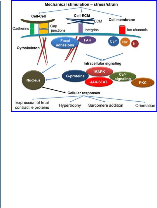
Mechanotransduction (biochemical response of cells to mechanical stimulation) is a very fundamental process in a living cell. The ability of cells to convert mechanical forces into biochemical regulatory information is crucial for normal physiology and pathological processes in many tissues. Mechanotransduction is mediated by various cellular and extracellular components, such as ECM components, cell-ECM adhesions (integrins, focal adhesions), cell-cell adhesions (cadherins and gap junctions), membrane components (ion channels, surface receptors), cytoskeletal components (microfilaments, microtubules), and, finally, intracellular and/or nuclear components (various kinases and transcription factors) that affect chromatin and gene expression [14, 15].
Cells of the myocardium are at home in one of the most mechanically dynamic environments in the body. Heart chamber filling and wall distention within diastole account for rapid changes in pressure and volume that are released by the wave of contraction that pumps blood through the body. At the cellular level, these pulsatile stimuli are experienced as cyclic strains (relative deformation) and stresses (force per unit area). Myocytes are stretched as the heart fills in diastole (preload) and actively contract against imposed load (afterload) in systole. Thus, mechanotransduction in cardiomyocytes occurs due to external tensile forces (outside in) and forces generated in cytoskeletons (inside out), including contractile forces that are produced through the sliding of actin and myosin filaments in sarcomeres [16, 17].
Cardiomyocytes employ numerous mechanisms to sense external forces and translate them into biochemical signals (Fig. 7.2). One of the most studied sensing/signaling pathways is ECM- integrin-cytoskeleton pathway. Binding of integrins on cell surface to ECM proteins promote assembly of “focal adhesion” anchoring complexes (though the activation of focal adhesion kinase and other signaling molecules). Focal adhesions play a central role in mechanotransduction. Force-dependent changes in shape and conformation of a subset of the load-bearing molecules in these adhesion plaques alter their biochemical activities, and thereby result in stress-dependent remodeling of the focal adhesion as well as associated changes in downstream signal transduction. Another mechanism of mechanical sensing is mediated by stretch-activated channels on the cell surface. These ion channels selectively regulate the permeability of the cell membrane to cations (Na+, K+, and Ca2+) in response to strain, that, in turn, affects signaling and physiological response [16, 18, 19]. Different mechano-sensing mechanisms lead to activation of intracellular signaling pathways, most prominently cascades of G-proteins, mitogen-activated protein kinases (MAPK, including extracellularregulated kinase (ERK), c-Jun N-terminal kinase ( JNK), and the p38 MAP kinase), Janus-associated kinase/signal transducers and activators of transcription ( JAK/STAT), protein kinase C (PKC), calcineurin, and intracellular calcium regulation (Fig. 7.2) [16, 19]. Multiple levels of cross-talk exist between these pathways.
94 7. PERFUSION BIOREACTORS AND STIMULATION PATTERNS
Figure 7.2: Major mechanotransduction signaling elements and responses. Stress and strain are sensed through various mechanisms, including cell-cell adhesions, cell-ECM interactions, and stretch-activated ion channels.This stimulation results in a series of intracellular events, including focal adhesion formation, cytoskeletal rearrangement and activation of various signaling cascades. These signals are translated to downstream responses, which culminates in changes in gene expression. The results of these changes include activation of fetal gene program, hypertrophy, sarcomere addition, and change in orientation and alignment. See text for more details. FAK, focal adhesion kinase; MAPK, mitogen-activated kinase; JAK/STAT, Janus-associated kinase/signal transducers, and activators of transcription; PKC, protein kinase C.
Cellular responses, that result from mechanical stimulation, are numerous and complex, and differ in kinetics and magnitude. Various parameters of stimulation, such as frequency, force magnitude and direction, are important factors that affect these responses, which are translated eventually to the physiological or pathological conditions. Mechanical stresses in the myocardium can be elevated by tissue damage following MI, excessive load (such as hypertension), or intrinsic defects, such as mutations in genes encoding contractile proteins. Cardiomyocytes respond to elevated loading conditions, in an attempt to adapt to new mechanical demands and restore wall stress to normal

7.3. INDUCTIVE STIMULATION PATTERNS IN CARDIAC TISSUE ENGINEERING 95
levels, with an increase in cell size, associated with transient and persistent molecular changes, a process generally referred as cellular hypertrophy. Changes in gene expression lead to an increased rate of contractile protein synthesis and cell mass. The long-term changes in gene expression also include the transition of some sarcomeric proteins to their fetal forms. Increase in protein mass lead to in-series or parallel addition of sarcomeres, depending on mechanical stress and overload nature (pressure or volume). Apart from changes in contractile protein mass, mechanical loading can also affect cytoskeletal or sarcomeric organization to regulate cell shape, alignment, and orientation. In pathological conditions, such as MI, hypertrophic response is initially beneficial, as it partially compensates for the decreased force generation and normalized wall stress. However, in the long term the hypertrophic response leads to cardiac failure. Local mechanics destabilization, altered calcium dynamics, structural disarray, and fibrosis are among the major factors that contribute to pathological remodeling and cardiac imbalance [17, 19].
The described processes and biomechanical dynamics are important in maturation and organization of native cardiac tissue. Thus, the ability of various engineering efforts to recreate those signals in vitro is critical for the formation of functional cardiac patches. As cardiac tissue is an excellent example of a tissue that constantly senses mechanostimulation in vivo, it may respond to a similar stimulation also in vitro.
The effect of mechanical stimulation on cells has been broadly investigated mainly in 2D cultivation systems. In 3D cell cultures, the homogenous distribution of these mechanical stimuli represents another challenge for bioreactor engineers.The most common examples for application of mechanical stimulation are bioreactors, developed to apply mechanical forces via piston/compression systems, substrate bending, hydrodynamic compression, and fluid shear [13, 20, 21, 22].
Zimmermann’s group pioneered the application of mechanical stimulation to induce cardiac tissue engineering [2]. They applied a phasic mechanical stretch on previously casted circular molds, comprised of cardiac myocytes from neonatal rats, mixed with collagen I and matrix factors. In addition to the cyclic mechanical stretch, the medium in the culture system was constantly mixed to improve nutrient transfer, resulting in enhanced cell metabolic activity. The ring-shaped engineered heart tissue (EHT) developed by this approach displayed the important hallmarks of differentiated cardiac muscle tissue, i.e., striated myofibers and contractility.
Akhyari et al subjected gelatin-based cell constructs, seeded with human pediatric heart cells (non-contractile cardiomyocyte-like cells, isolated from ventricular tissue removed during surgical repair of Tetralogy of Fallot in children) to a cyclic stretch. They showed that this stimulation increased cell proliferation and distribution in the construct. In addition, collagen matrix formation and organization were enhanced by this stretch, as well as maximal tensile strength and resistance to stretch [1].
Mol and colleagues, when engineering a heart valve, in vitro, utilized a different stimulation system [23]. The stimulation pattern mimicked the diastolic phase of the cardiac cycle as well as the opening and closing behavior of the leaflet. A bioreactor system, developed for this purpose, applied a dynamic pressure difference over the tissue-engineered valve, and in doing so induced dynamic

96 7. PERFUSION BIOREACTORS AND STIMULATION PATTERNS
strains within the leaflets. Exposure of the heart valve construct to this strain regime has led to the development of a tissue-engineered aortic human heart valve for replacement.
Nowadays, a leading approach in mechanostimulation of the cardiac constructs is via stimulation during construct cultivation within a perfusion bioreactor.The advantages of such a bioreactor as a mechanostimulator are obvious; the interstitial medium flow within the porous cell construct also creates a frictional force on the surface of the cells, termed shear stress. Mammalian cells respond to fluid shear stress in different ways, for example by enhancing proliferation or changing morphology and organization.
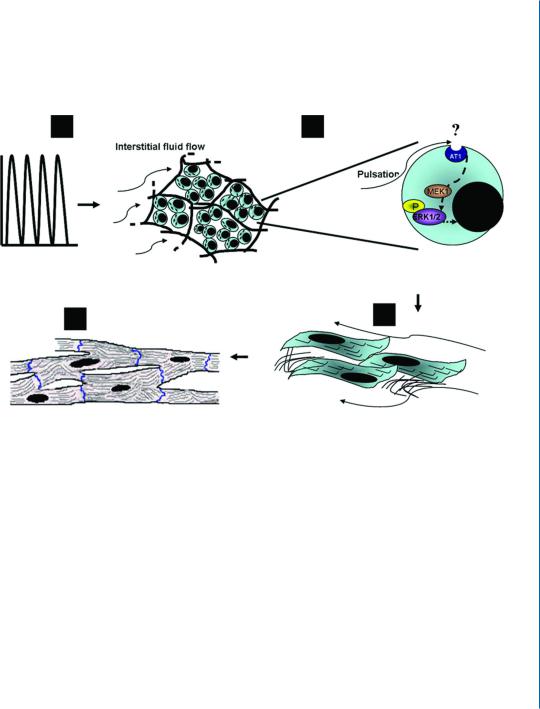
Our group has used the perfusion bioreactor described in Section 7.2.3 to investigate the effect of pulsatile, interstitial fluid flow stimulation on cardiac tissue engineering.The various cellular events taking place in the cell construct, from the cell signaling levels up to the ultrastructural phenotype of the regenerated cardiac muscle tissue, were elucidated in this study [13]. When the bioreactor was operated at a shear stress of 0.6 dynes/cm2, activation of the extracellular signal-regulated kinase (ERK) 1/2 signal transduction pathway was pronounced, followed by enhanced synthesis of the contractile proteins (Troponin T and sarcomeric α-actinin) and cell-cell interaction proteins (Cx-43 and N-cadherin). More importantly it was shown that long-term cultivation under the pulsatile flow regime like this promoted organization of thick, highly organized cardiac tissue, resembling the native heart. A model describing the proposed mechanism, translating mechanical cues to tissue regeneration, is presented in Fig. 7.3.
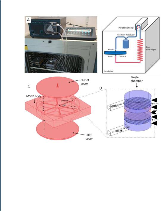
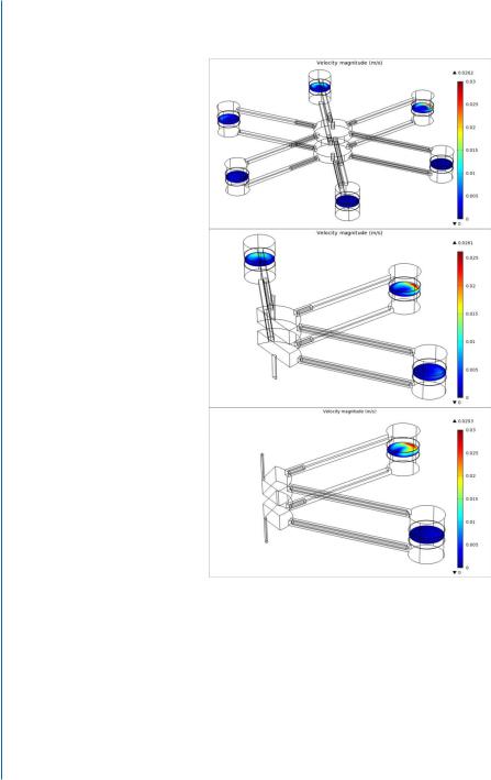
In a recent study, we described the micro-fabrication of a Multi-Shear Perfusion Bioreactor (MSPB), designed for cultivation of 3D cell constructs under differential fluid shear rates (Fig. 7.4) [24].
The MSPB device enables a simultaneous investigation of the effect of up to six different shear rates on the cell constructs. The multi-shear operation features of the bioreactor were modeled and then experimentally validated by measuring the effect of different shear rates on the behavior of human umbilical vein endothelial cells (HUVEC) seeded in macro-porous alginate scaffolds. Exposure of the constructs to three different levels of shear stress for 24 h resulted in constructs expressing three different levels of the membranal marker Intercellular Adhesion Molecule 1 (ICAM-1) and the phosphorylated endothelial nitric oxide synthetase (eNOS), markers known to be directly affected by shear stress. A longer period of cultivation, 17 days, under two different levels of shear stress, resulted in different lengths of cell sprouts within the constructs. Collectively, the HUVEC behavior within the different constructs confirms the feasibility of using the MSPB system for simultaneously imposing different shear stress levels, and for validating the flow regime in the bioreactor vessel as assessed by the computational fluid dynamic (CFD) model [24].
7.3. INDUCTIVE STIMULATION PATTERNS IN CARDIAC TISSUE ENGINEERING 97
$ |
% |
' |
& |
Figure 7.3: A proposed model describing the mechanistic effects of pulsatile interstitial fluid flow/shear stress on cardiac tissue regeneration. A. A pulsatile interstitial fluid flow, provided by the perfusion bioreactor subjects the scaffold-seeded cardiac cells to a physiological shear stress of 0.6 dynes/cm2. B. The applied shear stress activates the mechanoreceptor, AT1, thus initiating the ERK1/2 cascade. In the presence of the MEK inhibitor, U0126, the pulsatile flow cannot activate ERK1/2. C. Activation of ERK1/2 cascade signals for cardiac cell survival and hypertrophy, as revealed by the enhanced synthesis of contractile cardiac proteins and proteins associated with cell-cell contacts and gap junction responsible for electrical coupling. D. With the secretion of collagen and its arrangement in fibers around the cardiac myofibers, the cardiac tissue reaches full maturity [13].
98 7. PERFUSION BIOREACTORS AND STIMULATION PATTERNS
$
D |
E |
|
|
|
G |
F |
|
|
|
|
|
|
|
Figure 7.4: Multi-Shear Perfusion Bioreactor (MSPB) is designed for cultivation of 3D cell constructs under differential fluid shear rates. A. The MSPB design: (a) A picture of the MSPB system placed in the incubator; (b) The system and its flow circuit; (c) The bioreactor vessel has six chambers, located 30 mm from the center and connected via 22-mm long medium-leading channels to a medium flow splitter at the vessel center. The vessel is sealed by pressing top and bottom covers made of PDMS layer;
(d) The chamber dimensions are 8.2-mm height and 6.1-mm diameter and thinner where the construct is held (5.9 mm). The cellular construct (red, indicated by a chevron) and the two flow-distributing nets (indicated by dashed arrows) placed 1 mm upstream and downstream to the construct, are held in place by porous plastic discs (blue, indicated by arrow heads). The direction of the medium flow is upward. Reprinted with permission from [24].
7.3. INDUCTIVE STIMULATION PATTERNS IN CARDIAC TISSUE ENGINEERING 99
%
D |
|
E |
F |
|
|
|
|
G |
|
|
|
|
|
|
|
I |
|
|
H |
|||
|
|
|
|
|
|
|
|
|
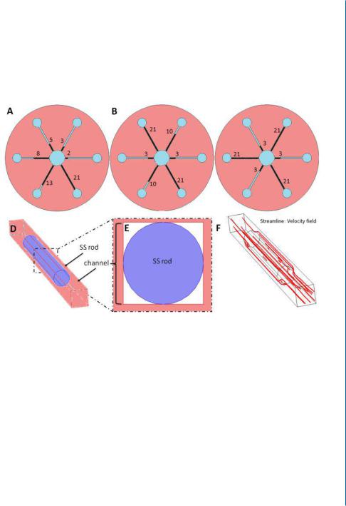
Figure 7.4: Multi-Shear Perfusion Bioreactor (MSPB) is designed for cultivation of 3D cell constructs under differential fluid shear rates. B. Illustration of the rods arrangement in the MSPB upper plane (as viewed from above) of three configurations of differential shear stress rates: The six-shear configuration
(a) are utilized by inserting 2, 3, 5, 8, 13, and 21 mm long rods. Three shear configurations (b) are utilized by inserting 3, 10, and 21 mm long rods.Two shear configurations (c) are utilized by inserting 3 and 21 mm long rods. The exact same arrangement of rods was set on the other side of the bioreactor. Insertion of the SS rod to the channel blocks most of its cross-sectional area for a medium flow (d, e), forcing the medium to flow around it as demonstrated by the red streamlines (f ). Reprinted with permission from [24].
100 7. PERFUSION BIOREACTORS AND STIMULATION PATTERNS
& D
E
F
Figure 7.4: Multi-Shear Perfusion Bioreactor (MSPB) is designed for cultivation of 3D cell constructs under differential fluid shear rates. C. Computational model of fluid velocity in the bioreactor operated in: (a) six-shear manner by placing 6 different lengths of SS rods (2, 3, 5, 8, 13, and 21 mm) in the channels; (b) 3-shear manner by placing three different lengths of SS rods (3, 10, and 21 mm) in the channels; (c) 2-shear manner by placing two different lengths of SS rods (3 and 21 mm) in the channels. Reprinted with permission from [24].