


120 9. ACELLULAR BIOMATERIALS FOR CARDIAC REPAIR
to the target tissue, including the heart. However, several technical issues are still to be resolved, such as the need for effective decellularization protocols, possible immunogenicity, preservation/storage, etc.
9.4INJECTABLE BIOMATERIALS
9.4.1INJECTABLE HYDROGELS BASED ON NATURAL OR SYNTHETIC POLYMERS
Various natural or synthetic polymers can form hydrogels, namely, solid networks of physically or chemically cross-linked polymer chains with varying water content, which can be directly injected into the infarcted heart. Such injectable hydrogels can recapitulate the microenvironment for inducing effective tissue repair, by replacing damaged ECM, providing temporary support for the infarct, and instructing tissue restoration.These effects could prevent the adverse remodeling and progression of heart failure. The application of injectable biopolymers is less invasive than implanting macroporous scaffolds, and is, therefore, more clinically appealing. Indeed, various natural or synthetic acellular injectable biomaterials have been found to improve the outcome after MI [16, 17, 18].
Christman et al pioneered the field of injectable biomaterials by exploring the effects of fibrin glue as a treatment strategy for MI. Fibrin forms a crosslinked 3D hydrogel in the myocardium upon injection with a dual-barreled syringe. One barrel contains fibrinogen and aprotinin (a fibrinolysis inhibitor), and the second barrel contains thrombin, factor XIIIa, and CaCl2. Following a similar mechanism to that involved in the normal clotting cascade in vivo, when fibrinogen and thrombin are mixed, fibrinogen is converted to fibrin which self-assembles and is crosslinked via the factor XIIIa. Fibrin hydrogel injection, in the ischemia/reperfusion (I/R) model, preserved cardiac function and scar thickness, five weeks after injection [19]. In another study, fibrin injection was associated with reduced infarct size, increased angiogenesis (shown by the increase in microvessel density), compared to BSA injection [20]
Utilizing a large animal swine model, Mukherjee et al injected composite hydrogels containing both fibrin and alginate to prevent geometric LV remodeling. One week post-MI, 200 μL each of the 25 injections were applied to the infarct area via a double-barreled injection device; one component was comprised of fibrinogen, fibronectin, factor XIII, plasminogen, and gelatin-grafted alginate dissolved in an aprotinin solution, while the second consisted of the cross-linking agents, thrombin and CaCl2. The therapeutic outcomes of this treatment included increased posterior wall thickness 1 week postinjection and a reduction in infarct expansion 21 and 28 days post-MI; however, no functional improvements were observed [21]
Yu et al investigated and compared the therapeutic effects of fibrin and alginate in a rodent model of chronic ischemic cardiomyopathy. The researchers found that both polymers can augment left ventricular wall thickness, resulting in reconstruction of left ventricular geometry and improvement of cardiac function. Echocardiography results at five weeks after injection of alginate demonstrated persistent improvement of left ventricular fractional shortening (FS) and prevention of a continued enlargement of left ventricular dimensions, whereas fibrin glue demonstrated no

9.4. INJECTABLE BIOMATERIALS 121
progression of left ventricular negative remodeling. There was increased arteriogenesis in both the alginate and fibrin glue groups compared with that seen in the phosphate-buffered saline control group. Infarct size was significantly reduced in the fibrin group, and there was a trend toward smaller infarcts in the alginate group.
Chitosan is a linear polysaccharide that is biocompatible and biodegradable and therefore has been used in a wide variety of tissue engineering applications. Chitosan hydrogels can be formed upon mixing of commercially produced chitosan with a glycerol phosphate and glyoxal solution. These gels exhibit a thermoresponsive gelation that is tuned to occur at 37◦C by changing the glyoxal concentration, while hydrogel degradation is controlled by the degree of deacetylation [18]. In a rat infarct model, a thermally responsive chitosan was injected one week post-MI [22]. Four weeks after hydrogel injection, the myocardium thickness was significantly increased compared to PBS controls, even though the amount of chitosan presented in the myocardium after four weeks had substantially decreased due to hydrogel degradation. There were also significant improvements in infarct size, FS, EF, end systolic diameter (ESD), end diastolic diameter (EDD), and microvessel density.
Collagen is a natural ECM protein that has been applied for LV remodeling therapies due to the ability to inject as a liquid, which subsequently gels at 37◦C [18]. Collagen injections (95% type I, 5% type III) in one week-old rat infarcts substantially increased infarct thickness, stroke volume (SV), and EF compared to saline injection controls; there was also a trend for smaller end systolic volume (ESV) and larger end-diastolic volumes (EDV) in biomaterial-treated animals [23].
Hyaluronan (HA) is a natural un-sulfated polysaccharide that is abundant in the body and plays a major role in several biological processes that include angiogenesis cell migration, and it is also a lubricant material. In most, if not all of the tissue engineering related strategies, only modified HA has been used; the modifications used to improve the material mechanical strength and adding functional groups for inducing photopolymerization leading to scaffold formation [18]. A study by Ifkovits et al tested therapeutics effects of an injectable hydrogel made from modified HA (methacrylated HA (MeHA)) with a high (43 kPa) and low (7.7 kPa) compression moduli in an ovine model of MI [24]. Treatment with both hydrogels significantly increased the wall thickness in the apex and basilar infarct regions compared with the control infarct. However, only the highermodulus (MeHA High) treatment group had a statistically smaller infarct area compared with the control infarct group. Moreover, reductions in normalized end-diastolic and end-systolic volumes were observed for the MeHA High group.This group also tended to have better functional outcomes (cardiac output and EF) than the low-modulus (MeHA Low) and control infarct groups. This study emphasizes the importance of the rational material design and mechanical properties on therapeutic outcome post-MI.
Synthetic materials provide additional potential in engineering a variety of gelation mechanisms and physical properties. One synthetic thermosensitive polymer, comprised of dextran (Dex) grafted poly(caprolactone)-2-hydroxyethyl methacrylate (PCL-HEMA) and copolymerized with poly(N-isopropylacrylamide) (PNIPAAm) termed Dex-PCL-HEMA/PNIPAAm, was developed

122 9. ACELLULAR BIOMATERIALS FOR CARDIAC REPAIR
to gel in situ. MI was induced in rabbits by coronary artery ligation; four days later, 200 μL Dex- PCL-HEMA/PNIPAAm of gel solution was injected into the infarcted myocardium. Injection of phosphate-buffered saline served as control. Thirty days after treatment, histological analysis indicated that injection of the biomaterial prevented scar expansion and wall thinning compared with controls. Echocardiography studies showed that injection of hydrogel increased left ventricular ejection fraction and attenuated left ventricular systolic and diastolic dilatation. Haemodynamic analysis demonstrated improved cardiac function following implantation of the hydrogel [25]
9.4.2INJECTABLE DECELLULARIZED ECM MATRICES
Decellularized ventricular and pericardial ECM can be processed to create a solubilized liquid with the ability to gel via self-assembly at physiological temperature both in vitro and in vivo upon injection into myocardial tissue [26, 27]. For this purpose, ventricular or pericardial tissue was harvested and decellularized using sodium dodecyl sulfate detergent. The decellularized matrix was then lyophilized and milled to create a fine powder, which was then solubilized using enzymatic digestion to create a liquid matrix for catheter-based delivery. The decellularized ventricular ECM biochemical composition was shown to retain the complexity of proteins, peptides, and glycosaminoglycans of natural ECM. Initial in vivo feasibility testing showed that the solubilized myocardial matrix undergoes gelation in healthy rat myocardial tissue upon direct epicardial injection. The in situ-formed hydrogel had a pore size of 30 μm and its nano-fibrillar structure resembled that of the intact decellularized tissue, prior to processing. The myocardial decellularized matrix was shown to promote the migration of human coronary artery endothelial cells and rat aortic smooth muscle cells in vitro as well as to promote the infiltration of vascular cells and the formation of arterioles in vivo [27]. The researchers also created pig and human pericardium-derived decellularized ECM injectable matrices by a similar procedure. The matrices induced neovascularization and promoted cell mobilization, suggesting that this type of constructs can represent an injectable scaffold for cardiac tissue engineering. Moreover, as the pericardium can be surgically resected by minimally invasive thoracoscopic pericardiectomy, this tissue layer represent a potentially autologous source of scaffold material, while sacrificing the properties of ECM derived from myocardium [26].
In a recent study, using a similar fabrication procedure, Syngelin et al prepared an injectable myocardial hydrogel from decellularized porcine ventricular ECM [28]. Rats underwent ischemia/reperfusion followed by intramyocardial injection of the hydrogel or saline two weeks later. Immunohistochemical assessment showed an increase in the size of cardiomyocyte islands surviving in the infarct area of the hydrogel-treated animals, suggesting that the matrix injection may act to salvage remaining cardiomyocytes in the infarct. The injection of myocardial matrix hydrogel was also associated with preserved cardiac function, as shown by preserved values of EF, end-diastolic and systolic volumes (evaluated by MRI). The authors also assessed arrhythmia inducibility using programmed electrical stimulation in vivo by burst and extra stimulus pacing protocols in rats one week post-injection of myocardial matrix compared to saline. In vivo pacing protocols did not show statistical difference when comparing the average incidence of ventricular tachycardia between
9.5. FIRST-IN-MANTRIAL OF INTRACORONARY DELIVERY OF ALGINATE BIOMATERIAL 123
hydrogel and saline-treated animals. Finally, the clinical feasibility of the myocardial hydrogel injection in healthy and infarcted pig hearts was shown by using minimally invasive percutaneous transendocardial catheter, under the guidance of unipolar electromechanical mapping (NOGA). A biotinylated matrix was delivered into 15 injection sites in the infarct and border zones, with no signs of pericardial effusion. Biotinylated matrix staining confirmed the presence of the material in the myocardial wall, as well as gelation of the matrix in vivo; the presence of the matrix was not observed in the satellite organs, confirming the lack of leakage of the material into the ventricle [28]. Collectively, this study demonstrates the feasibility and efficacy of decellularized ventricular ECM-derived in situ-forming hydrogel for the treatment of MI. These promising results await confirmation in a large animal model.
9.5FIRST-IN-MAN TRIAL OF INTRACORONARY DELIVERY OF ALGINATE BIOMATERIAL
Our group developed an injectable alginate biomaterial which can be delivered by intracoronary injection as a solution. At the infarct, due to the high calcium ion concentration immediately after acute MI, the solution undergoes gelation forming a hydrogel [29, 30].
The injectable solution is a partially cross-linked alginate network, prepared by mixing a 1% (w/v) solution of 30-50 kDa sodium alginate (having high G to M ratio), and 0.3% (w/v) D- gluconic acid/hemicalcium salt, and is capable of flowing due to its relatively low apparent viscosity ( 10cP) [31]. The unique mechanical properties of the partially cross-linked alginate solution were exemplified by rheology, the mechanical spectra revealing that the storage (G’) and loss (G”) moduli of the solution are closely related or sharing a cross-point. This type of physical behavior usually characterizes cross-linked material in the verge of phase transition from its liquid state into a hydrogel, and such transition can occur by increasing local cation concentration [31].
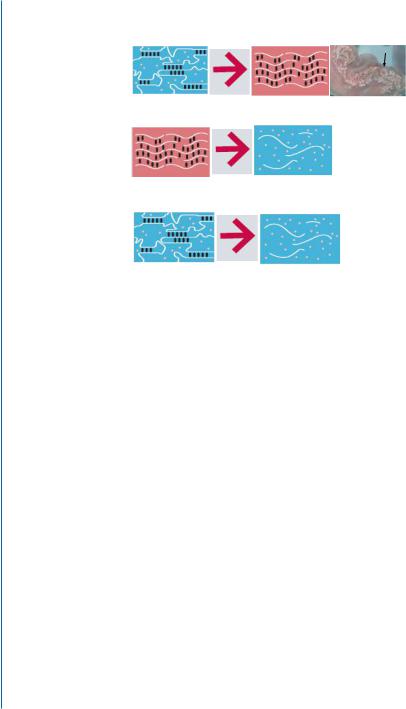
At the acute infarct, the partially cross-linked alginate solution undergoes a rapid gelation and phase transition into a hydrogel due to the additional cross-linking, in response to the elevated concentrations of calcium ions at the infarct after MI and due to water diffusion from the injectable solution to the surrounding tissue (Fig. 9.1A) [29, 30, 31]. The alginate hydrogel degrades and disappears from the infarct zone with time; 6 weeks after administration into the infarct, only remnants of biotinylated alginate material remained at infarct (Fig. 9.1B) and it was replaced by a host tissue composed of myofibroblasts and enriched with blood capillaries (Fig. 9.1C). The alginate hydrogel dissolution occurs via an exchange reaction between the crosslinking calcium ions by sodium ions from the surrounding tissue; a process occurring with time at the healing infarct due to the reduction in calcium ion concentration (Fig. 9.1A):
2N aAlg + Ca |
2 |
→ |
|
+ ← 2N a+ + Ca(Alg)2 |
The beneficial therapeutic effects of this novel in situ-forming alginate hydrogel on MI repair have been proven in recent and chronic models of MI in rats and in acute model in pigs [29, 30]. In an acute MI model in rats, myocardial injection of the partially cross-linked alginate solution into
124 9. ACELLULAR BIOMATERIALS FOR CARDIAC REPAIR
$
+LJK &DOFLXP ,RQV
/RZ ZDWHU FRQWHQW
'DPDJHG |
|
/LTXLG WR JHO |
7LVVXH |
|
WUDQVLWLRQ |
/LTXLG |
$OJLQDWH JHO |
,Q WLVVXH |
|
/RZ &DOFLXP |
|
+HDOHG
*HO WR OLTXLG DOJLQDWH FKDLQV
7LVVXH GLIIXVH DQG DUH FOHDUHG E\ NLGQH\
$OJLQDWH JHO /LTXLG 1D$OJLQDWH
/RZ &DOFLXP
1R GHSRVLWLRQ 7KH VROXWLRQ
1RUPDO LV GLOXWHG E\ a ORJV LQ
7LVVXH
EORRG DQG LV UHPRYHG
/LTXLG /LTXLG 1D$OJLQDWH
Figure 9.1: Beneficial therapeutic effects of injectable alginate biomaterial on LV remodeling after MI in rats (B-E) and pigs (G-I). A. A schematic model describing the three possible occurrences after intracoronary injection of the partially cross-linked alginate solution. In a damaged tissue after acute MI, the partially cross-linked alginate solution undergoes gelation at the infarct due to elevated calcium ion concentration. During healing, calcium ion concentration decreases in the tissue, leading to hydrogel dissolution via an exchange reaction of calcium ions with sodium ions. The water-soluble alginate molecules can diffuse and be excreted via the urine due to their low molecular weight (less than 50 kDa). In normal healthy tissue, no gelation occurs, and the solution is diluted in systemic circulation and is removed via urine. Reprinted with permission from [29, 30].
the infarct zone resulted in increased scar thickness and improved LV dimensions compared with saline, two months after injection. These results were similar or superior to injection of suspension of neonatal cardiomyocytes (Fig. 9.1C-D).
In a large animal model, we showed that intracoronary injection of the partially cross-linked alginate solution into the infarcted hearts of pigs is feasible and safe. The deposition of alginate biomaterial at the infarct zone (Fig. 9.1E) prevented and even reversed LV enlargement and increased scar thickness by 53% compared with saline, two months after the injection (Fig. 9.1F) [30]. These beneficial effects of the alginate hydrogel were dose-dependent (Fig. 9.1G) and are likely due to temporary replacement of the functions of the damaged ECM, followed by increased cellular infiltration during hydrogel erosion (Fig. 9.1H) [29, 30]. The in situ-forming alginate hydrogel was shown to undergo slow erosion and dissolution over a period of six weeks, during healing of the infarct.
9.5. FIRST-IN-MANTRIAL OF INTRACORONARY DELIVERY OF ALGINATE BIOMATERIAL 125
%
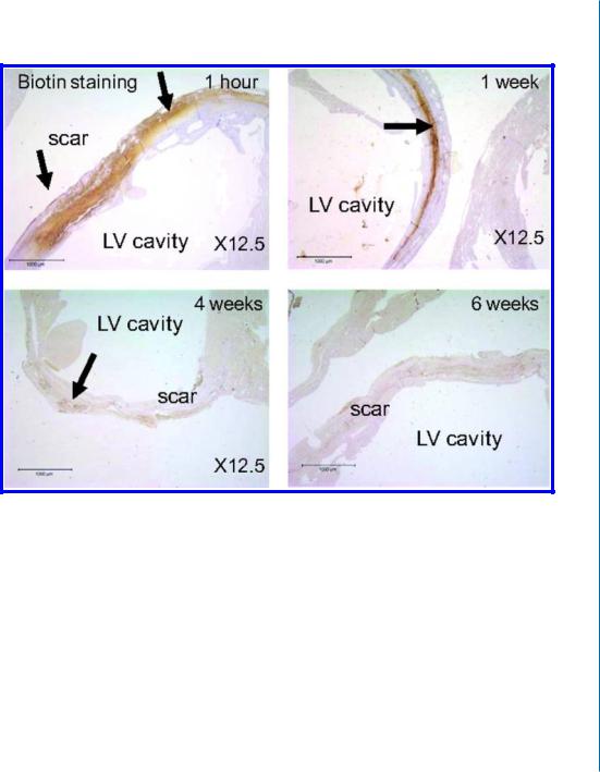
Figure 9.1: Beneficial therapeutic effects of injectable alginate biomaterial on LV remodeling after MI in rats (B-E) and pigs (G-I). B. The deposited alginate hydrogel dissolves and disappears from the infarct within six weeks. Distribution of biotinylated-labelled alginate biomaterial at the infarct after intramyocardial injection of calcium cross-linked alginate solution, in rat. The areas of positive biotin staining, as a percentage of the scar area, were decreased significantly at 1, 4, and 6 weeks after injection. Reprinted with permission from [29, 30].
These encouraging results have led to a first-in-man clinical trial, proving the safety of intracoronary injection of alginate biomaterial in acute MI patients [32]. Due to the localized effect of the biomaterial, it received FDA approval for a regulatory path as a medical device, intended to be injected to patients following acute myocardial infarction, for prevention of ventricular remodeling
126 9. ACELLULAR BIOMATERIALS FOR CARDIAC REPAIR
&
D |
F |
E |
G |
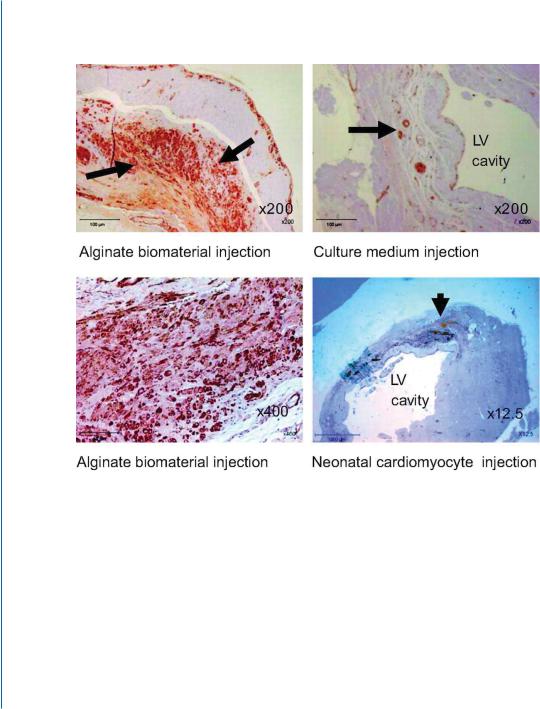
Figure 9.1: Beneficial therapeutic effects of injectable alginate biomaterial on LV remodeling after MI in rats (B-E) and pigs (G-I). C. Representative micrographs of infarcted hearts treated with biomaterial or cardiomyocyte transplantation (immunostaining for α-smooth muscle actin (SMA)). (a) Examination of the scar tissue 8 weeks after alginate biomaterial injection revealed extensive positive brown staining (arrows). (b) Higher magnification showed that the biomaterial-treated scar is populated with numerous SMA-positive cells, probably myofibroblasts, and with small vessels (marked by arrows). The scaffoldtreated scars were thicker than the scars treated with culture medium (c). d, Neonatal cardiac cell implant (arrow) at the border of the infarct zone, 8 weeks after cell suspension injection. Engrafted cells appeared yellow-brown, were undifferentiated, and were isolated from the host myocardium. Reprinted with permission from [29, 30].
9.5. FIRST-IN-MANTRIAL OF INTRACORONARY DELIVERY OF ALGINATE BIOMATERIAL 127
'
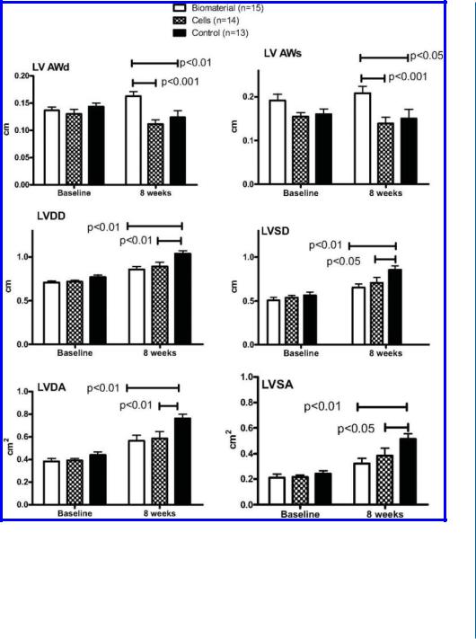
Figure 9.1: Beneficial therapeutic effects of injectable alginate biomaterial on LV remodeling after MI in rats (B-E) and pigs (G-I). D. Comparison of the functional effects of alginate biomaterial vs. cardiomyocytes vs. saline injection into recent (seven-day-old) scars in rats. Individual values and mean (±SE) are shown.The probability values are Bonferroni-adjusted for three comparisons. AWd indicates anterior wall diastolic thickness; AWs, anterior wall systolic thickness; LVDD, LV end-diastolic dimension; LVSD, LV end-systolic dimension; LVDA, LV end-diastolic area; and LVSA, LV end-systolic area. Reprinted with permission from [29, 30].
128 9. ACELLULAR BIOMATERIALS FOR CARDIAC REPAIR
( $OJLQDWH ELRPDWHULDO ZKLWH LQ LQIDUFW K DIWHU LQMHFWLRQ
)
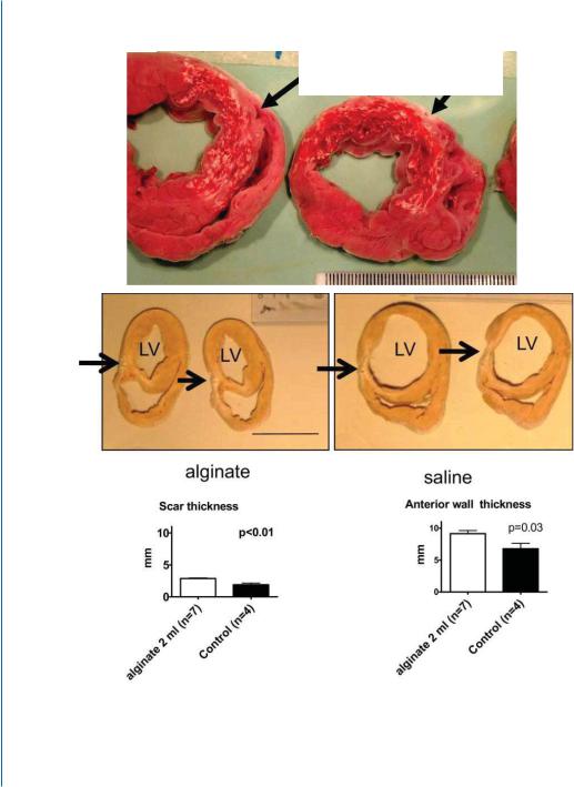
Figure 9.1: Beneficial therapeutic effects of injectable alginate biomaterial on LV remodeling after MI in rats (B-E) and pigs (G-I). E and F. Reprinted with permission from [29, 30]. (Continues. Caption on
the next page.)

9.5. FIRST-IN-MANTRIAL OF INTRACORONARY DELIVERY OF ALGINATE BIOMATERIAL 129
Figure 9.1: (Continued.) E. Macroscopic views of heart sections, 2 hours after intracoronary injection of calcium cross-linked alginate solution, reveals the in situ deposition of the alginate hydrogel (white areas, arrows), nicely distributed in the infarct. F. Post-mortem morphometry at 60 days after MI indicates that the alginate implant increases scar and arterial wall thicknesses as compared to saline treatment. Representative sections of heart treated with alginate (upper left panel) or saline (upper right panel). Morphometry shows that injection of alginate solution (2 ml) increases scar thickness (arrows) as well as anterior wall thickness (lower panels).
* |
+ |
||
|
|
|
|
|
|
|
D |
D |
|
||
|
|
|
|
|
|
|
|
|
E |
|
E |
|
|
|
|
|
Figure 9.1: Reprinted with permission from [29, 30]. (Continues. Caption on the next page.)
and subsequent congestive heart failure. At present, the PRESERVATION I pivotal clinical trial is running for the material, now known as Bioabsorbable Cardiac Matrix (BCM).