


BIBLIOGRAPHY 5
its efficacy for controlled multiple factor delivery and as a treatment for inducing active regeneration in various diseases and MI.
BIBLIOGRAPHY
[1]Langer R, Vacanti JP. Tissue engineering. Science. 1993;260:920–6. 1
[2]Ruvinov E, Dvir T, Leor J, Cohen S. Myocardial repair: from salvage to tissue reconstruction. Expert Rev Cardiovasc Ther. 2008;6:669–86. 1
[3]Rane AA, Christman KL. Biomaterials for the treatment of myocardial infarction a 5-year update. J Am Coll Cardiol. 2011;58:2615–29. DOI: 10.1586/14779072.6.5.669 1
[4]Vunjak-Novakovic G, Lui KO,Tandon N, Chien KR. Bioengineering heart muscle: a paradigm for regenerative medicine. Annu Rev Biomed Eng. 2011;13:245–67.
DOI: 10.1146/annurev-bioeng-071910-124701 1, 2
[5]Nunes SS, Song H, Chiang CK, Radisic M. Stem Cell-Based Cardiac Tissue Engineering. J Cardiovasc Transl Res. 2011. DOI: 10.1007/s12265-011-9307-x 1
[6]Martinez EC, Kofidis T. Adult stem cells for cardiac tissue engineering. J Mol Cell Cardiol. 2011;50:312–9. DOI: 10.1016/j.yjmcc.2010.08.009 1
[7]Segers VF, Lee RT. Biomaterials to enhance stem cell function in the heart. Circ Res. 2011;109:910–22. DOI: 10.1161/CIRCRESAHA.111.249052 1
[8]Ye KY, Black LD, 3rd. Strategies for Tissue Engineering Cardiac Constructs to Affect Functional Repair Following Myocardial Infarction. J Cardiovasc Transl Res. 2011.
DOI: 10.1007/s12265-011-9303-1 1
[9]Tous E, Purcell B, Ifkovits JL, Burdick JA. Injectable acellular hydrogels for cardiac repair. J Cardiovasc Transl Res. 2011;4:528–42. DOI: 10.1007/s12265-011-9291-1 1
[10]Singelyn JM, Christman KL. Injectable materials for the treatment of myocardial infarction and heart failure: the promise of decellularized matrices. J Cardiovasc Transl Res. 2010;3:478– 86. DOI: 10.1016/j.jacc.2011.10.888 1
[11]Segers VF, Lee RT. Local delivery of proteins and the use of self-assembling peptides. Drug discovery today. 2007;12:561–8. DOI: 10.1016/j.drudis.2007.05.003 2
[12]Ruvinov E, Harel-Adar T, Cohen S. Bioengineering the infarcted heart by applying bioinspired materials. J Cardiovasc Transl Res. 2011;4:559–74.
DOI: 10.1007/s12265-011-9288-9 2

6BIBLIOGRAPHY
[13]Burridge PW, Keller G, Gold JD, Wu JC. Production of de novo cardiomyocytes: human pluripotent stem cell differentiation and direct reprogramming. Cell Stem Cell. 2012;10:16–
28.DOI: 10.1016/j.stem.2011.12.013 2
[14]Radisic M, Marsano A, Maidhof R, Wang Y, Vunjak-Novakovic G. Cardiac tissue engineering using perfusion bioreactor systems. Nat Protoc. 2008;3:719–38. DOI: 10.1038/nprot.2008.40
[15]Shachar M, Cohen S. Cardiac tissue engineering, Ex-vivo: Design principles in biomaterials and bioreactors. Heart Fail Rev. 2003;8:271–6. DOI: 10.1023/A:1024729919743 2
[16]Lutolf MP, Gilbert PM, Blau HM. Designing materials to direct stem-cell fate. Nature. 2009;462:433–41. DOI: 10.1038/nature08602 2
[17]Ma PX. Biomimetic materials for tissue engineering. Adv Drug Deliv Rev. 2008;60:184–98. DOI: 10.1016/S0142-9612(03)00339-9 2
[18]Davis ME,Hsieh PC,Grodzinsky AJ,Lee RT.Custom design of the cardiac microenvironment with biomaterials. Circ Res. 2005;97:8–15. DOI: 10.1161/01.RES.0000173376.39447.01 2
[19]Zhang B, Xiao Y, Hsieh A, Thavandiran N, Radisic M. Microand nanotechnology in cardiovascular tissue engineering. Nanotechnology. 2011;22:494003.
DOI: 10.1088/0957-4484/22/49/494003 2
[20]Dvir T, Timko BP, Kohane DS, Langer R. Nanotechnological strategies for engineering complex tissues. Nature nanotechnology. 2011;6:13–22. DOI: 10.1038/nnano.2010.246 2
[21]Kelleher CM, Vacanti JP. Engineering extracellular matrix through nanotechnology. J R Soc Interface. 2010;7 Suppl 6:S717–29. DOI: 10.1098/rsif.2010.0345.focus 2
[22]Vunjak-Novakovic G, Tandon N, Godier A, Maidhof R, Marsano A, Martens TP, et al. Challenges in cardiac tissue engineering. Tissue Eng Part B Rev. 2010;16:169–87.
DOI: 10.1089/ten.teb.2009.0352 2

7
C H A P T E R 2
The Heart—Structure,
Cardiovascular Diseases, and
Regeneration
CHAPTER SUMMARY
A major task of cardiac tissue engineering is to provide functional myocardial tissues for replacement of a damaged myocardium. The engineered tissue features have to mimic the natural myocardial tissue to ensure its successful integration, regeneration, and functional recovery in a reasonable amount of time. This chapter presents a detailed description of the heart and its muscle structure to reveal the complexity with which the cardiac tissue engineer has to cope in the process. Then, two major pathophysiological conditions of the heart, myocardial infarction and congenital heart defects, are introduced, for which cardiac tissue engineering potentially can be an ideal solution. Finally, we discuss the regeneration capability of the adult heart and introduce the targets and possible therapeutic strategies and interventions aimed at inducing myocardial regeneration.
2.1INTRODUCTION
The heart is a central organ in the circulatory system, and is indispensable for normal organism homeostasis by providing a constant supply of blood to tissues, which carries oxygen and nutrients and removes carbon dioxide and waste products. The heart pumps blood through blood vessels, and to accomplish this, the heart beats about 100,000 times every day, which adds up to 35 million beats in a year and about 2.5 billion times in an average lifetime, pumping 5 liters of blood each minute. The unique heart structure ensures a continuous blood supply, at rest and in various stressful conditions. Due to the central function of the heart in sustaining life and normal homeostasis of the body, the diseases of the heart are a major concern in public health. The insufficient ability of the heart to self-regenerate after damage, such as after myocardial infarction, results in a progressive deterioration in heart function. During the course of this process, the heart undergoes significant structural and biochemical changes, leading to reduced contractility and heart failure. There is no cure for myocardial infarction, and the goal of cardiac tissue engineering is to provide solutions for this devastating disease.
82. THE HEART—STRUCTURE, CARDIOVASCULAR DISEASES, AND REGENERATION
2.2THE HEART AND CARDIAC MUSCLE STRUCTURE
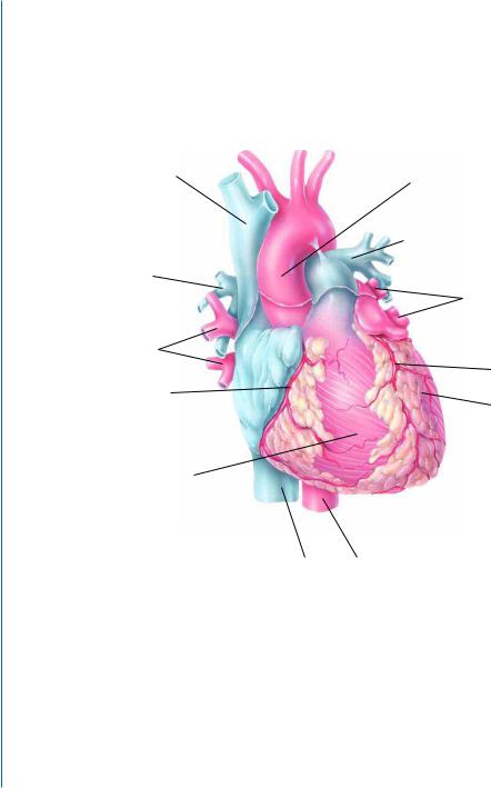
The heart has four chambers (Fig. 2.1A-B). The two superior receiving chambers are the atria, and the two inferior pumping chambers are the ventricles. The left ventricle of the heart pumps oxygenated blood into the systemic circulation to all tissues of the body except the air sacs (alveoli) of the lungs. The right ventricle of the heart pumps deoxygenated blood into the pulmonary circulation to the alveoli of the lungs.
D 6XSHULRU YHQD FDYD |
$VFHQGLQJ DRUWD |
|
|
|
/HIW SXOPRQDU\ DUWHU\ |
5LJKW |
|
SXOPRQDU\ |
/HIW SXOPRQDU\ |
DUWHU\ |
|
|
YHLQV |
5LJKW |
|
SXOPRQDU\ |
/HIW FRURQDU\ |
YHLQV |
|
5LJKW FRURQDU\ |
DUWHU\ |
DUWHU\ |
/HIW YHQWULFOH |
5LJKW DWULXP
5LJKW YHQWULFOH
,QIHULRU YHQD 'HVFHQGLQJ FDYD DRUWD
Figure 2.1: Heart and cardiac muscle structure. A. Anterior external view of the heart showing major surface features. Reprinted with permission of John Wiley & Sons, Inc. [1].
The wall of the heart consists of three layers: the epicardium (external layer), the myocardium (middle layer), and the endocardium (inner layer). Epicardium, the thin, transparent outer layer of the heart wall, is composed of mesothelium and delicate connective tissue that imparts a smooth, slippery texture to the outermost surface of the heart. The middle myocardium, which is the cardiac muscle tissue, constitutes about 95% of the heart mass and is responsible for its pumping action. The

2.2. THE HEART AND CARDIAC MUSCLE STRUCTURE 9
Figure 2.1: Heart and cardiac muscle structure. B. Major internal features of the heart. Blood vessels that carry oxygenated blood are colored red, whereas those that carry deoxygenated blood are colored blue. Reprinted with permission of John Wiley & Sons, Inc. [1].
cardiac muscle fibers swirl diagonally around the heart in bundles. The innermost endocardium is a thin layer of endothelium overlying a thin layer of connective tissue. It provides a smooth lining for the chambers of the heart and covers the valves of the heart. The endocardium is continuous with the endothelial lining of the large blood vessels attached to the heart, and it minimizes surface friction as blood passes through the heart and blood vessels.
The human myocardium consists of 2 to 3 billion cardiomyocytes (75% by volume, 30% by number), the striated muscle cells found only in the heart that can be distinguished from the skeletal and smooth muscle cells. Apart from muscle cells, the heart tissue is mainly composed of fibroblasts (about two thirds in terms of numbers) and endothelial cells. Unlike skeletal muscle cells, cardiomyocytes are controlled by the autonomic (involuntary) rather than the somatic (voluntary) nervous system. Furthermore, cardiomyocytes can generate their own excitatory impulses, functioning as a biological pacemaker.
The myocardium assumes a unique structure, enabling it to synchronously contract (Fig. 2.1C- D). Compared with skeletal muscle fibers, the cardiac muscle fibers are shorter in length and less

10 2. THE HEART—STRUCTURE, CARDIOVASCULAR DISEASES, AND REGENERATION
F
,QWHUFDODWHG GLVFV
2SHQLQJ RI WUDQVYHUVH WXEXOH
'HVPRVRPHV
*DS MXQFWLRQV
0LWRFKRQGULRQ
&DUGLDF PXVFOH ILEHU
1XFOHXV
6DUFROHPPD
Figure 2.1: Heart and cardiac muscle structure. C. Overall organization of cardiac muscle fibers. Sarcolemma is plasma membrane of a muscle cell. Sarcoplasmic reticulum (SR) is membranous sacs encircling each myofibril. In a relaxed muscle fiber, the sarcoplasmic reticulum stores calcium ions. Release of Ca2+ from the terminal cisterns of the SR triggers muscle contraction. Reprinted with permission of John Wiley & Sons, Inc. [1].
circular in the transverse section. They also exhibit branching, which gives the individual cardiac muscle fibers a “stair-step” appearance. A typical cardiac muscle fiber is 50–100 μm long and has a diameter of about 14 μm. The cardiomyocyte has one centrally located nucleus, although an occasional cell may have two nuclei. The ends of cardiac muscle fibers connect to neighboring fibers by irregular transverse thickenings of the sarcolemma called intercalated discs. The discs contain desmosomes, which hold the fibers together, and gap junctions, which allow the muscle action potentials to conduct from one muscle fiber to its neighbors. Gap junctions allow the entire myocardium of the atria or the ventricles to contract as a single, coordinated unit.
Myofibrils, the contractile structure of cardiomyocytes, are composed of repeating single contractile units known as sarcomeres (Fig. 2.1D). Electrical excitation of cardiomyocytes leads to contraction of the heart through the process of excitation-contraction coupling (ECC). The ubiquitous second messenger, Ca2+, is essential for cardiac electrical activity and is the direct activator of the myofilaments, which cause contraction [2]. Myocyte mishandling of Ca2+ is a central cause of both contractile dysfunction and arrhythmias in pathophysiological conditions [2]. The cardiomyocyte contraction machinery is based on two main proteins, myosin and actin, that build thick and thin filaments, respectively. During muscle contraction, actin fibers move toward the inner space of the sarcomere by sliding along the fixed myosin fibers. Each sarcomere is bounded by Z-lines formed by

2.2. THE HEART AND CARDIAC MUSCLE STRUCTURE 11
G |
$UUDQJHPHQW RI FRPSRQHQWV LQ D FDUGLDF PXVFOH ILEHU |
||||
|
|
7UDQVYHUVH |
0LWRFKRQGULRQ |
6DUFRSODVPLF |
|
|
|
UHWLFXOXP |
|||
6DUFROHPPD |
WXEXOH |
|
|
||
|
|
||||
|
|
|
|||
1XFOHXV |
|
|
|
|
7KLQ ILODPHQW |
|
|
|
|
||
|
|
|
|
|
|
7KLFN ILODPHQW
0 OLQH
= GLVF  = GLVF + ]RQH
, EDQG $EDQG , EDQG
6DUFRPHUH
Figure 2.1: Heart and cardiac muscle structure. D. Internal arrangement of the cardiac fiber showing basic sarcomere structure. The assembly of contractile proteins into sarcomeres is a complex process that requires coordinate synthesis of the constituent proteins, the polymerization of actin and myosin (and many associated proteins) into thin and thick filaments, respectively, and the association of the two filament systems into highly organized sarcomeres. Newly assembled sarcomeres consist of parallel arrays of 1.0 μm-long thin filaments that interdigitate with laterally aligned 1.6 μm-long thick filaments. Narrow, plate-shaped regions of dense protein material called Z discs separate one sarcomere from the next. Thus, a sarcomere extends from one Z disc to the next Z disc. The thick and thin filaments overlap one another to a greater or lesser extent, depending on whether the muscle is contracted, relaxed, or stretched. The pattern of their overlap, consisting of a variety of zones and bands (I, A, and H), creates the striations that can be seen both in single myofibrils and in whole muscle fibers. The M line marks the middle of the sarcomere. Reprinted with permission of John Wiley & Sons, Inc. [1].
protein aggregates, situated at the edge of the sarcomere.Together, the protein complexes comprising the sarcomere enable the macroscopic movement associated with contractile activity (Fig. 2.1D) [3].