

Учебники / Head_and_Neck_Cancer_Imaging
.pdf24 |
P. Delaere |
with limitation in vertical mobility, but later become attached to deeper structures in the prevertebral region with absolute fixation.
Lymphomas on the other hand have a rubbery consistency and are generally larger and multiple with matting together of adjacent nodes. Cystic degeneration in a metastatic jugulodigastric node from a squamous carcinoma of the oropharynx may have a similar presentation to a brachial cyst but the latter is a far less likely diagnosis in the older patient.
2.7 Larynx
The larynx communicates with the oropharynx above and the trachea below. Posteriorly it is partly surrounded by the hypopharynx. It may be functionally divided into three important areas. The supraglottis contains epiglottis, aryepiglottic folds, arytenoids, false cords and includes the laryngeal ventricle. The glottis includes the vocal cords and anterior com-
Epiglottis
Ventricle
True vocal cord
Thyroid cartilage |
Cricoid cartilage |
a
Epiglottis
False vocal fold
True vocal fold
Apex pyriform sinus
b |
c |
Fig. 2.7. a Indirect laryngoscopy with Hopkins rod telescope. Sagittal view. b View during quiet breathing. The arytenoid cartilages (1) articulate with facets on the superior surface of the posterior arch of the cricoid cartilage (2). A small mass of cartilage, the corniculate cartilage (3), usually articulates with the apex of the arytenoid and is located within the inferomedial part of the aryepiglottic fold (4). In the midline the mucosa forms a shallow notch between the two corniculate cartilages, known as the posterior commissure (5). On the lateral aspect of the corniculate cartilages, within the aryepiglottic folds, are the cuneiform cartilages (6). During laryngoscopy the corniculate and cuneiform cartilages appear as small paired swellings in the aryepiglottic folds lying on either side of the posterior commissure. c View during phonation. The aryepiglottic folds (1) define the anteromedial border of the pyriform fossae (2)

Clinical and Endoscopic Examination of the Head and Neck |
25 |
missure and posterior commissure. The subglottis is limited by the undersurface of the true cords to the inferior margin of the cricoid cartilage (Fig. 2.7a).
Patients with primary tumors of the larynx usually present with complaints of hoarseness of voice, discomfort in the throat, dysphagia, odynophagia, sensation of something stuck in the throat, occasional respiratory obstruction, hemoptysis, or with referred pain in the ipsilateral ear. Hoarseness is an early symptom of glottic cancer but may be seen later in advanced supraglottic or subglottic tumors indicating spread to the vocal cord, arytenoid or cricoarytenoid joint. Submucosal spread within the paraglottic space can occur from these sites to produce hoarseness without mucosal irregularity. Dyspnea and stridor occur with bulky supraglottic tumors or in the presence of vocal cord fixation. In most instances the diagnosis is made by a thorough clinical examination which includes mirror examination of the larynx for adequate assessment of the surface extent of the primary tumor and mobility of the vocal cords.
Examination must be carried out carefully to identify the possible spread of tumor beyond the larynx either directly or by metastasis to the regional lymph nodes.A neck mass almost always indicates lymphatic metastasis but may result from direct extension of the tumor into the soft tissues of the neck. The most frequent site of secondary deposits is the ipsilateral deep cervical chain, usually in the upper/middle region (level II, III). Glottic tumors rarely metastasize, while deposits in the lymph nodes are more frequent from supraglottic lesions. Examination must include
an assessment of the number, mobility and level of the lymph nodes. Some anterior swelling of the larynx, by widening or by penetration of tumor through the cricothyroid membrane, may be felt.
The use of the 70° or 90° Hopkins rod telescope (Fig. 2.7a) allows a high resolution view of the larynx. It allows assessment of vocal cord function, high quality photography, and is the ideal instrument for videostroboscopy of the larynx. The clinical appearance of a normal larynx seen through a rigid telescope is shown in Figure 2.7b. This view of the normal larynx provides adequate visualization of all the anatomic sites of the supraglottic and glottic larynx as well as the hypopharynx. The dynamic function of the larynx should also be observed and recorded by asking the patient to phonate. During phonation, the vocal cords adduct while the pyriform sinuses are opening up, revealing their apices (Fig. 2.7c). Stroboscopy is useful for the differentiation of functional from anatomical defects (Sercarz et al. 1992) and has been employed in the early detection of glottic cancer. In the latter setting, preservation of the mucosal wave suggests that a lesion is not invasive (Zhao 1992).
Technological advance is producing increasingly smaller diameter fiberoptic endoscopes for examination of the human body. The flexible nasendoscope can be used to examine the postnasal space, pharynx and larynx, down to the level of the vocal cords. Flexible nasolaryngoscopy (Fig. 2.8) is generally carried out in a normal anatomical position and during normal respiration, unlike the rather distorted posi-
Fig. 2.8. Flexible laryngoscopy. Fiberoptic laryngeal nasendoscopy provides a clear image of the larynx, laryngopharynx and base of tongue

26 |
P. Delaere |
tion achieved by indirect laryngoscopy or the use of the Hopkins rods. Additionally, flexible endoscopy can be used to directly observe the pharyngeal phase of swallowing, giving complementary information to that obtained by videofluoroscopy. Test swallows of milk or coloured food can be examined (Logemann 1983).
Direct laryngoscopy under general anaesthesia is the only reliable way to assess mucosal lesions of the larynx and pharynx (Phelps 1992; Parker 1992),and more often enables adequate biopsies to be sampled than with flexible techniques (Ritchie et al. 1993). If a tumor is detected, its limits in all directions should be determined both by sight and palpation.
The introduction of the operating microscope has facilitated detailed examination of the larynx (Kleinsasser 1965) (Fig. 2.9). Use of various telescopes (0°, 30°, 70° and 120°) provide an excellent and detailed view of the lesion.
Clinical examination is limited by the fact that certain areas of the larynx are inaccessible to both visualization and palpation; nevertheless involvement of these structures has an important bearing on staging as well as on management. Information from radiological imaging and operative endoscopy must be utilized in conjunction with physical findings to obtain an accurate pretreatment TNM staging record. Particularly supraglottic tumors are frequently understaged because the pre-epiglottic and paraglottic spaces cannot be assessed clinically.
2.8
Hypopharynx and Cervical Esophagus
The hypopharynx links the oropharynx superiorly to the larynx and oesophagus below. Its boundaries are roughly the hyoid and valleculae above and the cricoid below. Common sites for squamous cell cancers are the pyriform sinuses, the posterior pharyngeal wall and the postcricoid space.
Patients with primary tumors of the hypopharynx usually present with the complaints of discomfort in the throat, dysphagia, odynophagia, sensation of something stuck in the throat, referred pain in the ipsilateral ear, hemoptysis, hoarseness of voice or shortness of breath. In most instances, diagnosis is made by a thorough clinical examination including mirror examination of the hypopharynx and larynx, as well as either rigid telescopic or fiberoptic nasolaryngopharyngoscopic examination for adequate clinical assessment of the primary tumor.
While clinical examination permits the diagnosis of a primary tumor of the hypopharynx, direct laryngoscopy and esphagoscopy under general anesthesia are essential for accurate assessment of the tumor extent and to obtain a biopsy for histologic diagnosis.
The important features to be assessed during endoscopic examination under anesthesia include the site of origin of the primary tumor, and its local extension to the other sites within the hypopharynx and adjacent regions.
Fig. 2.9. The arrangement for stable microlaryngoscopy. After placement of the laryngoscope the laryngostat is attached and the tension tightened until the view is just adequate. The microscope is then brought into position and focused
Clinical and Endoscopic Examination of the Head and Neck |
27 |
In patients with a malignant tumor of the upper respiratory or upper digestive tract, it is advisable to perform flexible oesophagogastroduodenoscopy; the detection rate of a synchronous primary tumor is about 3%–13% (Levine and Nielson 1992).
2.9
Salivary Glands
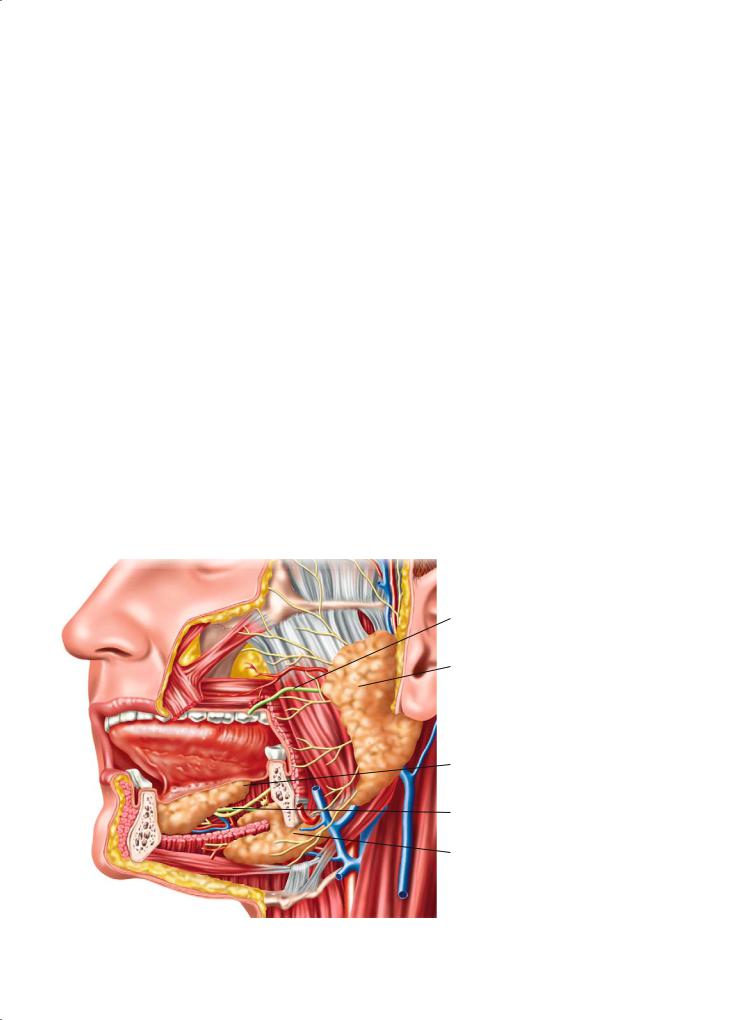
The parotid glands are located in close proximity to the cartilage of the external auditory canal.Anteriorly the gland abuts both the lateral and posterior border of the ramus of the mandible and the overlying masseter muscle, while inferiorly it rests medially on the posterior belly of the digastric muscle, as well as the sternocleidomastoid muscle laterally. Medially the parotid is adjacent to the parapharyngeal space, while superiorly it reaches the arch of the zygoma. The facial nerve courses through the parotid gland. The parotid gland is arbitrarily divided into a ‘superficial’ and ‘deep’ lobe by the plane of the facial nerve. Numerous lymph nodes are localized within, and adjacent to, the capsule of the parotid gland, serving as the first echelon drainage for the temporal scalp, portions of the cheek, the pinna, and the external auditory canal. For this reason, the parotid
gland may harbour metastatic cutaneous malignancy from these sites.
The submandibular glands are located in the anterior triangle of the neck, and are bounded superiorly and laterally by the body of the mandible. The mylohyoid muscle is located anterior to the gland, while the hyoglossus muscle lies medial to the gland. The submandibular (Wharton’s) duct exists the gland medial to the mylohyoid muscle, then courses anteriorly and superiorly into the anterior floor of mouth (Fig. 2.10).
Located beneath the mucosa of the floor of the mouth, the small sublingual glands drain directly into the oral cavity through numerous small ducts.
The majority of neoplastic lesions of salivary glands appear as a lump without other symptoms.
Swellings in the retromandibular sulcus, the immediate preauricular region, and over the masseter are, in most cases, of parotid gland origin. Although about 10% of parotid gland tumors arise medial to the plane of the facial nerve in the deep ‘lobe’ of the gland, more than three-fourths of these deep lobe tumors will present as a typical parotid mass.
In the parotid gland pleomorphic adenomas present as round, firm, reasonable well-demarcated tumors, with a tendency to nodularity as they grow. Their site of election is between the ascending ramus of the mandible anteriorly, and the mastoid process and sternomastoid posteriorly, usually in
Stenon’s duct
Parotid gland
Sublingual gland
Wharton’s duct
Submandibular gland
Fig. 2.10. Anatomic relations of the parotid, submandibular, and sublingual salivary gland

28 |
P. Delaere |
the tail of the gland. Occasionally they arise in the immediate preauricular region, where they tend to be small. Whartin’s tumors lie almost invariably in the lower pole of the gland, are ovoid in shape, and vary in consistency between soft and firm, depending on whether or not they have been exposed to previous inflammation; these tumors can occur bilaterally.
Weakness or paralysis of the facial nerve in a previously untreated patient almost always indicates that a tumor is malignant (Spiro et al. 1975; Borthne et al. 1986). Careful assessment should be made of the facial nerve and the nerves traversing the nearby carotid space (cranial nerve IX and XII) if a deep lobe or parapharyngeal space tumor is suspected.
It is often difficult to distinguish between a tumor arising within the submandibular gland or an enlarged node close to the gland or on its outer surface. Bimanual palpation is essential to differentiate between the two, since a node lying on the outer surface of the salivary gland is unlikely to be palpated by a finger in the mouth, whereas a tumor of the gland itself is more readily compressible bimanually. Pleomorphic adenomas of the submandibular gland are usually large, quite hard, and nodular, but may be confused with a slowly growing malignancy such as an adenoid cystic carcinoma. Submandibular gland neoplasms also need evaluation of the lingual and hypoglossal nerves.
The assessment of intraoral minor salivary gland neoplasms depends on the location of the tumor. Palatal lesions are the most common, usually giving the appearance of being fixed, whether they are benign or malignant, because of the tight adherence of the mucous membrane to bone. Tumors of the hard or soft palate are often fusiform, firm to hard in consistency and nodular. Again the distinction between mixed tumor and adenoid cystic carcinoma may be difficult to make.
A salivary gland tumor arising from the deep lobe of the parotid gland, or from a minor salivary gland in the parapharyngeal space, may cause secondary displacement of the palatotonsillar region.
Swelling detectable both in the pharynx and parotid region indicates a very bulky tumor originating from within the deep lobe of the parotid gland. This parotid swelling can be visible externally, but the technique of bimanual palpation will elicit the characteristic sign of ballottement between the examining fingers, typical of masses occupying such a wide area. The absence of both a visible swelling in the parotid gland and ballottement suggests an origin exclusively in the parapharyngeal space.
2.10
Thyroid Gland
The thyroid gland lies within the pretracheal fascia in the front of the neck, and consists of two symmetrical lobes united in the midline by an isthmus that overlies the second to fourth tracheal rings (Fig. 2.1). There is often a pyramidal lobe, which may extend as high as the top of the thyroid cartilage.
The incidence of palpable thyroid nodules is estimated at only 4%–7% of the general adult population. They occur more frequently in women and they are increasing with age (Mazzaferri et al. 1988). Slightly less than 5% of thyroid nodules are found to be malignant (Gharib and Goellner 1993). Most patients with differentiated carcinoma present with a palpable nodule in the thyroid gland of varying size, consistency and local extent. The primary tumor may present as a solitary, well-defined, intrathyroidal discrete palpable nodule or it may manifest with diffuse involvement of the thyroid gland with or without extrathyroid extension and fixation to the structures in the central compartment of the neck, or it may present as multiple palpable nodules. The most common location of palpable metastatic lymph nodes from thyroid cancer is at levels III, IV or V in the lateral neck. Procedures commonly used for the initial evaluation of thyroid nodules are: ultrasound, radionuclide imaging, and fine-needle aspiration biopsy (FNAB).
Anaplastic carcinoma of the thyroid gland usually manifests in the older population with a very short history of a rapidly enlarging thyroid mass. Physical examination reveals a diffusely enlarged firm to hard ill-defined thyroid mass with significant extrathyroid extension to adjacent soft tissues. The mass appears fixed and inseparable from the laryngotrachealesophageal complex.
2.11
Role of Imaging Studies
The clinical evaluation allows to appreciate the mucosal layer of the head and neck region quite well. However, the deep extent of potentially infiltrating lesions can only be judged indirectly. Some regions, such as the base of the skull, pterygopalatine and infratemporal fossa, orbits and brain are beyond clinical evaluation, but critical management decisions have to be made based on the involvement of these structures; imaging findings are of the utmost impor-

Clinical and Endoscopic Examination of the Head and Neck
tance in such cases. Perineural and/or perivascular spread, eventually leading to tumor progression or recurrences at distance from the primary tumor can often only be detected by imaging.
Metastatic adenopathies can be identified, sometimes still in a subclinical stage or at places not accessible for clinical examination, such as the retropharyngeal or paratracheal lymph nodes. Also, information on extranodal tumor spread and the relation to critical structures such as the carotid arteries, is necessary for determining the optimal patient management, and can be deduced from imaging studies.
Imaging is needed in submucosal lesions, covered by an intact mucosa. The origin and extent of such lesions is often difficult to determine based on the clinical evaluation alone. Imaging may provide important clues to the diagnosis, as representative biopsies may be difficult to obtain in deep-seated lesions.
All these findings can profoundly influence the staging and management of the patient with head and neck cancer. Finally, imaging may be used to monitor tumor response and to try to detect recurrent or persistent disease before it becomes clinically evident, possibly with a better chance for successful salvage.
The single most important factor in the optimal use of all this information is the mutual co-operation between the radiologist and the physicians in charge of patient care.
References
Benninger MS, Enrique RR, Nichols RD (1993) Symptomdirected selective endoscopy and cost containment for evaluation of head and neck cancer. Head Neck 15:532–536
Bolger WE, Kennedy DW (1992) Nasal endoscopy in the outpatient clinic. Otolaryngol Clin North Am 25:791–802
Borthne A, Kaalhus O, Vermund H (1986) Salivary gland malignant neoplasms: treatment and prognosis. Int J Radiat Oncol Biol Phys 12:747–754
Gharib H, Goellner JR (1993) Fine needle aspiration biopsy of the thyroid: an appraisal. Ann Intern Med 118:282–289
29
Hordijk GJ, Bruggink T, Ravasz LA (1989) Panendoscopy: a valuable procedure? Otolaryngol Head Neck Surg 101:426– 428
Kleinsasser O (1965) Weitere technische Entwicklungen und erste Ergebnisse der “endolaryngealen Mikrochirurgie”. Laryngol Rhinol Otol 44:711–727
Levine B, Nielson EW (1992) The justifications and controversies of panendoscopy - a review. Ear Nose Throat J 71:335–343
Levine HL (1990) The office diagnosis of nasal and sinus disorders using rigid nasal endoscopy. Otolaryngol Head Neck Surg 102:370–373
Lindberg RD (1972) Distribution of cervical lymph node metastases from squamous cell carcinoma of the upper respiratory and digestive tracts. Cancer 29:1446–1450
Logemann JA (1983) Evaluation and treatment of swallowing disorders. College Hill Press, San Diego
Mazzaferri EL, de los Santos ET, Rofagha-Keyhani S (1988) Solitary thyroid nodule: diagnosis and management. Med Clin North Am 72:1177–1211
Merritt RM, Williams MF, James TH et al (1997) Detection of cervical metastasis. A meta-analysis comparing computed tomography with physical examination. Arch Otolaryngol Head Neck Surg 123:149–152
Parker R (1992) Laryngoscopy, microlaryngoscopy and laser surgery. In: McGregor IA, Howard DJ (eds) Rob and Smith’s operative surgery: head and neck, part 2, 4th edn. Butterworth, Oxford, pp 451–463
Phelps PD (1992) Carcinoma of the larynx - the role of imaging in staging and pre-treatment assessments. Clinical Radiology 46:77–83
Ritchie AJ, McGuigan J, Stenvenson HM et al (1993) Diagnostic rigid and flexible oesophagoscopy in carcinoma of the oesophagus: a comparison. Thorax 48:115–118
Sercarz JA, Berke GS, Ming Y et al (1992) Videostroboscopy of human vocal fold paralysis. Ann Otol Rhinol Laryngol 101:567–577
Shah JP (1990) Patterns of nodal metastases from squamous carcinomas of the upper aerodigestive tract. Am J Surg 160:405–409
Spiro RH, Huvos AW, Strong EW (1975) Cancer of the parotid gland: a clinicopathologic study of 288 primary cases. Am J Surg 130:452–459
Stell PM, Maran AGD (eds) (1972) Pre-operative considerations. In: Head and neck surgery. William Heinnemann Medical, London, p 6
Zhao R (1992) Diagnostic value of stroboscopy in early glottic carcinoma. Chung Rua Ehr Pi Yan Hou Ko Tsa Chih 27:175–176
Watkinson JC, Johnston D, James D et al (1990) The reliability of palpation in the assessment of tumors. Clinical Otolaryngology 5:405–410

Imaging Techniques |
31 |
3Imaging Techniques
Robert Hermans, Frederik De Keyzer, and Vincent Vandecaveye
CONTENTS
3.1Introduction 31
3.2 |
Plain Radiography 31 |
3.3Ultrasonography 31
3.4Computed Tomography and Magnetic Resonance Imaging 32
3.4.1Computed Tomography 32
3.4.1.1Patient Positioning 32
3.4.1.2 |
Contrast Agent Injection 33 |
3.4.1.3 |
Data Acquisition and Image Reconstruction 35 |
3.4.1.4Dynamic Maneuvers 36
3.4.1.5 |
Three-Dimensional Image Reformatting 37 |
3.4.2 |
Magnetic Resonance Imaging 38 |
3.4.2.1Patient Positioning 39
3.4.2.2Coils 39
3.4.2.3Standard Sequences 39
3.4.2.4Contrast Agents 40
3.4.2.5 Additional MRI Techniques 40
3.5Nuclear Imaging 42 References 42
3.1 Introduction
Various imaging techniques are used in the evaluation of patients with head and neck cancer, before, during and after treatment. Each of these imaging techniques has its own advantages and disadvantages.
Many head and neck neoplasms arise from the mucosal lining; when a patient is referred for imaging,the histological diagnosis often was already established by endoscopic biopsy. Therefore, imaging should primarily supply information on the submucosal extension depth of the primary tumor, including its relation to surrounding structures, as well as on the presence of regional and/or distant metastasis.
The purpose of this chapter is to describe the various techniques available for imaging the head and
R. Hermans, MD, PhD
Professor, Department of Radiology, University Hospitals Leuven, Herestraat 49, 3000 Leuven, Belgium
F. De Keyzer, MSc; V. Vandecaveye, MD
Department of Radiology, University Hospitals Leuven, Herestraat 49, 3000 Leuven, Belgium
neck cancer patient, and to provide general rules for their use. Specialized imaging applications are described in the following chapters where appropriate.
3.2
Plain Radiography
In the past, a variety of conventional methods were applied to stage head and neck cancer, including soft tissue views of the neck, plain films of the facial skeleton, xeroradiography, plain film tomography, laryngography and barium swallow. The value of these studies to stage head and neck cancer is very limited; these techniques are now replaced by cross-section- ing imaging modalities.
Plain radiography is still widely used in radiotherapy planning (see Chap. 18). Barium swallow remains an indispensable method in the early phase after pharyngeal surgery, to rule out or confirm the presence of fistulae. This technique is also essential in the evaluation of functional disorders (such as bolus retention, delayed passage and aspiration) after surgery or radiotherapy.
3.3 Ultrasonography
Ultrasonography is a widely used technique for the evaluation of the thyroid gland (see Chap. 14), neck lymph nodes (see Chap. 15) and salivary glands, as it offers visualization of these structures with high spatial resolution, at a low cost and without using ionizing radiation.
Ultrasonography in combination with fine needle aspiration cytology (FNAC) is the most accurate method for neck nodal staging in most head and neck cancers (van den Brekel et al. 1991). However, execution of this procedure is time consuming, and the obtained results are operator dependent (Takes

32 |
R. Hermans et al. |
et al. 1996). Also, in a multicenter study where both computed tomography and ultrasound of the neck were applied for staging of head and neck cancer, the addition of ultrasound guided FNAC did not provide significant additional value (Takes et al. 1998).
3.4
Computed Tomography and Magnetic Resonance Imaging
Nowadays, in most patients computed tomography (CT) or magnetic resonance imaging (MRI) is performed for pretherapeutic staging of a head and neck malignancy. Both techniques can supply the information needed by the clinician for adequate treatment planning.
A common question is which of these techniques should be used in a particular patient. The most widely used technique is CT, as it has a number of important advantages over MRI:
•Wide availability
•Relative low cost
•Easy to execute, and this in a reproducible way
•Short examination time, resulting in less image quality degradation caused by motion, such as swallowing and respiration
making the technique sensible to motion artifacts which cause a non-diagnostic study (Fig. 3.3). It is also somewhat more difficult with MRI to properly stage both primary tumor and neck nodal disease in a single study. The lower availability of MRI, resulting in a longer waiting list, and its higher cost should also be taken into consideration.
In many institutions, CT is the preferred imaging method for evaluation of laryngeal and hypopharyngeal cancer, as well as of oral cavity and oropharyngeal cancer. These cancer sites constitute about 80%–85% of all head and neck malignancies (excluding skin cancer and lymphoma) in Europe and the USA. In most cases, a dedicated CT study will provide all answers needed by the clinician; in such a setting, MRI is used as a complementary tool to solve remaining questions.
Because of its higher contrast resolution, MRI is the preferred imaging method in rarer head and neck malignancies, such as nasopharyngeal cancer and sinonasal cancer. There is no consensus regarding the use of CT or MRI as primary imaging tool in salivary gland cancer, although there is a tendency among head and neck radiologists to choose MRI.
3.4.1
Computed Tomography
•Superior bone detail
•High quality multiplanar imaging on multidetecCT can be regarded as the ‘workhorse’ of head and
tor CT systems
•Easy extension of the study into the upper thoracic cavity or intracranial cavity, if needed
•Easier interpretation, especially regarding nodal involvement (Curtin et al. 1998)
However, CT also has a number of disadvantages compared to MRI:
•Relative low soft tissue contrast resolution
•Administration of iodinated contrast agent is necessary
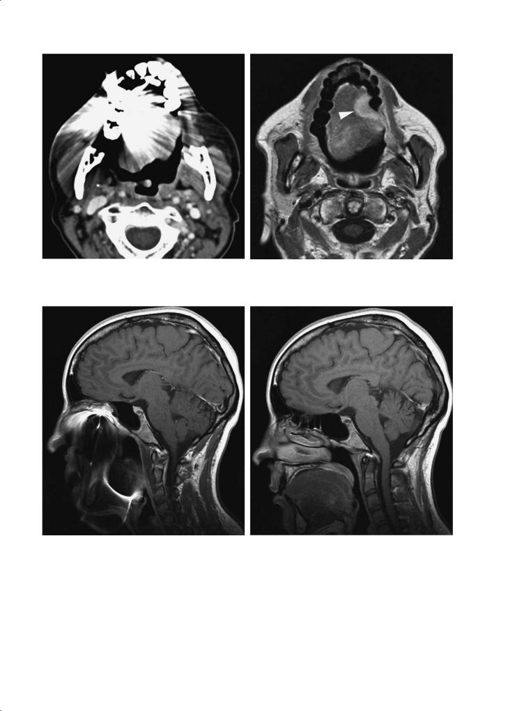
•Severe image quality degradation by dental fillings or other metallic foreign objects (Fig. 3.1)
•Radiation exposure
The advantages of MRI over CT in the evaluation of head and neck cancer are its superior soft tissue contrast resolution, and the absence of radiation exposure. Overall, the image quality is not or less hampered by the presence of dental fillings than in CT, but also MRI studies may be severely jeopardized by metallic implants (Fig. 3.2). The disadvantages of MRI are mainly related to the long acquisition time,
neck cancer imaging. It is not possible to define the ideal imaging protocol, as available equipment varies. The minimal requirements for an optimal diagnostic study will be outlined.
3.4.1.1
Patient Positioning
The images are obtained with the patient supine and during quiet respiration. The neck should be in slight extension. The head is aligned in the cephalocaudal axis in order to make it possible to compare symmetric structures. Malposition may result in an appearance that simulates disease. Every effort should be made to make the patient feel comfortable; this will help the patient dropping the shoulders to a position as low as possible.
This patient-friendly position is applicable for all indications if multidetector spiral CT (MDCT) is used, as this modality allows retrospective high quality reformatting in every spatial plane. In case an incremental or single spiral CT technique has to be used, additional direct coronal imaging is needed
Imaging Techniques |
33 |
a |
b |
Fig. 3.1a,b. Patient suffering left-sided oral tongue cancer. a Axial contrast-enhanced CT image. As the image quality is severely hampered by artifacts arising from dental fillings, the primary tumor is not visible.A complementary MR study was advised. b Gadoliniumenhanced T1-weighted image, not affected by presence of dental fillings, clearly shows the primary tumor (arrowhead)
a |
b |
|
Fig. 3.2a,b. Patient referred for MR study of the maxillofacial region and skull base because of unilateral facial pain. a Initial |
|
MR study was inconclusive, as artifacts caused by fixed orthodontic material severely degrade image quality. b After removal |
|
of the orthodontic material by the dentist, optimal image quality was achieved |
in the evaluation of sinonasal and skull base neoplasms. This can be realized by hyperextension of the neck, either in supine or prone position, and tilting the gantry to a position perpendicular to the hard palate.
3.4.1.2
Contrast Agent Injection
While evaluating a patient suffering head and neck cancer, a proper injection method of iodinated con-

34 |
R. Hermans et al. |
a |
b |
|
Fig. 3.3a–c. Patient suffering tongue base cancer. a,b Axial and |
|
sagittal reformatted contrast-enhanced MDCT images clearly |
|
show tumor extent into tongue base and involvement of free |
|
epiglottic rim (arrowheads), as well as the relationship of the |
|
tumor to the pre-epiglottic space (asterisk). c For study pur- |
|
poses, an MR study was performed in this patient 1 day later. |
|
Because of motion artifacts, the tumor is not confidently dis- |
c |
cernible. This MR study was considered non-diagnostic |
trast agent is crucial to obtain state of the art CT images. Optimal tissue enhancement, allowing correct discrimination of tumoral from normal tissue, and a high neck vessel density must be realized at the same time. Several contrast agent injection protocols have been described, some of them being fairly complicated. For all practical purposes, a single bolus technique with an injection rate of 1 cc/s is appropriate on modern CT machines (Keberle et al. 2002).
A total amount of 100 ml is sufficient in MDCT; a somewhat higher volume (up to 150 ml) may be required when an incremental or single slice spiral CT technique is used.
It is essential to wait 60 s before starting the acquisition, as the contrast agents needs some time to diffuse in the normal and pathologic soft tissues. If re-angulation of the gantry at the oral level is performed (see below), the contrast injection needs not