

Учебники / Head_and_Neck_Cancer_Imaging
.pdfMasticator Space Neoplasms |
177 |
10 Masticator Space Neoplasms
Thierry P. Duprez and Emmanuel E. Coche
CONTENTS |
|
|
10.1 |
Masticator Space Anatomy 177 |
|
10.2 |
Introduction to Masticator Space Malignancies |
177 |
10.3 |
Practical Issues for Masticator Space Imaging |
178 |
10.4Radiological Patterns of Neoplastic Masticator Space Involvement 179
10.4.1Extrinsic Impingement on the MS by Benign Neoplasm 180
10.4.2Extrinsic Infiltration of the MS by a Malignant
|
Neoplasm 181 |
10.4.3 |
Intrinsic Benign Neoplasm of the MS 181 |
10.4.4 |
Intrinsic Malignancies of the MS 182 |
10.4.5Pseudo-neoplastic Benign Hypertrophy of MS Muscles 183
10.5Special Concerns 183
10.5.1Synergy Between CT and MRI in Radiological
Work-Up of MS Neoplasms 183
division of the fifth cranial nerve (mandibular trigeminal branch or V3). The V3 nerve gives motor innervation to the mastication muscles and relays sensory information from the inferior teeth, gums and lower lip/chin region through the inferior alveolar nerve. The nerve emerges from the endocranium to the MS through the foramen ovale. The space is easy to identify on both CT and MR images (Figs. 10.1, 10.2) because of easily recognizable shape and location of the mastication muscles (Harnsberger and
Osborn 1991; Mukherji and Castillo 1998). The inferior limit of the MS is the attachment of the medial pterygoid muscle to the mandible. The space has two distinct superior margins. The base of the skull is the superior limit of the “infratemporal fossa” or “na-
10.5.2Imaging of the Tumoral Spread Along the V3 Cranial sopharyngeal masticator space” which encompasses
Nerve 186
10.5.3 Treatment and Post-treatment Issues 186
10.5.3.1Role of Initial Imaging Findings in Optimizing Treatment Strategy 186
10.5.3.2Potential Roles for Imaging Monitoring During and After Treatment 187
10.5.3.3 Imaging of Treatment Complications 189
References 189
10.1
Masticator Space Anatomy
The masticator space (MS) is a deep facial space delineated by a splitting of the deep cervical fascia which encloses the four muscles of mastication: the medial and the lateral pterygoid, the masseter, and the temporalis muscles – hence the denomination of “masticator space” (Harnsberger 1995; Mukherji and Chong 2004). The MS also contains the ramus and posterior body of the mandible and the third
T. P. Duprez, MD; E. E. Coche, MD, PhD
Department of Radiology and Medical Imaging, Université Catholique de Louvain, Cliniques Universitaires Saint-Luc, Avenue Hippocrate, 10, 1200 Brussels, Belgium
all soft tissue below the foramen ovale. The second superior margin is the attachment of the temporalis muscle to the outer table of the skull; this part is called the “temporal fossa”, or the “suprazygomatic masticator space” because it is above the zygomatic arch (Fig. 10.2). The MS is separated from the parapharyngeal space by a fascial layer extending from the medial pterygoid muscle to the skull (Curtin 1987). The fascia is also attached to the anterior aspect of the mandibular ramus, has an interface with the oral cavity, and reaches the posterior margin of the ramus, where the masticator space constitutes the anterior border of the parotid space.
10.2
Introduction to Masticator Space Malignancies
Tumors in the MS may arise from each major component of the space (muscles, bone, V3), or may be the extension of a tumor arising from adjacent spaces. Muscular malignancies are of mesenchymal origin in the vast majority of the cases. Imaging features of the different sarcomas are poorly specific. Metastases must be included in the differential diagnosis of MS

178
Fig. 10.1. Masticator space anatomy. Transverse T1-weighted image: masticator space (MS) has been highlighted on the left side, excluding the parotid gland posteriorly and the buccinator muscle anteriorly. Critical contents of MS are (right side): M, masseter muscle; MP, medial pterygoid muscle; V3, mandibular branch of the fifth cranial nerve, and ramus of the mandible. Observe close relation to the fatty parapharyngeal space (PPS)
Fig. 10.2. Masticator space anatomy. Coronal T1-weighted image: similar to Fig. 10.1, the masticator space (MS) has been highlighted on the left side. Observe the presence of a second more cranial compartment containing the belly of the temporalis muscle (T), i.e. the suprazygomatic masticator space (SZMS). Other critical contents of the MS include: LP, lateral pterygoid muscle; MP, medial pterygoid muscle; M, masseter muscle; V3, mandibular branch of the fifth cranial nerve. The fatty parapharyngeal space and bone marrow of the ramus disclose hypersignal intensity
masses, but primitive osteosarcoma is the most frequent malignancy arising from the mandibular bone component of the space (Chong and Fan 1996). The V3 cranial nerve which courses medial to the lateral pterygoid muscle before penetrating the base of the skull through the foramen ovale constitutes a “high-
T. P. Duprez and E. E. Coche
way” for tumoral spreading of malignant neoplasms from the MS to the base of the skull and the endocranium. The MS may be invaded by infiltrative malignancies arising from adjacent anatomical spaces, the most frequent one being primary squamous cell carcinoma (SCC) of the pharynx or of the retromolar trigone. Infectious mass-occupying processes are the main alternative hypothesis to benign/malignant neoplastic diseases of the MS. Since the imaging findings of MS neoplasms have low specificity, the diagnosis requires the knowledge of clinical/biological data and ultimately relies on the anatomopathological examination of biopsy or surgical specimens (Eskey et al. 2000).
10.3
Practical Issues for Masticator Space Imaging
•Changes in muscle volumes. The four mastication muscles account for the majority of the MS volume. Many processes involving the space may have a dramatic – direct or indirect – impact on muscular trophicity, thereby affecting the total MS volume (Hardin et al. 1985):
a)Space-occupying processes lead to MS enlargement.
b)Infiltration along the V3 nerve may lead to MS volume shrinkage by denervation atrophy of the muscles.
Comparison between both sides on MR images is therefore warranted regarding intrinsic signal intensity characteristics of muscles on both T2and post-contrast T1-weighted images, and regarding muscles’ volumes. Benign masseteric muscle hypertrophy is a common benign condition in which muscles are symmetrically or asymmetrically enlarged, with normal signal intensities when compared to unenlarged muscles (see also Sect. 10.4.5) (Waldhart and Lynch 1971).Denervation atrophy with increased contrast-enhancement of the masticator muscles at early phase may be seen when the V3 nerve has been infiltrated by a malignant process without true neoplastic infiltration of the muscles (Davis et al. 1995; Russo et al. 1997).
It may be difficult to distinguish tumoral muscle infiltration from the early phase of muscle denervation related to the neoplastic invasion of the V3 nerve. Long-standing denervation leads to delayed muscle fatty atrophy which is

Masticator Space Neoplasms |
179 |
easily distinguishable from radiation-induced muscular fibrosis (Parsons 1994).
•Pathways of tumor spread to the skull base. Because of the attachment of the MS muscles to the base of the skull and the presence of the V3 nerve, infiltrative processes may readily reach and destroy the base of the skull. Careful assessment of the skull base must be regarded as a part of comprehensive radiological work-up for MS abnormalities (Harnsberger 1995).
•Preferential spreading of MS processes to the oral cavity. Infections or malignancies involving the MS may spread early to the oral cavity (OC) because of the close contiguity of the two areas and because of the absence of a firm fascial layer which could act as a barrier against tumor progression. Anatomical structures of the OC being adjacent to the MS are the distal portion of the parotid duct, and the buccinator muscle and space, an assessment of which must be included in the checklist of radiological MS work-up (Harnsberger 1995).
•Clinical symptoms of MS involvement with malignancies. Pain or numbness of the ipsilateral chin and jaw are the main ‘neurologic’ symptoms of infiltration of the third branch of the trigeminal nerve (V3).
Trismus, a reactive spasm of masticator muscles resulting in mouth opening impairment, is the typical symptom for muscular involvement by malignancies or infections (Schellhas 1989; Russo et al. 1997).
The clinical tripod of MS muscles atrophy, mandibular dental pain, and serous otitis media secondary to dysfunction of the tensor muscle of the soft palate has been regarded almost pathognomonic for malignant infiltration of the V3 nerve (Caldemeyer et al. 1998).
ÆEmpirical rules for radiological examination of the masticator space:
a)Cover the entire course of the cranial nerve V3 from the root entry zone in lateral pons to the mental foramen of the mandible, especially when symptoms suggesting involvement of the nerve are present.
b)Cover the full extent of all masticator muscles, including the suprazygomatic area (temporalis muscle) since infiltrative processes rapidly involve the whole muscular corpus and dystrophic processes are often diffuse.
c)Image both mandible and skull base which are osseous boundaries of the MS from/to which many processes either originate or infiltrate
10.4
Radiological Patterns of Neoplastic Masticator Space Involvement
The first step for accurately proposing diagnostic hypotheses for MS masses is to determine whether the lesion appears ‘extrinsic’ (originating from outside) or ‘intrinsic’ (originating from inside) to the MS. Four criteria can be used to assess the MS origin of a spaceoccupying process (Harnsberger 1995):
•The epicentre of the mass is located within the masticator muscles or the mandible.
•The mass is anterior to the fatty parapharyngeal space.
•The mass invades the parapharyngeal space from anterior to posterior.
•The fat of the parapharyngeal space is displaced posteriorly.
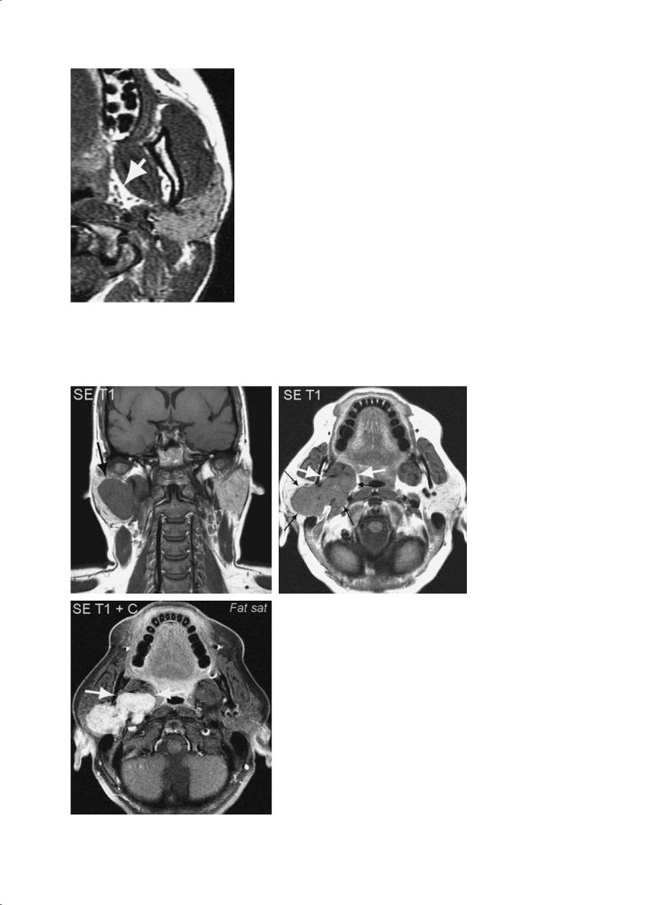
The displacement pattern of a mass arising from the MS is anterior to posterior with slight internal obliquity (Fig. 10.3). Posterior bowing of the anterior margin of the parapharyngeal space is often seen. The benign or malignant nature of a neoplasm often remains speculative until histopathological analysis is performed on biopsy or surgical specimens. Usually, malignancy is suspected in the presence of a heterogeneous and infiltrative process, and benignity is hypothesized in the presence of a sharply and well delineated process. But these criteria have low accuracy in MS examination since malignancies may have sharply defined margins (such as seen in many sarcomas originating either from bone or soft tissues), and benign neoplasms may have a poorly vascularized or fibrous core resulting in a heterogeneous texture of the lesion. Clinical data including the history of the growth rate of the tumor and the associated signs of pain, trismus and visible inflammatory changes, together with white blood cell count and inflammatory markers at routine blood analysis are crucial. The presence of mandibular or skull base bone destruction in a space-occupying process without inflammatory signs is strongly suggestive for malignancy. Perineural spreading is almost pathognomonic for a malignant neoplasm and is frequently associated with pain or dysaesthesias. Extrinsic/intrinsic and presumptively malignant/benign features of the le-
180 |
T. P. Duprez and E. E. Coche |
Fig. 10.3. Direction of mass effect from masticator space (MS). Transverse T1-weighted close-up on MS: the arrow shows the direction of the mass effect from within the MS. Impingement on the fatty parapharyngeal space with posterior bowing of its anterior margin is frequently seen
a
sion may result in the semiological categorization of the following five patterns of neoplasms involving the MS.
10.4.1
Extrinsic Impingement on the MS by Benign Neoplasm
A benign mass-occupying process originating from adjacent area(s) displaces the MS muscles without infiltrating them. The impingement on the MS is often asymptomatic or causes few symptoms. Complaints of mechanical impairment of mastication due to the mass effect of the lesion on muscle and/or mandibula, or both, are rarely present, as the displaced muscles are not involved by secondary inflammatory or tumoral change, and the V3 nerve is not invaded. Similarly, mandibular/dental pain is absent since the integrity of the V3 nerve is not compromised (Fig. 10.4).
b
|
Fig. 10.4a–c. Pattern of extrinsic impingement on the masticator space by |
|
benign neoplasm. A 34-year-old woman with surgically-proven pleomorphic |
|
adenoma of the parotid gland. a Coronal unenhanced T1-weighted spin-echo |
|
(SE) MR image showing large, well-delineated, hypointense lesion in both the |
|
superficial and deep lobe of the right parotid gland (arrows). b Transverse |
|
unenhanced T1-weighted SE MR image showing the same lesion as a ‘diab- |
|
olo-like’ well delineated and homogeneous mass (thin black arrows). The |
|
medial pterygoid muscle is pushed forward but does not show infiltration. |
|
A demarcation between tumor and muscle is present (between thick white |
|
arrows). Observe internal displacement of the fatty parapharyngeal space. |
|
c Transverse post-contrast T1-weighted SE MR image with fat saturation (FS) |
|
showing intense and homogeneous enhancement by abnormal tissue. Lesion |
|
is clearly delineated from surrounding structures and impinges on pterygoid |
|
muscle which does not display intrinsic changes. Clear-cut interface is more |
c |
obvious than on previous illustration (between arrows) |

Masticator Space Neoplasms |
181 |
10.4.2
Extrinsic Infiltration of the MS by a Malignant Neoplasm
Extrinsic malignancies arising from areas adjacent to the MS often have infiltrative potentialities which may result in functional impairment such as dental/mandibular pain due to V3 nerve invasion, and trismus due to mastication muscle invasion and V3 invasion. The invasion of bone, muscles and nerve may occur concomitantly or not.
Nasopharyngeal carcinoma is a common cause of malignant infiltration of mastication muscles (Chong 1997) (Fig. 10.5). The retromolar trigone also constitutes a common site of origin of squamous cell cancer invading the MS; mandibular bone invasion is commonly present (Rosai 1996) (Fig. 10.6).
10.4.3
Intrinsic Benign Neoplasm of the MS
Only a few benign neoplasms may arise from within the MS. The paradigmatic one is the schwannoma of the V3 nerve which mainly occurs in cases of type 2 neurofibromatosis (NFB type 2) (Smirniotopoulos and Murphy 1996; Weber et al. 2000). The VIIIth cranial nerve is the most frequently involved in this condition, but the Vth nerve is the second preferentially involved pair. Clinical background (autosomal dominant inheritance) and imaging workup showing multiple masses arising from cranial nerves together with multiples meningiomas and ependymomas are strongly suggestive of NFB type 2, which is ten times less frequent than NFB type 1 and can be confirmed by genetic analysis
a 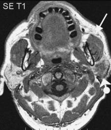
 b
b
|
Fig. 10.5a–c. Pattern of masticator space muscle infiltration by extrinsic |
|
malignant neoplasm. A 58-year-old man with adenoid cystic carcinoma |
|
of the parotid gland. a Unenhanced transverse T1-weighted SE image |
|
showing hypointense area within the left parotid gland, resulting from fat |
|
replacement by the tumoral process (ball-arrowhead). Apparently normal |
|
masseter (arrowhead). b Transverse T2-weighted SE image demonstrat- |
|
ing diffuse abnormal hyperintensity within left masseter (arrow), together |
|
with shrinkage and hypointense areas within left parotid (ball-arrowhead). |
|
c Transverse contrast-enhanced T1-weighted SE image with fat saturation |
|
demonstrating strong enhancement within both left parotid (ball-arrow- |
|
head) and masseter muscle (arrow). Anatomopathological examination of |
|
biopsy specimens demonstrated malignant infiltration of both the V3 nerve |
c |
and the masseter muscle |

182 |
T. P. Duprez and E. E. Coche |
a 
 b
b
d
c
Fig. 10.6a–d. Pattern of masticator space (MS) muscle infiltration by extrinsic malignant neoplasm. A 48-year-old man with squamous cell carcinoma (SCC) of the retromolar trigone (RMT) invading MS. a Unenhanced transverse T1-weighted MR image showing tumoral epicentre in RMT (arrow) and tumoral spreading into the mandibular ramus (ball-arrowhead). b Post-contrast transverse T1-weighted MR image with fat suppression showing enhancement of both extra- (arrow) and intra- (ball-arrow- head) osseous components of the tumor. c Transverse T2-weighted FSE MR image with fat suppression option showing hypersignal intensity within both components of the diseased area. d Unenhanced transverse T1-weighted image shows enlargement of the left mandibular canal on left side, compatible with perineural tumor spread
(Kyriakos and El-Mofty 1999) (Fig. 10.7). Other benign MS neoplasms are rare; they include synovial chondromatosis and benign minor salivary gland tumors.
10.4.4
Intrinsic Malignancies of the MS
Malignancies which may arise from the MS are:
•Osteosarcoma (OS)
•Rhabdomyosarcoma (RMS)
•Malignant fibrous histiocytoma
•Non-Hodgkin’s lymphoma
•Fibrosarcoma
•Hemangiosarcoma
•Malignant minor salivary gland (adenoid cystic, mucoepidermoid, adenocarcinoma, low-grade polymorphous adenocarcinoma)
OS and RMS are the most frequently observed intrinsic tumors of the MS.
Mandibular ameloblastoma and desmoid fibromatosis are usually benign conditions but may exhibit local aggressiveness in a small subset of patients (Cole and Guiss 1969; Delbaso 1998).

Masticator Space Neoplasms |
183 |
Fig. 10.7. Pattern of intrinsic benign neoplasm of the masticator space. A 26-year-old woman with genetically-proven neurofibromatosis type 2 presenting with multiple schwannomas of the endocranium, the base of the skull and the spinal canal. Transverse FSE T2-weighted image showing neurofibroma within the left masticator space (between black arrows). Numerous similar lesions are observed within spinal canal and left carotid space (paired black arrows). Observe severe atrophy of the right masseter and right lateral pterygoid when compared to the left side (ball-arrowheads)
Osteosarcoma (OS) of the mandible. In contrast to long bone OS, which are most commonly diagnosed during childhood, mandibular OS are found in adults and constitute the majority of head and neck OS (Marcus et al. 1994; Chong and Fan 1996). Known risk factors for developing OS are Paget’s disease, fi- brous dysplasia, and irradiation. CT may significantly underestimate the true extraosseous and bone marrow extension and MRI, therefore, is mandatory for a comprehensive work-up of the tumoral extension (see Sect. 10.5.1) (Fig. 10.8).
Rhabdomyosarcoma (RMS) of mastication muscles.
RMS is most often seen in the paediatric age where it accounts for 50%–70% of all childhood sarcomas (Cunningham et al. 1996). Half of the patients are diagnosed before the age of 5 years. Multimodal therapeutic strategies, including surgery, chemotherapy and radiation therapy, provide the best survival rate. Prognosis has significantly improved over the past decade with a current 5-year disease-free rate of about 75%. RMS should always be considered in the presence of a primary tumor from the MS arising in childhood, showing bone destruction (Fig. 10.9).
10.4.5
Pseudo-neoplastic Benign Hypertrophy of MS Muscles
In its typical form, the muscles are symmetrically increased in size in an adolescent or young adult, without further complaints (Waldhart and Lynch 1971). Bruxism or excessive chewing tendency has been described in association with the condition. Enlarged muscles display normal tissue features on both CT and MR images, except for their increase in size. The bilateralism of the megaly is strongly suggestive for benign hypertrophy; however, hypertrophy of the masseter muscle may also occur unilaterally. Unilateral enlargement of the masseter muscle can also be due to the presence of a vascular malformation (Fig. 10.10).
10.5
Special Concerns
10.5.1
Synergy Between CT and MRI in Radiological Work-Up of MS Neoplasms
The superiority of CT in depicting subtle bone changes is evident. Since the MS contains mandibular bone, CT is useful for detecting, delineating and characterizing cortical bone involvement. But the true extent of the infiltrative process within the trabecular bone network is poorly assessed by the technique. For this purpose, MRI is clearly superior. The replacement of the normal fatty marrow (displaying bright T1 signal intensity) by the neoplastic proliferation (displaying low signal) allows precise delineation of the full extent of the tumoral process within the bone marrow space. Moreover, the exquisite soft tissue contrast obtained by MRI also enables precise and accurate delineation of the lesion into the adjacent extra-osseous soft tissues.
Isotopic bone scanning still remains a valuable additional technique by screening the whole skeleton in a one-step procedure, which is of major value in malignancies with a high tendency to spread to the bones, such as OS (Fig. 10.11). Nowadays, positron emission tomography using fluoro-deoxy-glucose as tracer (FDG-PET) is routinely performed in cases of MS malignant neoplasms because of the precise and sensitive delineation of the primary tumor and the high sensitivity for detection of distant metastatic foci and possibly a second concomitant synchronous malignant neoplasm.

184 |
T. P. Duprez and E. E. Coche |
a 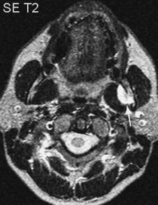
 b
b
d
c
Fig. 10.8a–d. Pattern of intrinsic malignancies arising within the masticator space. A 44-year-old man presenting with isolated dysaethesias of the left V3 territory, suffering osteosarcoma. a Transverse T2-weighted SE image showing strongly hyperintense tumoral mass arising from the medial aspect of the ascending ramus of the mandible and impinging on the lateral pterygoid muscle (arrow). b Transverse unenhanced T1-weighted SE image showing disappearance of the normal brightness of the fatty bone marrow of the ascending ramus of the mandible (white arrow). Tumoral mass (black arrow) is isointense to muscle. c Transverse contrast-enhanced T1-weighted SE image in a similar plane to Fig. 10.7 showing enhancement of the tumoral mass (black arrow), clearly delineating enhanced tumor from adjacent pterygoid muscle. The bone marrow of the ascending ramus also enhances (white arrow). d Axial unenhanced T1-weighted SE image shows extensive infiltration of the mandibular bone marrow: the normal bright fatty marrow has been replaced by material displaying low signal intensity; the anterior border of the bone marrow infiltration is indicated by white arrows

Masticator Space Neoplasms |
185 |
a 
 b
b
c |
d |
Fig. 10.9a–d. Pattern of intrinsic malignancies arising within the MS. Axial contrast-enhanced T1-weighted SE images (a–c), and coronal T2-weighted SE image (d). Rhabdomyosarcoma (courtesy of Robert Hermans, MD, PhD, Leuven, Belgium). A 20- year-old woman presenting with progressive swelling of the left cheek. A large contrast-enhanced soft tissue mass is present within the left infratemporal fossa. Lesion involves the mandibular ramus and masticatory muscles with anterior extension into the cheek. Superiorly the mass grows behind the maxillary sinus with pterygopalatine fossa involvement. Perineural tumoral spread along the V2 and V3 cranial nerve reaching the cavernous sinus can be recognized. More cranially, involvement of the superior orbital fissure and extension into the extracranial temporal fossa is seen

186 |
T. P. Duprez and E. E. Coche |
a 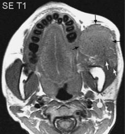
 b
b
|
Fig. 10.10a–c. Pattern of pseudo-neoplastic hypertrophy of the MS. |
|
A 15-year-old girl with hemi-mandibular developmental hypertrophy |
|
due to abnormalities of feeding/draining vasculature. a Transverse |
|
unenhanced T1-weighted SE image showing nodular venous malforma- |
|
tion of the left cheek (between black arrows) and encroachment on the |
|
anterior aspect of the masseter by the lesion (white arrows). Secondary |
|
hypertrophy of the left ramus is present. b Transverse T2-weighted FSE |
|
image showing enlargement of the left ramus (between black arrows) |
|
with preservation of global anatomical architecture (e.g. the symmetry |
|
of the V3 penetration points (white arrows). c Coronal T2-weighted FSE |
|
image showing unilateral enlargement of the left ramus (thick black |
|
arrow), encroachment on the masseter, and infiltration of the left tem- |
c |
poral muscle by the process |
10.5.2
Imaging of the Tumoral Spread Along the V3 Cranial Nerve
Assessment of tumoral invasion of the mandibular branch of the trigeminal nerve (V3) is crucial because all head and neck tumors with tumoral nerve involvement carry a bad prognosis,and because nerve infiltration requires appropriate local treatment (including excision of the diseased nerve if possible, and the inclusion of its entire course into radiation therapy portals). Perineural spread is most often seen in adenoid cystic and squamous cell carcinoma which are the most ‘neurophilic’ malignant neoplasms of the head and neck. V3 perineural infiltration symptoms and signs are usually overshadowed by the primary disease. Denervation atrophy is the common consequence of malignant nerve involvement. Atrophy of the temporalis and masseter muscles may be visible
at clinical examination. Facial neuralgia may be the prominent symptom. Inclusion of the full course of the V3 nerve into the field of view of the MR examination is mandatory when imaging suprahyoid head and neck neoplasms, especially in those arising from the MS (Fig. 10.12).
10.5.3
Treatment and Post-treatment Issues
10.5.3.1
Role of Initial Imaging Findings in Optimizing Treatment Strategy
Imaging findings have a crucial impact on the therapeutic strategy by precisely defining the size of the lesion and the involvement of the adjacent non-mu- cosal structures such as bones, muscles, nerves and