

Учебники / Head_and_Neck_Cancer_Imaging
.pdfNeoplasms of the Sinonasal Cavities
a
b
c
197
The pterygopalatine fossa may become involved by tumor growth through the posterior wall of the maxillary sinus, or via perineural tumor spread.
Perineural tumor spread usually occurs along the nerve sheath, not within the nerve itself. Such perineural spread may be discontinuous, with skip areas. Although adenoid cystic carcinoma is notorious for its tendency towards perineural spread, such extension pattern is also seen in other tumor types, such as squamous cell carcinoma. Perineural tumor spread occurs more frequently in case of tumor recurrence. Perineural tumor spread is associated with a decreased survival rate. Symptoms include pain, paresthesias and muscle weakness and atrophy, but about 40% of patients do not show particular symptoms. Imaging diagnosis is important to avoid tumor ‘recurrence’ from unrecognized perineural spread; however,microscopic perineural tumor spread cannot be recognized by CT and MRI (Nemzek et al. 1998).
Imaging findings in perineural tumor spread include (Fig. 11.9):
•Thickening and/or enhancement of one or more nerve branches
•(Often small) tumoral lesions at some distance from the primary site, in a neural 'crossroad' such as the pterygopalatine fossa or Meckel's cave
•Widening, destruction or enhancement of a skull base neural foramen or canal (e.g. foramen ovale, vidian canal)
•Denervation atrophy of muscles supplied by the affected nerve
Fig. 11.5a–c. a Coronal unenhanced CT image (bone window) in a patient complaining of frontal headache and a single episode of epistaxis. A large right-sided nasoethmoidal mass (asterisks) is seen, growing through the superior part of the nasal septum and destroying the bony ethmoidal walls and middle turbinate. Erosions in the roof of the nasal cavity and ethmoidal cells are noted (arrowheads).Biopsy revealed squamous cell carcinoma. b Coronal T2-weighted spin echo image. The tumor mass appears with intermediate signal intensity, and is clearly growing through the nasal septum. Subtle brain hyperintensity is seen in the anterior cranial fossa (arrowheads). At this moment, the patient has retro-obstructive inflammation in the right maxillary sinus. c Coronal gadolinium-enhanced T1-weighted spin echo image. The enhancing tumor mass is seen to extend into the anterior cranial fossa (white arrowheads), and can not be delineated from the overlying brain parenchyma. Neighbouring enhancing dural thickening (arrows), probably due to reactive inflammation; some extradural tumor extension is suspected at the right side. Note the subtle enhancement against the orbital side of the lamina papyracea (black arrowhead), representing early orbital invasion. The brain invasion, the reactive dural changes and the intraorbital extension were pathologically proven in this patient

198 |
R. Hermans |
a |
b |
|
Fig. 11.6a–c. a Coronal T2-weighted spin echo image. Large, bilat- |
|
eral nasoethmoidal soft tissue mass, involving the anterior cranial |
|
fossa. The lesion can not be clearly delineated from the brain paren- |
|
chyma. Perilesional edema (arrowheads) is seen in the right fron- |
|
tal lobe. No orbital involvement. b Coronal gadolinium-enhanced |
|
T1-weighted spin echo image. Moderate enhancement of the mass |
|
lesion is seen. c Sagittal gadolinium-enhanced T1-weighted spin |
|
echo image. Poor delineation between the intracranial tumor com- |
|
ponent and the surrounding brain tissue (arrowhead). Posteriorly, |
|
the lesion invades the anterior portion of the sphenoid sinus; retro- |
|
obstructive inflammation (asterisk). Biopsy revealed undifferenti- |
|
ated carcinoma. The lesion was considered to be inoperable due to |
|
the extensive intracranial involvement. The mass completely disap- |
|
peared during radiation therapy. At present, the patient is followed- |
c |
up for 3 years, without evidence of tumor recurrence |
Several neural pathways exist towards the pterygopalatine fossa, such as the infraorbital nerve, the posterior superior alveolar neurovascular bundle in the posterolateral wall of the maxillary sinus, and the neurovascular bundle in the greater and lesser palatine foramina (Ginsberg and DeMonte 1998; Mancuso et al. 1988).
From the pterygopalatine fossa, the tumor may grow superiorly into the inferior orbital fissure and infiltrate the orbital apex. Further superior extension will lead to infiltration of the superior orbital fissure, from where the tumor has easy access to the cavernous sinus. The tumor may grow posteriorly along the maxillary nerve in the fora-
men rotundum, eventually also leading to the cavernous sinus.
11.5
Tumor Types
The focus of this section is on malignant neoplasms of the sinonasal region. However, a number of histologically benign lesions are included, as they may mimick the radiological appearance of a malignant lesion, and/or because they may show clinically an aggressive behaviour.

Neoplasms of the Sinonasal Cavities |
199 |
a |
b |
c |
d |
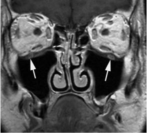
Fig. 11.7a–d. Patient presenting with nasal obstruction and progressive loss of vision on left side. a,b Axial gadolinium-enhanced T1-weighted spin echo images (a caudal to b). Moderately enhancing soft tissue mass in the left nasal cavity, growing into the posterior ethmoidal cells and sphenoid sinus. Extension into the pterygopalatine fossa (indicated by arrowhead on opposite side) and into the cavernous sinus (arrow), encasing the internal carotid artery, and reaching the anterior margin of Meckel’s cave (curved arrow). At the level of the superior orbital fissure (b), the tumor grows into the orbital apex. c,d Coronal gado- linium-enhanced T1-weighted spin echo images (c posterior to d). Extension into the maxillary sinus (asterisk). Thickening and increased enhancement of the medial and inferior rectus muscle (arrows), corresponding to neoplastic involvement. On the more anterior image (d), apart from the muscular thickening, some infiltration of the peri-orbital fat is also noticed (arrowhead). Biopsy revealed undifferentiated carcinoma. The patient was treated by a combination of irradiation and chemotherapy. No evidence of tumor recurrence 3 years after treatment
11.5.1
Epithelial Tumors
11.5.1.1
Inverted Papilloma
Inverted papilloma is an uncommon epithelial tumor of the nose and paranasal sinuses. Essentially,
this is a benign tumor, but recurrence after resection is fairly common, and in 7%–15% of patients an associated squamous cell carcinoma is found in the resection specimen. Even in the absence of frank malignancy, an inverted papilloma may behave aggressively, with rapid recurrences and invasion of surrounding structures (Pasquini et al. 2004).
200 |
R. Hermans |
The name refers to the characteristic invagination of the proliferating epithelium beneath the surface. Inverted papillomas typically arise from the lateral nasal wall, in the region of the middle turbinate. At presentation, they often involve both the nasal cavity and the adjacent maxillary cavity; the ethmoids may also be involved, but extension to the frontal and sphenoid sinus is not often seen. A polypoid mass is
seen during clinical examination, sometimes resembling an antrochoanal polyp.
Apart from showing the location of the tumor mass at the junction of the nose and maxillary antrum, usually with a well defined bone defect at the maxillary ostium, CT may demonstrate the presence of intratumoral calcifications (Fig. 11.10). Sometimes the bony walls adjacent to the tumor appear sclerotic.
a |
b |
Fig. 11.8a–c. Patient suffering from sinonasal squamous cell carcinoma. a Axial contrast-enhanced CT image. The tumor
|
mass is invading the sphenoid sinus (asterisk), lower part of |
|
the orbit, upper part of the maxillary sinus and pterygopala- |
|
tine fossa. The mass extends from the pterygopalatine fossa |
|
into the infratemporal fossa through the pterygomaxillary fis- |
|
sure (arrow), and into the middle cranial fossa through the |
|
foramen rotundum (arrowhead). b Section somewhat cranial |
|
to (a). The tumor is infiltrating the orbital apex. From here, |
|
the tumor is extending through the superior orbital fissure |
|
(arrow) into the cavernous sinus. c Section somewhat caudal |
|
to (a). The mass is massively invading the maxillary sinus, and |
|
growing through the posterolateral bony wall of the maxillary |
|
sinus (arrowheads) into the infratemporal fossa. Anterior to |
|
the tumor mass, the sinus content shows a homogeneous at- |
c |
tenuation, suggestive of retro-obstructive fluid (asterisk) |
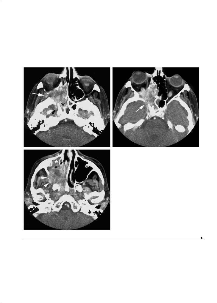
Fig. 11.9a–f. Patient with history of recurrent upper lip skin cancer, treated at another institution, now presenting with left-sided facial pain. a-c Axial gadolinium-enhanced T1-weighted spin echo images. Soft tissue swelling is seen in the upper lip (arrow, a and b), reaching the infra-orbital foramen (curved arrow, b). At a level slightly more cranial, enhancement of the infra-orbital nerve (white arrowheads, c) and the maxillary nerve, passing through the pterygopalatine fossa and foramen rotundum (black arrowhead, c) is seen. Thickening and increased enhancement of the cavernous sinus (curved arrow, c). d-f Coronal gadoliniumenhanced T1-weighted spin echo images. Enhancement of left infra-orbital nerve (compare to opposite side, arrows), maxillary nerve in foramen rotundum (arrowheads) and cavernous sinus (curved arrows). These findings are consistent with recurrent cancer in the upper lip, spreading perineurally along the left infra-orbital and maxillary nerve, into the cavernous sinus

Neoplasms of the Sinonasal Cavities |
201 |
a |
b |
 d
d
c
e |
f |

202 |
R. Hermans |
a |
b |
Fig. 11.10a,b. Coronal CT images (a anterior to b) showing expansile mass lesion in left nasal cavity, frontal sinus and ethmoidal cells (asterisks). The lesion contains rough calcifications (arrows). Inverted papilloma; the soft tissue thickening in the left maxillary sinus corresponded to inflammatory secretions
Some inverted papillomas are radiologically indistinguishable from an antrochoanal polyp. Because of the sclerosis of the sinus wall, it may also mimic chronic sinus infection. The MR characteristics of the soft tissue mass are nonspecific, not allowing differentiation from other tumor types.
11.5.1.2
Squamous Cell Carcinoma
Squamous cell carcinoma is the most common type of sinonasal carcinoma, constituting about 60% of all cases. Most sinonasal squamous cell carcinomas arise in the nose or the maxillary sinus, but when first seen the tumor usually is already involving the nose, ethmoidal cells and maxillary antrum. Primary frontal or sphenoidal squamous cell carcinoma is rare.
Sinonasal squamous cell carcinoma does not show specific CT or MR characteristics. The main goal of imaging is to determine the submucosal extent of sinonasal neoplasms. Both surgical and radiotherapeutic management require an accurate mapping of tumor extent (Maroldi et al. 1997; Hermans et al. 1999). The prognosis is affected by involvement of critical anatomical areas (Suarez et al. 2004).
11.5.1.3 Adenocarcinoma
Adenocarcinoma is a malignant neoplasm of epithelial cells, arranged in a glandular or gland-like pattern. About 10% of all sinonasal neoplasms have
a glandular origin. Adenocarcinomas have a pronounced predilection for the ethmoid sinuses and the superior part of the nasal cavity.
The term ‘adenocarcinoma’ is used for those glandular malignancies that cannot be placed within another more definable class of tumor, such as adenoid cystic carcinoma, acinic cell carcinoma or mucoepidermoid carcinoma. Some sinonasal adenocarcinoma may present with a histological pattern closely resembling adenocarcinoma of the colon, villous adenoma or even normal intestinal mucosa; the term ‘intestinal-type adenocarcinoma (ITAC)’ is used for these tumors. The risk to develop ITAC is related to exposure to wood dust. As the inhaled wood particles concentrate on the anterior part of the nasal septum and turbinates, the high incidence of a nasoethmoidal localisation in occupational adenocarcinoma can be explained (Barnes et al. 2001). Non-occupational ITAC has a predilection for the maxillary sinus, and this localisation is associated with a worse prognosis.
The imaging findings in sinonasal adenocarcinoma are similar to those in sinonasal squamous cell carcinoma.
11.5.1.4
Adenoid Cystic Carcinoma
Adenoid cystic carcinoma is a malignant epithelial tumor which develops in the major and minor salivary glands. Of all malignant paranasal sinus tumors, 5%–15% are adenoid cystic carcinomas (Kim et al. 1999).

Neoplasms of the Sinonasal Cavities
Adenoid cystic carcinoma of the sinonasal cavity is a slowly progressive, aggressive neoplasm that has a high incidence of both local recurrence and distant metastasis, regardless of treatment modality. Cervical lymph node metastasis of adenoid cystic carcinoma is rarely seen.
Recurrence and metastasis can occur decades after treatment of the primary tumor. This behaviour can be partially explained by its tendency to extend submucosally and perineurally along major and minor nerves. The complexity of the local anatomy can make treatment difficult. The proximity of the tumors to the skull base and the major nerves combined with the tumor’s tendency to spread perineurally contributes to the difficulty to obtain surgical margins free of disease (Wiseman et al. 2002). Therefore, accurate pre-operative imaging is required to select patients for surgery and to determine surgical borders. MRI is a sensitive method to detect perineural tumor spread; however, sensitivity for mapping the entire perineural tumor extent is only about 63% (Nemzek et al. 1998).
Most cases are ultimately fatal, although long dis- ease-free intervals are observed. Therefore, the presence of distant metastatic disease is considered not to be a contraindication to surgical treatment of the primary tumor in order to achieve local control. A combination of surgery and radiotherapy offers these patients the best chance for disease control (Wiseman et al. 2002).
11.5.1.5
Staging of Sinonasal Carcinomas
The staging of sinonasal carcinomas is based on the clinical examination and imaging findings. The anatomic subsites distinguished for staging are reported in Table 11.2. There is a different T-staging system for maxillary sinus carcinomas (Table 11.3) and ethmoidal carcinoma (Table 11.4). The N staging is similar to that of laryngeal and oro-hypopharyn- geal cancer.
11.5.1.6
Other Tumors From Epithelial Origin
11.5.1.6.1
Dental Tumors (Ameloblastoma)
Ameloblastoma is a benign odontogenic epithelial neoplasm that histologically mimics the embryonic enamel organ but does not differentiate to the point of forming dental hard tissue; it behaves as a slowly
203
Table 11.2. Anatomical sites and subsites in the nasal cavity and paranasal sinuses (UICC 2002)
Nasal cavity
Septum
Floor
Lateral wall
Vestibule
Maxillary sinus
Ethmoid sinus
Left
Right
Table 11.3. T staging of maxillary sinus cancer (UICC 2002)
T1 |
Tumor limited to the sinus mucosa, with no erosion or |
|
destruction of bone |
T2 |
Tumor causing bone erosion or destruction, including |
|
extension into the hard palate and/or middle nasal |
|
meatus, but not to posterior maxillary sinus wall and |
|
pterygoid plates |
T3 |
Tumor invades any of the following: posterior bony |
|
wall of maxillary sinus, subcutaneous tissues, floor or |
|
medial wall of orbit, pterygoid fossa, ethmoid sinuses |
T4a |
Tumor invades any of the following: anterior orbital |
|
contents, cheek skin, pterygoid plates, infratemporal |
|
fossa, cribriform plate, sphenoid or frontal sinuses |
T4b |
Tumor invades any of the following: orbital apex, dura, |
|
brain, middle cranial fossa, cranial nerves other than |
|
maxillary nerve, nasopharynx, clivus |
|
|
Table 11.4. T staging of nasal cavity and ethmoid sinus cancer (UICC 2002)
T1 |
Tumor restricted to one subsite of nasal cavity or |
|
ethmoid sinus, with or without bony invasion |
T2 |
Tumor involves two subsites in a single site or extends |
|
to involve an adjacent site within the nasoethmoidal |
|
complex, with or without bone invasion |
T3 |
Tumor extends to invade the floor or medial wall of |
|
the orbit, maxillary sinus, palate, or cribriform plate |
T4a |
Tumor invades any of the following: anterior orbital |
|
contents, skin of nose or cheek, minimal extension to |
|
anterior cranial fossa, pterygoid plates, sphenoid or |
|
frontal sinuses |
T4b |
Tumor invades any of the following: orbital apex, dura, |
|
brain, middle cranial fossa, cranial nerves other than |
|
maxillary nerve, nasopharynx, clivus |
|
|
growing expansile radiolucent tumor. This tumor occurs most commonly in the mandible (80%). Two basic histological forms are distinguished: the simple follicular form, and the plexiform form.
Distant metastasis may occur without cytologic evidence of malignancy, but this is rarely seen; such lesions are called ‘malignant ameloblastomas’. The term ‘ameloblastic carcinoma’ is used for ameloblastoma showing cytological evidence of malignancy in

204 |
R. Hermans |
the primary or recurrent tumor,regardless of whether it has metastasized.
There are no specific radiological signs for an ameloblastoma; it appears as a multilocular, osteolytic lesion, sometimes with bone trabeculas running through it. An ameloblastoma may also appear as a unilocular lesion. In about 10% of cases a tooth is retained within the lesion. The lesion is usually well demarcated and may thin the surrounding cortical bone. Cortical bone disruption occurs late in the disease; extension in neighbouring soft tissue is uncommon (Fig. 11.11).
Although slowly growing and nearly painless, maxillary ameloblastoma can reach a considerable size within the mid-facial structures, involving the orbit, skull base and even brain. Despite wide resection, recurrence is commonly seen due to invasion of the adjacent bone (Zwahlen and Grätz 2002). Some cases of recurrent ameloblastoma respond readily to radiotherapy (Miyamoto et al. 1991). Due to local progressive disease, maxillary ameloblastoma is a potential lethal disease.
Malignant variants of other odontogenic epithelial tumors exist, but are very rare. Malignant transformation may rarely occur in odontogenic cysts. About half of the cases of central mucoepidermoid carcinoma are associated with dental cysts; such tumors usually occur in the mandible (Simon et al. 2003). Several types of odontogenic sarcomas have been described (Verbin and Appel 2001).
11.5.1.6.2
Malignant Melanoma
Sinonasal malignant melanoma (MM) is uncommon, representing <7% of all malignant sinonasal neoplasms (Matias et al. 1988). Contrary to the increasing incidence of skin MM, the incidence of mucosal MM has remained stable over the last decades.
Sinonasal MM is presumed to originate from melanocytic precursors normally present in the mucosa. There are more often located within the nose (anterior part of nasal septum, lower and middle turbinate) than in the paranasal sinuses.
The tumor mass usually shows no specific findings, although sometimes it may show spontaneous hyperintensity on T1-weighted images, due to the presence of hemorrhage; melanin, a paramagnetic substance, is frequently lacking (Prasad et al. 2003). Bone destruction may or may not be present.
On light microscopy, mucosal MM may be confused with other malignancies, including sarcomas, plasmacytomas, and carcinomas. This diagnosis is
Fig. 11.11. Axial CT image (bone window) shows multicystic lesion in the left maxillary sinus invading the hard palate (black arrowhead). More laterally, the lesion appears less compartmentalized, and causes destruction of the lateral maxillary wall (white arrowheads). Plexiform ameloblastoma
confirmed by ancillary immunohistochemical studies (Medina et al. 2003). A metastasis from skin MM to the sinonasal tract should also be considered in the differential diagnosis.
Treatment is usually by a combination of surgery and irradiation (Patel et al. 2002).
Skin and also mucosal MM may, similarly to other neoplasms in this area, show perineural tumor spread along cranial nerve branches; this may occur after a long latent period following primary diagnosis (Chang et al. 2004).
11.5.1.6.3 Metastasis
Metastatic lesions in the sinonasal cavities are uncommon. The most common tumor causing sinonasal metastasis is renal cell carcinoma (Fig. 11.12). Other tumors causing sinonasal metastasis are lung and breast cancer.Also tumor originating from other organs have been reported to metastasize to the nose and paranasal sinuses, but this is very rare (Fig. 11.13).
These metastases cause aspecific symptoms, such as nasal obstruction. Renal cell cancer may cause epistaxis. Renal cell carcinomas are known for their tendency to early metastasis, and symptoms due to the metastatic sinonasal lesion may be the initial manifestation (Lee et al. 2005).

Neoplasms of the Sinonasal Cavities |
205 |
a
Fig. 11.12. Coronal gadolinium enhanced T1-weighted spin echo image in a patient with a history of renal cell cancer and bone metastases, now presenting with sinusitis. An enhancing soft tissue mass is seen in the left nasoethmoidal cavity (white arrowheads); possibly some signal voids are seen within the lesion (black arrowheads). Retro-obstructive secretions/inflammation in the left frontal and maxillary sinus (asterisks). Biopsy showed metastatic renal cell cancer
11.5.2
Non-epithelial Tumors
11.5.2.1
Neuro-ectodermal and Nervous System Tumors
11.5.2.1.1 Nasal Glioma
Nasal glioma is an uncommon lesion, consisting of a mass of heterotopic glial tissue at the root of the nose, either intraor extranasally, not communicating with the intracranial fluid spaces. The absence of such a communication differentiates it from a nasal encephalocele. Nasal glioma is not a neoplasm but a congenital malformation.
11.5.2.1.2
Olfactory Neuroblastoma
Olfactory neuroblastoma is a rare malignant tumor, occurring in all age groups, but with a peak incidence between the second and fourth decades. It is also called esthesioneuroblastoma. It arises from olfactory epithelium, a structure of neural crest origin.
b
Fig. 11.13a,b. Contrast-enhanced axial (a, soft tissue algorithm) and coronal (b, bone algorithm) CT images showing soft tissue mass centered on the anteromedial part of the right maxilla, causing osteolysis. Biopsy findings were compatible with metastatic prostatic cancer
It accounts for about 3% of all tumors of the nasal cavity and paranasal sinuses.
Radiologically, this tumor most often presents as an enhancing soft tissue mass with or without bony bowing and destruction, depending on the stage of the disease. In the initial stage, a small unilateral mass in the nasal vault with or without unilateral ethmoid involvement may be seen. At a later stage extensive nasal fossa involvement and widespread involvement of the paranasal sinuses and bony destruction may be recognized. Further growth leads to expansion of bony margins with orbital or intracranial involvement. Usually, no hyperostosis or new bone forma-

206 |
R. Hermans |
tion is recognized, although punctuate calcifications can occur. However, in rare cases gross calcifications may be seen (Fig. 11.14) (Vanhoenacker et al. 1993). Olfactory neuroblastoma associated with significant hyperostosis has also been reported (Regenbogen et al. 1988).
Histological diagnosis may prove to be difficult. Differentiation from lymphoma, sarcoma, melanoma and undifferentiated sinonasal cancer may be difficult.
Olfactory neuroblastoma is clinically staged according to the Kadish classification (Kadish et al. 1976) (Table 11.5). Like most malignant sinonasal tumors, olfactory neuroblastoma frequently is diagnosed in an advanced stage of disease. Another classification, according to Hyams, is based on histopathologic grading, distinguishing low-grade and high-grade tumors (Miyamoto et al. 2000). Hyams’ grading is an important prognostic factor and should be considered in therapeutic planning (Constantinidis et al. 2004).
The natural history of this tumor is variable and difficult to predict; some cases show slow progression, while others display a very aggressive course. Currently there is no recommended standardized therapy. Multimodal therapeutic strategies, combining surgery, radiotherapy and chemotherapy, have been developed in recent years and have lead to improved survival rates.
Fig. 11.14. Coronal CT image, bone window, obtained in a 14-year-old boy presenting with epistaxis and unilateral nasal obstruction. Large expansile mass in the right ethmoid, middle nasal meatus and maxillary sinus. Note deformation of the lamina papyracea and orbital floor. Bowing and irregular aspect of the anterolateral wall of the maxillary sinus. The lamina cribrosa and fovea ethmoidalis appear intact. Extensive intralesional calcifications are present. On MRI (not shown), the mass appeared inhomogeneous, without evidence of intracranial extension. The lesion turned out to be an olfactory neuroblastoma. Treatment was by radiotherapy and surgery
11.5.2.1.3
Neuro-endocrine and Undi erentiated Carcinoma
Sinonasal undifferentiated carcinoma (SNUC) is a highly malignant neoplasm, showing no glandular or squamous differentiation. Histologically, the differential diagnosis from other malignant neoplasms occurring within the sinonasal tract may be difficult. Also radiologically, no distinction can be made between SNUC and many other malignant sinonasal neoplasms. The prognosis of SNUC is poor; management is by aggressive multimodality treatment (Kim et al. 2004).
It is a matter of debate whether neuro-endocrine carcinoma is a separate entity. A variety of neoplasms showing neuro-endocrine differentiation may involve the sinonasal cavity, such as olfactory neuroblastoma, invasive pituitary adenoma and others (Barnes et al. 2001). Undifferentiated carcinoma has been described to be at the extreme end of the spectrum of sinonasal neuro-endocrine neoplasms, with olfactory neuroblastoma being on the other, differentiated end of the spectrum.
Table 11.5. Classification of olfactory neuroblastoma according to Kadish (1976)
Stage A Disease confined to nasal cavity
Stage B Disease confined to the nasal cavity and one or more paranasal sinuses
Stage C Disease extending beyond the nasal cavity or paranasal cavities
11.5.2.1.4
Peripheral Nerve Sheath Tumors (PNST)
Benign PNSTs have been divided into neurilemoma (schwannoma) and neurofibroma. Three types of neurofibromas are classically described: localized, diffuse, and plexiform. All three types of neurofibromas can be associated with neurofibromatosis type I. Plexiform neurofibromas are essentially pathognomonic of neurofibromatosis type I (Murphey et al. 1999).
Malignant PNST corresponds to a spindle cell sarcoma arising from nerve or neurofibroma, or dem-