Учебники / Head_and_Neck_Cancer_Imaging
.pdf
Introduction: Epidemiology, Risk Factors, Pathology, and Natural History of Head and Neck Neoplasms |
13 |
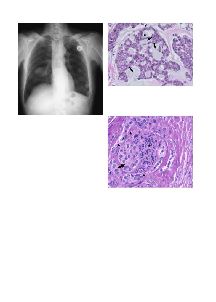
Fig. 1.17. Diffuse lung metastasis in a patient with subglottic adenoid cystic carcinoma diagnosed 10 years earlier
solitary, well circumscribed, multilobular masses. Microscopically typically acinar cells with cytoplasmic periodic acid Schiff ’s reagent positive glycogen granules are the main component of the tumour. A more aggressive [papillocystic (Spiro et al. 1978), microcystic (Colmenero et al. 1991)] subgroup is increasingly being distinguished, making up about 15% of acinic cell carcinomas (Hoffman et al. 1999) and requiring more aggressive treatment.
1.2.2.2.3.4
Adenocarcinoma Not Otherwise Specified (NOS)
Quite frequently a salivary gland adenocarcinoma lacks specific features allowing the pathologist to make a more specific diagnosis. About one in four salivary gland carcinomas cannot be accommodated in the other specific subtypes (Vander Poorten et al. 2003). Microscopically they range from well-differ- entiated and low grade to high grade, invasive lesions, displaying perineural growth (Fig. 1.19).
Acknowledgement
The author would like to express his sincere gratitude to Raf Sciot, MD PhD, for providing the illustrative microscopic images corresponding to the clinical examples displayed in this chapter.
Fig. 1.18. Adenoid cystic carcinoma of the tongue base. Strands of tumour cells (arrowheads) grow in a cribriform pattern on a mucinous background (arrows) thus shaped as pseudolumina. (Courtesy Raf Sciot, MD, PhD)
Fig. 1.19. Perineural growth in a high grade adenocarcinoma (not otherwise specified) of the parotid gland. Arrowheads demarcate perineurium, stretched by tumour cells (arrow), compressing the nerve bundles (asterisk). (Courtesy Raf Sciot, MD, PhD)
References
Acheson ED, Cowdell RH, Hadfield E et al (1968) Nasal cancer in woodworkers in the furniture industry. Br Med J 2:587– 596
Armstrong JG, Harrison LB, Spiro RH et al (1990) Malignant tumors of major salivary gland origin. A matched-pair analysis of the role of combined surgery and postoperative radiotherapy. Arch Otolaryngol Head Neck Surg 116:290–293
Batsakis JG (2003) Clinical pathology of oral cancer. In: Shah JP, Johnson NW, and Batsakis JG (eds) Oral cancer, 1st

14
edn. Martin Dunitz, Taylor and Francis Group, London, pp 77–123
Batsakis JG, el Naggar AK, Luna MA (1994) Hyalinizing clear cell carcinoma of salivary origin. Ann Otol Rhinol Laryngol 103:746–748
Blot WJ, McLaughlin JK, Winn DM et al (1988) Smoking and drinking in relation to oral and pharyngeal cancer. Cancer Res 48:3282–3287
Boffetta P, Trichopoulos D (2002) Cancer of the lung, larynx and pleura. In: Adami HO, Hunter D, Trichopoulos D (eds) Textbook of cancer epidemiology. Oxford University Press, Oxford, pp 268–272
Brugere J, Guenel P, Leclerc A et al (1986) Differential effects of tobacco and alcohol in cancer of the larynx, pharynx, and mouth. Cancer 57:391–395
Colmenero C, Patron M, Sierra I (1991) Acinic cell carcinoma of the salivary glands. A review of 20 new cases. J Craniomaxillofac Surg 19:260–266
Day GL, Blot WJ, Austin DF et al (1993) Racial differences in risk of oral and pharyngeal cancer: alcohol, tobacco, and other determinants. J Natl Cancer Inst 85:465–473
De Rienzo DP, Greenberg SD, Fraire AE (1991) Carcinoma of the larynx. Changing incidence in women. Arch Otolaryngol Head Neck Surg 117:681–684
De Stefani E, Ronco A, Mendilaharsu M et al (1999) Diet and risk of cancer of the upper aerodigestive tract – II. Nutrients. Oral Oncol 35:22–26
Delange F (2000) Iodine deficiency. In: Braverman LE, Utiger RD (eds) Werner and Ingbar’s the thyroid, 8th edn. Lippincott, Williams and Wilkins, Philadelphia, pp 295–296
Deveci MS, Deveci G, Gunhan O (2000) Oncocytic mucoepidermoid carcinoma of the parotid gland: report of a case with DNA ploidy analysis and review of the literature. Pathol Int 50:905–909
Ellis GL, Auclair PL (1996a) Salivary gland tumors: general considerations: site specific tumor differences. In: Ellis GL, Auclair PL (eds) Tumors of the salivary glands. Third Series. Armed Forces Institute of Pathology, Washington DC, p 32
Ellis GL, Auclair PL (1996b) The normal salivary glands. In: Ellis GL, Auclair PL (eds) Tumors of the salivary glands. Third Series. Armed Forces Institute of Pathology, Washington DC, pp 1–23
Eneroth CM, Zetterberg A (1974) Malignancy in pleomorphic adenoma. A clinical and microspectrophotometric study. Acta Otolaryngol 77:426–432
Franceschi S, Munoz N, Bosch XF et al (1996) Human papillomavirus and cancers of the upper aerodigestive tract: a review of epidemiological and experimental evidence. Cancer Epidemiol Biomarkers Prev 5:567–575
Gallo O (2001) Aetiology and molecular changes in salivary gland tumours. In: Mc Gurk M, Renehan AG (eds) Controversies in the management of salivary gland tumours, 1st edn. Oxford University Press, Oxford, pp 13–23
Gallo O, Bocciolini C (1997)Warthin’s tumour associated with autoimmune diseases and tobacco use. Acta Otolaryngol 117:623–627
Gnepp DR (1993) Malignant mixed tumors of the salivary glands: a review. Pathol Annu 28:279–328
Greenlee RT, Hill-Harmon MB, Murray T et al (2001) Cancer statistics, 2001. CA Cancer J Clin 51:15–36
Hay ID, Klee GG (1993) Thyroid cancer diagnosis and management. Clin.Lab Med 13:725–734
V. Vander Poorten
Healey WV, Perzin KH, Smith L (1970) Mucoepidermoid carcinoma of salivary gland origin. Classification, clinicalpathologic correlation, and results of treatment. Cancer 26:368–388
Heller KS, Attie JN (1988) Treatment of Warthin’s tumor by enucleation. Am J Surg 156:294–296
Hoffman HT, Karnell LH, Robinson RA et al (1999) National Cancer Data Base report on cancer of the head and neck: acinic cell carcinoma. Head Neck 21: 297–309
IARC (1997) Cancer incidence in five continents, vol.VII. IARC Sci Publ (143):i–xxxiv, 1–1240
IARC CancerBase N°4. EUCAN (1999) Cancer incidence, mortality and prevalence in the European Union 1997, version 4.0. IARC Press, Lyon
Jeannel D, Bouvier G, Hubert A (1999) Nasopharyngeal cancer. In: Newton R, Beral V, Weiss RA (eds) Infections and human cancer. Cold Spring Harbor Press, Plainview, New York, pp 125–156
Kane WJ, McCaffrey TV, Olsen KD et al (1991) Primary parotid malignancies. A clinical and pathologic review. Arch Otolaryngol Head Neck Surg 117:307–315
Kopp P, Kimura ET, Aeschimann S et al (1994) Polyclonal and monoclonal thyroid nodules coexist within human multinodular goiters. J Clin Endocrinol Metab 79:134–139
La Vecchia C, Tavani A, Franceschi S et al. (1997) Epidemiology and prevention of oral cancer. Oral Oncol. 33:302–312 Lefebvre J, Lartigau E, Kara A et al (2001) Oral cavity, pharynx
and larynx cancer. Environment-related prognostic factors. In: Gospodarowicz MK, Henson DE, Hutter RVP, O’Sullivan BO, Sobin LH,Wittekind Ch (eds) UICC prognostic factors in cancer, 2nd edn. Wiley-Liss, New York, pp 151–165
Lydiatt WM, Lydiatt DD (2001) The larynx: early stage disease. In: Shah JP (ed) Cancer of the head and neck, 1st edn. BC Decker Inc., Hamilton, London, pp 169–184
Maruya S, Kurotaki H, Fujita S et al (2001) Primary chondrosarcoma arising in the parotid gland. ORL J Otorhinolaryngol Relat Spec 63:110–113
Medina JE, Dichtel W, Luna MA (1984) Verrucous-squamous carcinomas of the oral cavity. A clinicopathologic study of 104 cases. Arch Otolaryngol 110:437–440
Moley JF (1995) Medullary thyroid cancer. Surg Clin North Am 75:405–420
Morse DE, Katz RV, Pendrys DG et al (1996) Smoking and drinking in relation to oral epithelial dysplasia. Cancer Epidemiol Biomarkers Prev 5:769–777
Parkin DM, Pisani P, Ferlay J (1999) Global cancer statistics. CA Cancer J Clin 49:33–64, 1
Pedersen EA, Hogetveit C, Andersen A (1973) Cancer of respiratory organs among workers at a nickel refinery in Norway. Int J Cancer 12:32–41
Russell WO, Ibanez ML, Clark RL (1963) Thyroid carcinoma. Classification, intraglandular dissemination, and clinicopathological study based upon whole organ sections of 80 glands. Cancer 16:1425–1460
Saku T, Hayashi Y, Takahara O et al (1997) Salivary gland tumors among atomic bomb survivors, 1950–1987. Cancer 79:1465–1475
Seifert G, Sobin LH (1992) The World Health Organization’s histological classification of salivary gland tumors. A commentary on the second edition. Cancer 70:379–385
Shaha AR, Loree TR, Shah JP (1995) Prognostic factors and risk group analysis in follicular carcinoma of the thyroid. Surgery 118:1131–1136

Introduction: Epidemiology, Risk Factors, Pathology, and Natural History of Head and Neck Neoplasms |
15 |
Silverman S Jr, Gorsky M, Kaugars GE (1996) Leukoplakia, dysplasia, and malignant transformation. Oral Surg Oral Med Oral Pathol 82:117
Spiro JD, Spiro RH (2001) Salivary tumors. In: Shah JP, Patel SG (eds) Cancer of the head and neck, 1st edn. Decker BC Inc, Hamilton, London, pp 240–250
Spiro RH (1986) Salivary neoplasms: overview of a 35-year experience with 2,807 patients. Head Neck 8:177–184
Spiro RH, Huvos AG (1992) Stage means more than grade in adenoid cystic carcinoma. Am J Surg 164:623–628
Spiro RH, Huvos AG, Strong EW (1978) Acinic cell carcinoma of salivary origin. A clinicopathologic study of 67 cases. Cancer 41:924–935
Tezelman S, Clark OH (1995) Current management of thyroid cancer. Adv Surg 28:191–221
UNSCEAR (2000) The United Nations Scientific Committee on the Effects of Atomic Radiation. Health Phys 79: 314
Vander Poorten VLM, Balm AJM, Hilgers FJM et al (1999a)
Prognostic factors for long term results of the treatment of patients with malignant submandibular gland tumors. Cancer 85:2255–2264
Vander Poorten VLM, Balm AJM, Hilgers FJM et al (1999b) The development of a prognostic score for patients with parotid carcinoma. Cancer 85:2057–2067
Vander Poorten VLM, Balm AJM, Hilgers FJM et al (2000). Stage as major long term outcome predictor in minor salivary gland carcinoma. Cancer 89:1195–1204
Vander Poorten VLM, Hart AA, van der Laan BF et al. (2003) Prognostic index for patients with parotid carcinoma: external validation using the nationwide 1985–1994 Dutch Head and Neck Oncology Cooperative Group database. Cancer 97:1453–1463
World Health Organization Collaborating Centre for Oral Precancerous Lesions (1978) Definitions of leukoplakia and related lesions: an aid to studies on oral precancer. Oral Surg Oral Med Oral Pathol 46:518–539

Clinical and Endoscopic Examination of the Head and Neck |
17 |
2Clinical and Endoscopic Examination of the Head and Neck
Pierre Delaere
CONTENTS
2.1Introduction 17
2.2Neck 17
2.3 |
Nose and Paranasal Sinuses 20 |
2.4Nasopharynx 21
2.5Oral Cavity 22
2.6Oropharynx 23
2.7Larynx 24
2.8 |
Hypopharynx and Cervical Esophagus 26 |
2.9Salivary Glands 27
2.10Thyroid Gland 28
2.11 |
Role of Imaging Studies 28 |
|
References 29 |
2.1 Introduction
Head and neck neoplasms present with variable signs and symptoms, depending on their site of origin and extension pattern. Thorough clinical examination, aided by modern endoscopic devices, is a cornerstone of the preand posttherapeutic evaluation of the patient suffering head and neck cancer. This chapter reviews the possibilities, but also the limitations of the clinical examination for each of the major subsites in the head and neck region.
2.2 Neck
Clinical examination still remains an important method of assessing regional lymph nodes. The presence of a clinically palpable, unilateral, firm, enlarged lymph node in the adult should be considered metastatic until proven otherwise. External examination
P. Delaere, MD, PhD
Professor, Department of Otorhinolaryngology, Head and
Neck Surgery, University Hospitals Leuven, Herestraat 49,
3000 Leuven, Belgium
of the neck represents an important starting point in the examination of the patient. It is important to remember that some cervical masses may escape the very best surgical palpation. It is essential that an orderly and systematic examination of the lymphatic fields on both sides of the neck is performed (Stell and Maran 1972).
Regional lymphatic drainage from the mucosa of the upper aerodigestive tract, salivary glands, and the thyroid gland occurs to specific regional lymph node groups (Shah 1990). They should be appropriately addressed in treatment planning for a given primary site. The major lymph node groups of the head and neck are shown in Fig. 2.1. Cervical lymph nodes in the lateral aspect of the neck primary drain the mucosa of the upper aerodigestive tract. These include the submental and submandibular group of lymph nodes located in the submental and submandibular triangles of the neck. Deep jugular lymph nodes include the jugulodigastric, jugulo-omohy- oid, and supraclavicular group of lymph nodes adjacent to the internal jugular vein. Lymph nodes in the posterior triangle of the neck include the accessory chain of lymph nodes located along the spinal accessory nerve and the transverse cervical chain of lymph nodes in the floor of the posterior triangle of the neck. The retropharyngeal lymph nodes are at risk of metastatic dissemination from tumors of the pharynx. The central compartment of the neck includes the delphian lymph node overlying the thyroid cartilage in the midline draining the larynx, and the perithyroid lymph nodes adjacent to the thyroid gland. Lymph nodes in the tracheoesophageal groove provide primary drainage to the thyroid gland as well as the hypopharynx, subglottic larynx, and cervical esophagus. Lymph nodes in the anterior superior mediastinum provide drainage to the thyroid gland and the cervical esophagus.
The localisation of a palpable metastatic lymph node often indicates the potential source of a primary tumor. In Fig. 2.1 the regional lymph node groups draining a specific primary site as first echelon lymph nodes are depicted.

18
Submandibular
Jugulodigastric (upper jugular)
Spinal accessory chain
Jugulo-omohyoid (mid-jugular)
Transverse cervical
Supraclavicular (lower jugular)
Tracheoesophageal groove
P. Delaere
Lower lip, fl oor of mouth, lower gum
Face, nose, paranasal sinuses, oral cavity, submandibular gland
Anterior scalp, forehead, parotid
Oral cavity, oropharynx, nasopharynx, hypopharynx, supraglottic larynx
Delphian node (larynx)
Thyroid, larynx, hypopharynx, cervical esophagus
Nasopharynx, thyroid, esophagus, lung, breast
Nasopharynx, thyroid, esophagus, lung, breast
Fig. 2.1. The regional lymph nodes of the head and neck region; the major regional lymphatic chains are annotated on the left. These regional lymph node groups drain a specific primary site as first echelon lymph nodes (indicated on right)
In order to establish a consistent and easily reproducible method for description of regional cervical lymph nodes, providing a common language between the clinician, the pathologist, and radiologist, the Head and Neck Service at Memorial Sloan-Kettering Cancer Center has described a leveling system of cervical lymph nodes (Fig. 2.2). This system divides the lymph nodes in the lateral aspect of the neck into five nodal groups or levels. In addition, lymph nodes in the central compartment of the neck are assigned levels VI and VII.
• Level I: Submental group and submandibular group. Lymph nodes in the triangular area bounded by the posterior belly of the digastric muscle, the inferior border of the body of the mandible, and the hyoid bone.
•Level II: Upper jugular group. Lymph nodes around the upper portion of the internal jugular vein and the upper part of the spinal accessory nerve, extending from the base of the skull up to the bifurcation of the carotid artery or the hyoid bone. Surgical landmarks: base of skull superiorly, posterior belly of digastric muscle anteriorly, posterior border of the sternocleidomastoid muscle posteriorly, and hyoid bone inferiorly.
•Level III: Mid-jugular group. Lymph nodes around the middle third of the internal jugular vein. Surgical landmarks: hyoid bone superiorly, lateral limit of the sternohyoid muscle anteriorly, the posterior border of sternocleidomastoid muscle posteriorly, and the caudal border of the cricoid cartilage inferiorly.
•Level IV: Lower jugular group. Lymph nodes around the lower third of the internal jugular. Surgical landmarks: cricoid superiorly, lateral limit of the sternohyoid muscle anteriorly, posterior border of the sternocleidomastoid muscle posteriorly, and clavicle inferiorly.
•Level V: Posterior triangle group. Lymph nodes around the lower portion of the spinal accessory nerve and along the transverse cervical vessels. It is bounded by the triangle formed by the clavicle, posterior border of the sternomastoid muscle, and the anterior border of the trapezius muscle.
•Level VI: Central compartment group. Lymph nodes in the prelaryngeal, pretracheal, (Delphian), paratracheal and tracheoesophageal groove. The boundaries are: hyoid bone to suprasternal notch and between the medial borders of the carotid sheaths.

Clinical and Endoscopic Examination of the Head and Neck |
19 |
Fig. 2.2. Level system of cervical lymph nodes: 7 levels are distinguished (labeled I to VII)
•Level VII: Superior mediastinal group. Lymph nodes in the anterior superior mediastinum and tracheoesophageal grooves, extending from the suprasternal notch to the innominate artery.
Some nodes in the neck are more difficult to palpate than others. Thus the retropharyngeal and highest parajugular nodes are almost impossible to detect by palpation until they are very large.
Structures in the neck which may be mistaken for enlarged lymph nodes are the transverse process of the atlas, the carotid bifurcation and the submandibular salivary gland.
Physical examination of the neck for lymph node metastasis has a variable reliability (Watkinson et al. 1990). A meta-analysis comparing computed tomography (CT) with physical examination (PE) yielded the following results: sensitivity, 83% (CT) vs 74% (PE); specificity, 83% (CT) vs 81% (PE); and accuracy, 83% (CT) vs 77% (PE). Overall, PE identified 75% of pathologic cervical adenopathy; this detection rate increased to 91% with addition of CT (Merritt et al. 1997).
The American Joint Committee on Cancer and the International Union against Cancer has agreed upon a uniform staging system for cervical lymph nodes. The exact description of each N stage of lymph node metastasis from squamous carcinomas of the head
and neck is described in Table 2.1. Squamous carcinomas of the nasopharynx and well-differentiated thyroid carcinomas have a different biology and cervical metastases from these tumors are assigned different staging systems.
An enlarged metastatic cervical lymph node may be the only physical finding present in some patients whose primary tumors are either microscopic or occult at the time of presentation. A systematic search for a primary tumor should be undertaken in these patients prior to embarking upon therapy for the metastatic nodes. If a thorough head and neck ex-
Table 2.1. N staging of lymph node metastasis from squamous cell carcinoma of the head and neck except nasopharynx and thyroid gland (AJCC/UICC, 2002)
Nx Regional lymph nodes cannot be assessed
N0 No regional lymph node metastasis
N1 Metastasis in a single ipsilateral lymph node, < 3 cm in greatest dimension
N2a Metastasis in single ipsilateral lymph node > 3 cm but < 6 cm in greatest dimension
N2b Metastasis in multiple ipsilateral lymph nodes, none > 6 cm in greatest dimension
N3 Metastasis in a lymph node > 6 cm in greatest dimension

20 |
P. Delaere |
amination, including fiberoptic nasolaryngoscopy, CT or MRI study, and PDG-PET scan, fails to show a primary tumor, then the diagnosis of metastatic carcinoma to a cervical lymph node from an unknown primary is established.
2.3
Nose and Paranasal Sinuses
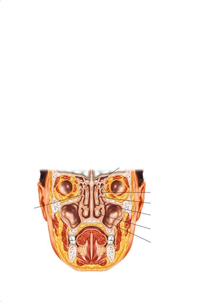
The nasal cavity is the beginning of the upper airway and is divided in the midline by the nasal septum. Laterally, the nasal cavity contains the nasal conchae, the inferior concha being part of the nasal cavity, and the superior and middle conchae being composite parts of the ethmoid complex. The nasal cavity is surrounded by air containing bony spaces called paranasal sinuses, the largest of which, the maxillary antrum, is present on each side. The ethmoid air cells occupy the superior aspect of the nasal cavity, and separate it from the anterior skull base at the level of the cribriform plate. Superoanteriorly, the frontal sinus contained within the frontal bone forms a biloculated pneumatic space. The sphenoid sinus at the superoposterior part of the nasal cavity is located at the roof of the nasopharynx. The adult ethmoid sinus is narrowest anteriorly in a section known as the os-
tiomeatal complex and this is the site of drainage of the maxillary and frontal sinuses (Fig. 2.3).
Since all of the paranasal sinuses are contained within bony spaces, primary tumors of epithelial origin seldom produce symptoms until they are of significant dimensions, causing obstruction, or until they have broken through the bony confines of the involved sinus cavity. Tumors of the nasal cavity often produce symptoms of nasal obstruction, epistaxis, or obstructive pansinusitis early during the course of the disease. Unilateral epistaxis, obstruction, or sinusitis should raise the index of suspicion regarding the possibility of a neoplastic process. Tumors of the maxillary antrum may present with symptoms of obstructive maxillary sinusitis. Swelling of the upper gum or loose teeth may be the first manifestation of a malignant tumor of the maxillary antrum. Locally advanced tumors may present with anesthesia of the skin of the cheek and upper lip, diplopia, proptosis, nasal obstruction, epistaxis, a mass in the hard palate or upper gum, or a soft tissue mass in the upper gingivobuccal sulcus. Advanced tumors may present with trismus and visible or palpable fullness of the check. Trismus usually is a sign of pterygoid musculature invasion. Epistaxis may be the first manifestation of tumors of the ethmoid or frontal sinus. This may be accompanied by frontal headaches or diplopia. Occasionally anosmia may be present in patients
Ostiomeatal complex
Frontal sinus
Ethmoid air cells
Uncinate process
Middle concha
Inferior concha
Maxillary antrum
Fig. 2.3. Coronal section through maxillofacial region, showing proximity of orbit and anterior cranial fossa to nasal cavity and paranasal sinuses. Disease of the sinuses and nasal cavity may spread directly into adjacent structures with catastrophic results

Clinical and Endoscopic Examination of the Head and Neck |
21 |
with esthesioneuroblastoma. Anesthesia in the distribution of the fifth cranial nerve or paralysis of the third, fourth or sixth cranial nerve may be the first manifestation of a primary tumor of the sphenoid sinus. Although sinonasal malignancy is rare, persistent nasal symptoms should always be investigated, particularly if unilateral. Tumors of the nasal cavity and paranasal sinuses are the most challenging to stage. Endoscopic evaluation of the nasal cavity is crucial in accurate clinical assessment of an intranasal lesion. Fiber optic flexible endoscopy provides adequate visualization of the lower half of the nasal cavity. Therefore, lesions presenting in the region of the inferior turbinate, middle meatus, and the lower half of the nasal septum can be easily visualized by office endoscopy.
Rigid endoscopic evaluation with telescopes generally requires adequate topical anesthesia as well as shrinkage of the mucosal surfaces of the interior of the nasal cavity with the use of topical cocaine. A set of 0°, 30°, 70° and 90° telescopes should be available for appropriate evaluation of the interior of the nasal cavity (Fig. 2.4). Diagnostic nasal endoscopy allows the characterization of intranasal anatomy and identification of pathology not otherwise visible by traditional diagnostic techniques, such as the use of a headlight, speculum, and mirror (Bolger and
Kennedy 1992; Levine 1990).
2.4 Nasopharynx
The nasopharynx is the portion of the pharynx bounded superiorly by the skull base and the sphe-
noid and laterally by the paired tori of the eustachian tubes, with the Rosenmüller’s fossa. Anteriorly the posterior choanae form the limit of the space, and inferiorly an artificial line drawn at the level of the hard palate delimits the nasopharynx from the oropharynx.
Presenting symptoms of nasopharyngeal cancer may include a neck mass, epistaxis, nasal obstruction, otalgia, decreased hearing, or cranial neuropathies. Approximately 85% patients have cervical adenopathy and 50% bilateral neck involvement (Lindberg 1972). Serous otitis media may occur due to Eustachian tube obstruction. Cranial nerve VI is most frequently affected but multiple cranial nerves may be involved.
Nasopharyngeal carcinoma has a tendency for early lymphatic spread. The lateral retropharyngeal lymph node (of Rouvier) is the first lymphatic filter but is not palpable. The common first palpable node is the jugulodigastric and/or apical node under the sternomastoid which are second echelon nodes. Bilateral and contralateral lymph node metastases are not uncommon.
Nasendoscopy (Fig. 2.5) using the flexible scope gives a good view of the nasal floor, the walls of the nasopharynx and the fossa of Rosenmüller. Nasopharyngeal tumors in any quadrant including the fossa of Rosenmüller can be seen and accurately biopsied. For the nasopharynx, also rigid 0° and 30° sinus endoscopes can be similarly used in the clinical setting. Under anaesthesia, should this be necessary, these are the scopes of choice for visual assessment and biopsy.
Evidence of lower cranial nerve deficits may be apparent from palatal or glossal paralysis and atrophy. A full evaluation of the remaining cranial nerves
Fig. 2.4. The rigid endoscope allows for detailed examination of the nasal cavity. The scope can be rotated laterally under the middle turbinate into the posterior aspect of the middle meatus (asterisk). An excellent view of the middle turbinate, uncinate process, and surrounding mucosa can be obtained

22 |
P. Delaere |
Sphenoid sinus
Torus tubarius
Opening of eustachian tube
Rosenmuller’s fossa
Fig. 2.5. Examination of nasopharynx with flexible scope
should include visual assessment and examination of the tympanic membranes.
2.5
Oral Cavity
The oral cavity extends from the vermilion borders of the lips to the junction of the hard and soft palates superiorly and to the line of the circumvallate papillae inferiorly.Within this area are the lips, buccal mucosa, alveolar ridges with teeth and gingiva, retromolar trigone, floor of mouth, anterior two-thirds of the tongue, and hard palate (Fig. 2.6).
All mucosal surfaces of the mouth require thorough and systematic examination. The oral cavity is lined by a mucous membrane which is a non-keratin- izing stratified squamous epithelium and is therefore pink. It contains taste buds and many minor salivary glands. All mucosal surfaces should be examined using tongue blades under optimal lighting conditions.
The clinical features of the primary tumors arising in the mucosal surface of the oral cavity are variable. The tumor may be ulcerative, exophytic or endophytic, or it may be a superficial proliferative lesion. Most patients with a mouth cancer present with a painful ulcer. Squamous carcinomas with excessive keratin production and verrucous carcinomas present as white heaped-up keratotic lesions with varying degrees of
keratin debris on the surface. Bleeding from the surface of the lesion is a characteristic for malignancy and should immediately raise the suspicion for a neoplastic process. Endophytic lesions have a very small surface component but have a substantial amount of soft tissue involvement beneath the surface.
Oral salivary tumors may present as a nodule, a non-ulcerative swelling or more usually as an ulcerative lesion. Metastatic tumors may also present as submucosal masses. Mucosal melanoma shows characteristic pigmentation.
Macroscopic lesions should be evaluated for mobility, tenderness and be palpated with the gloved finger to detect submucosal spread. This is particularly important in tongue lesions extending posteriorly into the posterior third and tongue base. The distance from the tumor to the mandible and the mobility of the lesion in relation to the mandible are critical elements in determining the management of perimandibular cancers. The indications for examination under anesthesia include an inadequate assessment of the extent of the disease by history and physical examination and imaging, or the presence of symptoms referable to the trachea, larynx, hypopharynx and esophagus that need endoscopic assessment. It is not cost-effective screening to perform panendoscopy on all patients with oral cavity cancer (Benninger et al. 1993; Hordijk et al. 1989).
Palpation of the neck is of course essential in the assessment of a patient with mouth cancer. Neck

Clinical and Endoscopic Examination of the Head and Neck |
23 |
nodal disease is the single most important factor de- |
|
termining the method of treatment, and also prog- |
|
nosis is determined by the presence of metastatic |
|
nodes. Full examination of the neck must be carried |
|
out to detect any lymph node metastases and each |
|
level must be carefully palpated, particularly the up- |
|
per and middle deep cervical nodes deep to the ster- |
|
nomastoid, from behind the patient, using the tips |
|
of the fingers. Carcinoma of the oral tongue has the |
|
greatest propensity for metastasis to the neck among |
|
all oral cancers. The primary echelon of drainage is |
|
level II but other levels may be also involved.
|
Circumvallate |
|
papillae |
2.6 |
Foramen caecum |
Oropharynx |
|
The oropharynx is that part of the pharynx which extends from the level of the hard palate above to the hyoid bone below. The anterior wall of the oropharynx is formed by the base or posterior third of the tongue bounded anteriorly by the v-shaped line of circumvallate papillae (Fig. 2.6). When present, the initial symptoms of oropharyngeal cancer are often vague and non-specific, leading to a delay in diagnosis. Consequently, the overwhelming majority of patients present with locally advanced tumors.
Presenting symptoms may include sore throat, foreign-body sensation in the throat, altered voice or referred pain to the ear that is mediated through the glossopharyngeal and vagus nerves. Over two-thirds of patients present with a neck lump. As the tumor grows and infiltrates locally, it may cause progressive impairment of tongue movement which affects speech and swallowing.
Most tumors of the oropharynx can be easily seen with good lighting, but those originating in the lower part of the oropharynx and tongue base are best viewed with a laryngeal mirror. The patient should be asked to protrude the tongue, to rule out injury to the hypoglossal nerve. Trismus is a sign of invasion of the masticator space. Sensory and motor function should be assessed, particularly mobility of the tongue as well as fixation. Fiberoptic nasopharyngeal endoscopy has greatly enhanced the ease of examination of these tumors, particularly in assessing the lower extent of the tumor and also the superior extent if the nasopharynx is involved. The extent of involvement is often underestimated on inspection, and bimanual palpation of the tumor must be undertaken in all patients. Careful palpation should be carried out to estimate the extent of infiltration, but this examina-
Fig. 2.6. Oral cavity and oropharynx. The posterior limits of the oral cavity are the anterior tonsillar pillars, the junction of the anterior two-thirds and posterior one-third of the tongue (i.e. the circumvallate papillae) and the junction of the hard and soft palate. The soft palate and the tonsil are therefore part of the oropharynx. Carcinoma of the anterior two-thirds of the tongue is the most frequent site for a mouth cancer and the lateral border (1) is the most common location. Carcinoma of the floor of the mouth most commonly occurs anteriorly either in the midline or more usually to one side of the midline (2). Carcinoma of the oropharynx most commonly occurs in the slit between tonsil and base of tongue, at the level of the anterior tonsillar pillar (3)
tion may be limited by patient tolerance; thorough palpation under general anaesthetic is advisable. Advanced tumors that cause trismus may also be better assessed under a general anesthetic. A detailed examination and biopsy under general anaesthetic may be the only accurate method of assessing the extent of tumors such as those of the tongue base that may be in a submucosal location.
Examination of the neck must be carried out systematically and each level must be carefully palpated to detect lymph node enlargement or deep invasion of the tumor.
Nodal metastases from squamous cell carcinomas are typically hard and when small are generally mobile. As they enlarge, those in the deep cervical chain initially become attached to the structures in the carotid sheath and the overlying sternomastoid muscle
