

Учебники / Head_and_Neck_Cancer_Imaging
.pdfIntroduction: Epidemiology, Risk Factors, Pathology, and Natural History of Head and Neck Neoplasms |
3 |
Fig. 1.2. Relative risk for oropharyngeal cancer for males according to amount of tobacco and alcohol use. (Based on data from Blot et al. (1988), with permission)
habits. There is also a lower level of oral hygiene. Once cancer is established, people in lower socioeconomic groups will seek medical help later, because of less education and more difficult access to the health system, and thus will present with more advanced stages of disease. Another reason for advanced disease at presentation lies in the fact that often a significant part of the intake of calories is provided for by alcohol and thus symptoms such as dysphagia for solid food will become problematic later. Stage at presentation is the strongest negative prognostic factor for outcome of treatment in HNSCC. Treatment of this disease, especially of the advanced stages, has often a serious impact on physical and psychological functioning of the patient. A great deal of effort is needed to become adapted to the resulting altered body image, to get integrated back into society, and also to achieve the change in lifestyle needed to prevent occurrence of second primary HNSCC. Unfortunately, immediately following treatment, patients in lower social classes are often isolated in facing this difficult challenge. Return to occupation thus becomes an illusion for the majority of these patients (Lefebvre et al. 2001). It is clear that a lower socioeconomic environment is a strong negative prognostic factor for the oncologic results following treatment, and also for
survival in general because of the impact of many of the comorbidities that result from the previous way of living (pulmonary insufficiency, atherosclerosis, liver disease, etc.).
Viral infections have also been implicated in the carcinogenesis of HNSCC (Franceschi et al. 1996). Human papilloma virus DNA is prevalent in about 50% of oral cancer case series. Relative risks up to 6.2 for development of oral cancer have been reported. Epstein-Barr virus (EBV) has also been strongly associated with nasopharyngeal cancer. EBV antibody titers are much higher in cases than in controls, and biopsy specimens of undifferentiated nasopharyngeal carcinoma patients are 100% EBV positive and monoclonal as to this virus (Jeannel et al. 1999). EBV titers following treatment are used to monitor patients for disease recurrence. Patients infected with the human immunodeficiency virus (HIV) are at higher risk of developing HNSCC and Kaposi’s sarcoma.
Among environmental factors, chronic sun exposure has been proven to induce development of skin and lip cancer. Occupational factors have been implied in HNSCC development. Working in industry that is associated with higher exposure to aromatic amines and phenoxy herbicides confides an elevated

4 |
V. Vander Poorten |
risk for all sites. A specific and strong association has been repeatedly described between working in specific industries and the development of sinonasal cancer. The rate of development of SCC of the sinonasal tract has been observed to be increased 250 times in workers exposed to nickel (Pedersen et al. 1973). Working with wood in environments where there is no aspiration system for the dust particles has been found to result in a 500to 1000-fold increase in the baseline incidence of sinonasal “intestinal type” adenocarcinoma and has led in several countries to the recognition of this cancer as an occupational disease and to the introduction of stringent safety precautions to minimize dust exposure (Acheson et al. 1968).
1.1.2.2
Risk Factors for Development of Glandular Neoplasms
Radiation exposure is the only firmly established environmental risk factor for the development of thyroid carcinoma. The information comes from scrutinized follow-up of atomic bomb survivors in Japan and atomic disaster survivors in Chernobyl (UNSCEAR: The United Nations Scientific Committee on the Effects of Atomic Radiation 2000) Typically a low dose exposure (e.g. about 3 Gy) results in the development of mainly papillary thyroid carcinoma, some 5–10 years later. The risk follows a linear dose– effect relationship and the incidence can be increased by more than 30 times.
Radiation exposure has also been observed to result in an increased incidence of both benign (Warthin’s tumour) and malignant (mucoepidermoid carcinoma) salivary gland tumours in followup studies (Saku et al. 1997) in the same cohorts. For Warthin’s tumour, also a doubled incidence has been observed in smokers versus non-smokers (Gallo and Bocciolini 1997). Epstein-Barr virus has been implicated in the genesis of bilateral Warthin’s tumours and undifferentiated carcinoma of the salivary gland (Gallo 2001).
1.2
Pathology and Natural History of Frequent Benign and Malignant Head and Neck Neoplasms
With regard to tumour pathology, tumour typing is the first important subject. Within the different tu-
mour types, the second subject is then detection of features with prognostic significance, such as grading, perineural or vascular invasion, and the assessment of radicality of resection margins. Regarding histological typing of head and neck neoplasms, it has already been mentioned that a great majority consists of primary epithelial neoplasms of the mucous membranes of the upper aerodigestive tract. A detailed discussion of all different tumour types that can be encountered in the head and neck area is beyond the scope of this introductory chapter and for this the reader is referred to surgical and pathological literature. What follows is an overview of the clinical course and pathological specificities of the most frequent tumour types.
1.2.1
Epithelial Neoplasms of the Mucous Membranes
1.2.1.1
Tumour Typing and Clinical Behaviour
1.2.1.1.1 Benign Lesions
Benign papillary lesions do occur, albeit that they are less frequently a reason for seeking medical attention than the malignant and premalignant lesions. Oral, pharyngeal and laryngeal sites can display squamous papillomas, which are white lesions with a wart-like appearance that have no signs of invasion of the deeper structures. A viral etiology in a part of these lesions (e.g. juvenile laryngeal papillomatosis) has been ascribed to HPV (type 6 and 11). Sinonasal papillomas are also called Schneiderian papillomas and can be exophytic, endophytic (inverted) or oncocytic in presentation. Especially the inverted type papilloma is considered a premalignant condition (Fig. 1.3). Most of the symptomatic epithelial neoplasms of the mucous membranes of the upper aerodigestive tract that bring patients to the doctor will turn out to be premalignant or malignant.
1.2.1.1.2 Premalignant Lesions
Premalignant lesions will often not be visualized on routine imaging studies. On the macroscopic level we consider leukoplakia and related lesions: homogeneous leukoplakia versus non-homogeneous leukoplakia (nodular leukoplakia, erythroplakia, proliferative verrucous leukoplakia; Fig. 1.4). On the

Introduction: Epidemiology, Risk Factors, Pathology, and Natural History of Head and Neck Neoplasms |
5 |
Fig. 1.3. Inverted papilloma (arrow) of the maxillary sinus being removed by Caldwell-Luc approach (antrostomy: arrowheads)
Fig. 1.4. Erythroplakia (arrowheads) with areas of nodular leukoplakia (arrows) of the tonsil, glossotonsillar sulcus, anterior tonsillar pillar, and hard and soft palate
microscopic level epithelial hyperplasia, dysplasia and carcinoma in situ can be discerned.
Leukoplakia is a descriptive clinical term use to describe “a white plaque or patch that cannot be characterized, clinically or histopathologically, as any other disease” (World Health Organization Collaborating Centre for Oral Precancerous Lesions 1978). Furthermore, in order to be designated as leukoplakia, the lesion should not be associated with any known physical (frictional keratosis, candidal leukoplakia) or chemical causal agent, except with the use of tobacco. It should also be impossible to scrape off the lesion.
Homogeneous leukoplakia is histologically either hyperorthoor hyperparakeratosis and rarely shows associated dysplasia. Less frequently we are dealing with non-homogeneous leukoplakia (nodular leukoplakia, erythroplakia, proliferative verrucous leukoplakia), which is usually associated with dysplasia and thus much more at risk for becoming real malignant disease (Batsakis 2003). Dysplasia can be “mild”, meaning there is an increased number of mitotic figures and an abnormal cytologic appearance (loss of an orderly nuclear mosaic pattern: decreased nuclear/cytoplasmatic ratio and an irregular random nuclear placement) only in the basal layer of the epithelium, whereas suprabasal mitosis and cytologic abnormality indicates “moderate” dysplasia. In “severe” dysplasia the atypical cells with mitotic activity can be observed everywhere from the basal to the most superficial layers. The yearly rate of malignant transformation of homogeneous leukoplakia is estimated to be between 2% and 6% in the Western world, and is higher as the patient is older, female, and as the lesion persists for a longer time. The rate of malignant transformation in non-homogeneous (speckled) leukoplakia and erythroplakia can be more than 50% (Silverman et al. 1996).
1.2.1.1.3 Malignant Lesions
Less frequent entities with a specific clinical behaviour are verrucous carcinoma, papillary SCC, basaloid squamous cell carcinoma and sarcomatous SCC, increasingly aggressive in that order. Verrucous carcinoma is an exophytic papillomatous SCC that is a low grade tumour, very well differentiated, without known potential for regional or distant metastasis (Medina et al. 1984). Papillary SCC displays an exophytic growth with a poorly differentiated cell layer lining a central fibrovascular core. The behaviour of this type of tumour is more aggressive than verrucous carcinoma in that metastasis is observed. Basaloid SCC and sarcomatoid SCC are highly aggressive variants of SCC.
Most of the malignancies of the mucous membranes are simply called “invasive squamous cell carcinoma” and can be graded into well-, moderatelyand poorly-differentiated SCC, paralleling the amount of keratin formation by cells. SCC cells by definition produce intercellular bridges (Figs. 1.5 and 1.6). Absence of these intercellular bridges is one of the features of undifferentiated carcinoma of the upper aerodigestive tract. This type of tumour occurs most frequently in the nasopharynx and is often

6 |
V. Vander Poorten |
Fig. 1.5. Hemiglossectomy for ulcerative and deeply invasive well differentiated squamous cell carcinoma of the lateral tongue (arrowhead). Specimen in continuity with radical neck dissection (arrow)
Fig. 1.6. Microscopical appearance of the same tumour. Note the diffuse infiltrative aspect of the tumour islets (arrows), the dense mononuclear inflammatory reaction, and the formation of keratin pearls (vertical arrow). (Courtesy Raf Sciot, MD, PhD)
diagnosed because of the massive neck lymph node metastasis already present at initial diagnosis, frequently bilaterally, and involving the posterior neck (region V).
1.2.1.1.4
Natural History Before and At Diagnosis
As pointed out under “risk factors”, many patients with oral and pharyngeal cancer will present at an advanced stage of their disease, because of the late occurrence of symptoms and the social situation with more difficult access to the medical system. Patients
with glottic laryngeal cancer tend to present at earlier stages, given the rapid effect of even a small vocal cord lesion on the voice quality. Early glottic laryngeal cancer is also not very likely to result in regional metastasis and thus often has a good prognosis following radiotherapy or surgery, with 5-year survival rates of 70%–100% (Lydiatt and Lydiatt 2001). Advancing stage and origin of SCC in most other anatomical subsites of the upper aerodigestive tract, are associated with lesser chances for successful treatment, and for the specifics the reader is referred to specific head and neck oncological literature.
1.2.1.1.5
Natural History Following Diagnosis and Successful Treatment of Malignant HNSCC
The annual incidence of second primary cancer following successful treatment of an index SCC in the head and neck area is 3%–7%. A known feature in HNSCC is the observation of field cancerization of the upper aerodigestive tract: several synchronous and also metachronous primary carcinomas and areas of moderate to severe dysplasia – carcinoma in situ are observed with areas of normal mucous membranes in between. This is caused by exposure of the entire upper aerodigestive tract to the same carcinogens – usually combined alcohol and tobacco. Patients are especially at risk of developing lung cancer, esophageal and gastric cancer, and a new localization of HNSCC. As already discussed under “risk factors”, a change of lifestyle is essential to decrease the incidence of second primaries, but this is often difficult to achieve given the social context of the patient.
1.2.1.1.6
Microscopical Findings with Negative Prognostic Impact for Malignancy of the Mucous Membranes
The most important findings that are to be determined following resection of a primary head and neck SCC and the regional lymph nodes are listed in Table 1.1. They are routinely determined during microscopical analysis because they are known to carry a worse prognosis and thus contribute to decision making regarding the need for further therapy, i.e. postoperative radiotherapy. Many of these parameters (cTNM classification, perineural growth, tumour thickness, extracapsular spread in metastatic lymph nodes) can already be strongly suspected based on a high quality imaging study before the resection.

Introduction: Epidemiology, Risk Factors, Pathology, and Natural History of Head and Neck Neoplasms |
7 |
Table 1.1. Histopathological negative prognostic factors in
HNSCC
(p)TNM classification (size of primary tumor, number/laterality of positive nodes, size of largest node)
Vascular invasion
Perineural growth
Resection margins (e.g. less than 5 mm is considered “close margins” in oral cancer)
Thickness
Invasive front
Differentiation
Exophytic versus endophytic growth pattern
Field cancerization
Mitotic index
Presence of extracapsular spread in metastatic lymph nodes
Fig. 1.7. Large multinodular goitre with pharyngeal, esophageal and tracheal compression
1.2.2
Glandular Neoplasms
1.2.2.1
Thyroid Neoplasia
1.2.2.1.1
Benign Disease: Multinodular Enlargement
Benign multinodular enlargement (multinodular goitre) is very frequently observed and is estimated to affect almost one in three persons worldwide (Delange 2000). Iodine deficiency is the most frequent contributory factor. In areas where iodine supply is sufficient, the prevalence of clinically detectable goitres is usually less than 4%, and thought to result from elevated thyroid stimulating hormone (TSH) levels or from elevated stimulation of the TSH receptor (such as in Graves’ disease and non-atrophic Hashimoto’s goitres). Most patients with this condition are asymptomatic, and medical concerns mostly arise when compressive symptoms appear (Fig. 1.7), when autonomous hyperfunction becomes apparent, or when presence of malignancy is feared. The latter is the case in rapidly enlarging goitres, suspicion of enlarged lymph nodes,a history of prior radiotherapy to the neck, or when fine needle aspiration cytology (FNAC) of dominant nodules indicates papillary carcinoma or a microfollicular lesion. A microfollicular lesion can be follicular carcinoma in about one in ten patients.
Macroscopically, following thyroidectomy, we usually see a polynodular, soft, and globally enlarged thyroid gland (Fig. 1.8). There may be one or more dominant larger nodules which deserve subsequent
Fig. 1.8. The same goitre as in Fig. 1.7, following resection.
microscopical analysis. Up to 70% of hyperplastic nodules are clonal, neoplastic proliferations (Kopp et al. 1994). Microscopically, within the different nodules, there is a varied pattern of large and small follicles, usually with abundant colloid. There is an often oedematous stroma with fibrosis, macrophages, hemosiderin, and calcifications.
1.2.2.1.2
Benign Disease: Uninodular Enlargement – the Solitary Thyroid Nodule
Any clinically visible or palpable nodule, and“any discrete macroscopic intrathyroidal lesion that is clearly distinguishable from the adjacent normal thyroid parenchyma” (Hay and Klee 1993) on ultrasonography or Technetium scanning, should lead to action to estimate the chance of malignancy and to determine

8 |
V. Vander Poorten |
subsequent action. The next step will be to do an ultrasound guided FNAC. A solitary thyroid nodule can be due to degenerative lesions such as cysts or degenerative colloid nodules on one hand, or to neoplastic lesions on the other. The latter can be benign or malignant. The global incidence of cancer in patients with a thyroid nodule is 10%, but this increases to 30% for women older than 50 and to 45% for men older than 50 (Tezelman and Clark 1995).
The rest will be benign lesions, where nine out of ten will be follicular adenomas, the remainder being mostly Hürthle cell adenomas. Macroscopically, adenomas are well demarcated from the adjacent parenchyma, and fleshy and pale, sometimes cystic or hemorrhagic on cut surface. The microscopic appearance of a solitary adenomatous nodule displays large and small follicles with a lot of colloid and a stromal component with hemosiderin, macrophages, fibrotic changes and often calcifications. A Hürthle cell variant displays oncocytic cells, with an intensely eosinophylic cytoplasm due to a lot of abnormal mitochondria, and large vesicular nuclei.
1.2.2.1.3 Malignant Disease
this group). Overall women are affected three times as frequently as men. The clinical picture is usually a symptomless swelling in the thyroid area, although enlarged lymph nodes may be the primary presenting feature (Fig. 1.9). Indeed, regional metastasis to the paratracheal (level VI) and cervical (level II, III, and IV) lymph nodes is observed in one out of two patients at presentation (Fig. 1.10). Distant metastasis occurs usually late in the disease course.
Following thyroidectomy, macroscopically typical features are multifocality and bilaterality, which may
An important issue in suspected malignant thyroid disease is the avoidance of iodine containing contrast medium in imaging studies for thyroid lesions. “Differentiated thyroid cancer” (vide infra) are tumours that retain the ability to concentrate iodine, and hence can be effectively and very selectively treated with radioactive iodine. This treatment, however, will be delayed by 3 months following an imaging study using iodine contrast medium, due to saturation of the iodine binding capacity of the targeted thyroid cancer cells.
Generally a distinction is made between “differentiated thyroid cancer” with a relatively good (papillary carcinoma, follicular carcinoma, mixed papillary follicular carcinoma) to intermediate (Hürthle cell carcinoma) prognosis, and “other” cancers, with worse (medullary thyroid cancer) to fatal prognosis (anaplastic thyroid cancer). Of human malignant tumours, the thyroid harbours both the tumours with the best (papillary carcinoma) and the worst (anaplastic carcinoma of the thyroid) prognosis.
1.2.2.1.4
Papillary Thyroid Cancer
Papillary thyroid cancer is the most frequent thyroid cancer (four out of five thyroid cancers belong to
Fig. 1.9. Thyroidectomy specimen showing papillary carcinoma on cut surface in the left lobe and the isthmus. Posterior view. The specimen is inked to assess resection margins
Fig. 1.10. Functional neck dissection specimen showing a typical black cystic metastatic neck node of papillary thyroid carcinoma (arrow)

Introduction: Epidemiology, Risk Factors, Pathology, and Natural History of Head and Neck Neoplasms |
9 |
occur in up to 87% of thyroid specimens (Russell et al. 1963). Lymph node metastasis is often cystic and dark bluish in appearance (Fig. 1.10).
Microscopically only 3% is true papillary carcinoma and 97% is “follicular variant of papillary carcinoma”. Both forms have an equally good prognosis, with overall up to 95% of patients surviving 20 years following treatment (Hay and Klee 1993).
Essential for the diagnosis are the papillae with a central fibrovascular core and an epithelial lining showing the typical nuclear features with overlapping nuclei and nuclear grooves, making it possible to get the diagnosis from a fine needle aspiration cytology. Psammoma bodies, calcific concretions with concentric laminations, can be observed in about one in two of these tumours, and when present can also already be recognized on FNAC (Fig. 1.11).
1.2.2.1.5
Follicular Thyroid Cancer
About one in ten thyroid malignancies are follicular carcinomas. These are macroscopically solitary, encapsulated tumours. The features discriminating them from follicular adenomas, their benign counterparts, are microscopical: the presence of vascular invasion and full thickness capsular invasion into the adjacent normal thyroid parenchyma. To be able to search the entire capsule for areas of invasion all solitary nodules, where FNAC results in the diagnosis “follicular lesion”, should be completely excised with capsule and surrounding thyroid tissue. The degree of capsular invasion allows for the definition of a subgroup of minimally invasive follicular carcinomas, behaving essentially as follicular adenomas. The tendency for vascular invasion in invasive follicular carcinoma explains that metastasis is primarily haematogenous to the lungs and the bones, rather than to the cervical lymph nodes, as observed in papillary thyroid cancer. Prognosis is somewhat less than for papillary carcinoma, with an overall 20-year survival of 81% (Shaha et al. 1995).
1.2.2.1.6
Hürthle Cell Carcinoma
Hürthle cell carcinomas are also solitary, encapsulated tumours that are distinguished from their benign counterparts by the presence of capsular and vascular invasion. They have an intermediate prognosis of about 65% 20-year survival (Shaha et al. 1995).
Fig. 1.11. Microscopical appearance of papillary thyroid cancer. Psammoma body (arrow) and papillary growth pattern (arrowheads). (Courtesy Raf Sciot, MD, PhD)
1.2.2.1.7
Medullary Thyroid Cancer
Somewhat less than one in ten thyroid carcinomas are medullary thyroid carcinomas (MTC). MTC is a malignant tumour of the calcitonin-secreting parafollicular C cells of the thyroid.
These cells are embryologically maximally located in the upper two thirds of the thyroid gland and this explains that tumours are usually found in that upper part of the gland.
The tumours can occur in a sporadic form, which is usually unifocal, and presents in the age group of 40–60 years. This sporadic form is responsible for about 80% of MTC. An autosomal dominant hereditary form, due to a mutation in the retinoblastoma (RET) proto-oncogen, can occur within the framework of multiple endocrine neoplasia syndromes (MEN 2a: MTC, phaeochromocytoma, and parathyroid hyperplasia and MEN 2b: MTC, phaeochromocytoma and multiple mucosal neurinomas) or as familial medullary thyroid cancer (FMTC) without associated endocrinopathy. These hereditary forms usually occur earlier in life and are often multifocal and present in both thyroid lobes. In patients with MTC, lymph node metastasis, often bilateral, is frequently present at diagnosis and has a negative prognostic impact.
Microscopically the diagnosis is suggested by the presence of amyloid and confirmed by immunostaining for calcitonin, chromogranin an carcinoembryonic antigen (CEA). The 20-year survival following adequate treatment of MTC is about 65% (Moley 1995).

10 |
V. Vander Poorten |
1.2.2.1.8 |
1.2.2.2 |
Anaplastic Thyroid Cancer |
Salivary Gland Neoplasia |
About 5% of thyroid cancers are anaplastic carcinomas. This is a highly lethal variant of thyroid cancer which is rapidly progressive and almost universally fatal. Patients are usually 60to 75-years-old and present with a suddenly appeared and rapidly enlarging mass in the neck. Frequently at presentation there are already signs of local invasion of the surrounding structures: hoarseness due to recurrent laryngeal nerve paralysis, respiratory obstruction following tracheal compression or invasion, dysphagia due to esophageal invasion. Surgical treatment is almost never satisfactory and can only exceptionally be considered in the rare patient where disease is still intrathyroidal. Most patients are treated with radiotherapy with or without chemotherapy and survival is usually measured in months (Fig. 1.12).
The clinical diagnosis can sometimes be confirmed by FNAC, but often an incisional biopsy under local anaesthesia will be performed to rule out thyroid lymphoma. Macroscopically, the surgeon performing an incisional biopsy sees a grey, hard, necrotic and hemorrhagic tumour. Microscopically there is a high mitotic index, marked cellular pleomorphism, necrosis and tumour extension in blood vessels.
Fig. 1.12. Anaplastic thyroid carcinoma, growing through the dehiscent incision of the previous biopsy, during the radiotherapy. Note the tattoo on the skin of the patient demarcating the radiation field (arrows)
A distinction is made between the paired major salivary glands (parotid, submandibular, and sublingual) and the minor salivary glands. The latter are the seromucous glands that are found throughout the entire upper aerodigestive tract: 500–1000 of these glands are located in the oral cavity including lips, floor of mouth, cheek mucosa, tongue, soft and posterior hard palate, but also the nasal cavity, paranasal sinuses, nasopharynx, middle ear, Eustachian tube, oropharynx, hypopharynx and even trachea (Ellis and Auclair 1996a). The majority of tumours (64%–80%) arise in the parotid glands, 15%–32% of which are malignant. Between 7% and 11% arise in the submandibular glands, 41%–45% being malignant. Less than 1% of salivary gland tumours occur in the sublingual gland, most of these (70%–90%), however, are malignant. Minor salivary gland tumours form 9%–23% of the entire group, one in two being malignant (Ellis and Auclair 1996b). This observation has been the basis for the didactic rule “the smaller the salivary gland, the less frequent a tumour arises in it, but the more frequently malignancy is involved”.
1.2.2.2.1
Tumour Typing and Clinical Behaviour
The extensive list of tumour types that can occur in the salivary glands is listed in Table 1.2, that is based on the 1991 World Health Organization Classification (Seifert and Sobin 1992). With time, the number of consistently identifiable light microscopically diverse types is still increasing. Among other new entities are hyalinizing clear cell adenocarcinoma (Batsakis et al. 1994), oncocytic mucoepidermoid carcinoma (Deveci et al. 2000) and even primary chondrosarcoma (Maruya et al. 2001). The key features of the most frequent benign and malignant types are shortly presented.
1.2.2.2.2 Benign Tumours
1.2.2.2.2.1 Pleomorphic Adenoma
Pleomorphic adenoma is definitely the most frequently occurring salivary gland tumour and accounts for up to 70% of parotid tumours, 50% of submandibular salivary gland tumours, 35% of the minor salivary gland tumours, and 6% of sublingual tumours (Ellis and Auclair 1996a).

Introduction: Epidemiology, Risk Factors, Pathology, and Natural History of Head and Neck Neoplasms |
11 |
|||
Table 1.2 The WHO 1991 histologic classification of benign |
|
|
||
and malignant salivary gland tumors (Seifert and Sobin |
|
|
||
1992) |
|
|
|
|
|
|
|
|
|
Adenomas |
|
|
||
|
|
|
|
|
1. Pleomorphic adenoma |
|
|
||
2. Myoepithelioma (myoepithelial adenoma) |
|
|
||
3. Basal cell adenoma |
|
|
||
4. Warthin’s tumor (adenolymphoma) |
|
|
||
5. Oncocytoma (oncocytic adenoma) |
|
|
||
6. Canalicular adenoma |
|
|
||
7. Sebaceous adenoma |
|
|
||
8. Ductal papilloma |
|
|
||
8.1. Inverted ductal papilloma |
|
|
||
8.2. Intraductal papilloma |
|
|
||
8.3. Sialadenoma papilliferum |
|
|
||
9. Cystadenoma |
|
|
||
9.1. Papillary cystadenoma |
Fig. 1.13. Typical picture of a long-standing |
symptomless |
||
9.2. Mucinous cystadenoma |
||||
swelling in the left parotid region, following excision the di- |
||||
|
|
|||
Carcinomas |
agnosis of pleomorphic adenoma was confirmed |
|||
|
|
|
|
|
1. Acinic cell carcinoma |
|
|
||
2. Mucoepidermoid carcinoma |
|
|
||
3. Adenoid cystic carcinoma |
|
|
||
4. Polymorphous low-grade adenocarcinoma (terminal duct |
|
|
||
adenocarcinoma) |
|
|
||
5. Epithelial myoepithelial carcinoma |
|
|
||
6. Basal cell adenocarcinoma |
|
|
||
7. Sebaceous carcinoma |
|
|
||
8. Papillary cystadenocarcinoma |
|
|
||
9. Mucinous adenocarcinoma |
|
|
||
10. Oncocytic carcinoma |
|
|
||
11. Salivary duct carcinoma |
|
|
||
12. Adenocarcinoma |
|
|
||
13. Malignant myoepithelioma (myoepithelial carcinoma) |
|
|
||
14. Carcinoma in pleomorphic adenoma (malignant mixed |
Fig. 1.14. Pleomorphic adenoma of the submandibular gland |
|||
tumor) |
||||
with a mainly mesenchymal – chondroid differentiation. Grey |
||||
15. Squamous cell carcinoma |
||||
to white and lobulated on cut surface. Specimen inked for as- |
||||
16. Small cell carcinoma |
||||
sessment of resection margins. Note the tumour looks easy |
||||
17. Undifferentiated carcinoma |
to “shell out” (arrowheads demarcating the normal subman- |
|||
18. Other carcinomas |
dibular gland parenchyma that spontaneously retracts upon |
|||
bisection of the gland) |
|
|||
|
|
|
||
Patients typically present with a long-standing,pain- less swelling that is usually well circumscribed towards the surrounding structures (Fig. 1.13). Macroscopically the tumours is well delineated from the normal salivary tissue, and this explains the old, bad surgical habit of thinking the tumour can be shelled out.It is grey to white and lobulated on cut surface (Fig. 1.14).Microscopically, a “mixture” of epithelial and mesenchymal (stromal) components in a varying combination are observed, an
observation explaining the name of the tumours,“pleomorphic” adenoma or “mixed” tumour (Fig. 1.15). The tumour is notorious for its capacity to recur, often in a multinodular way, following inadequate surgical treatment. A 2%–23% rate of becoming malignant, the socalled carcinoma ex pleomorphic adenoma, has been reported (Gnepp 1993). The rate of malignant degeneration increases with time of presence of the lesion (Eneroth and Zetterberg 1974).
12
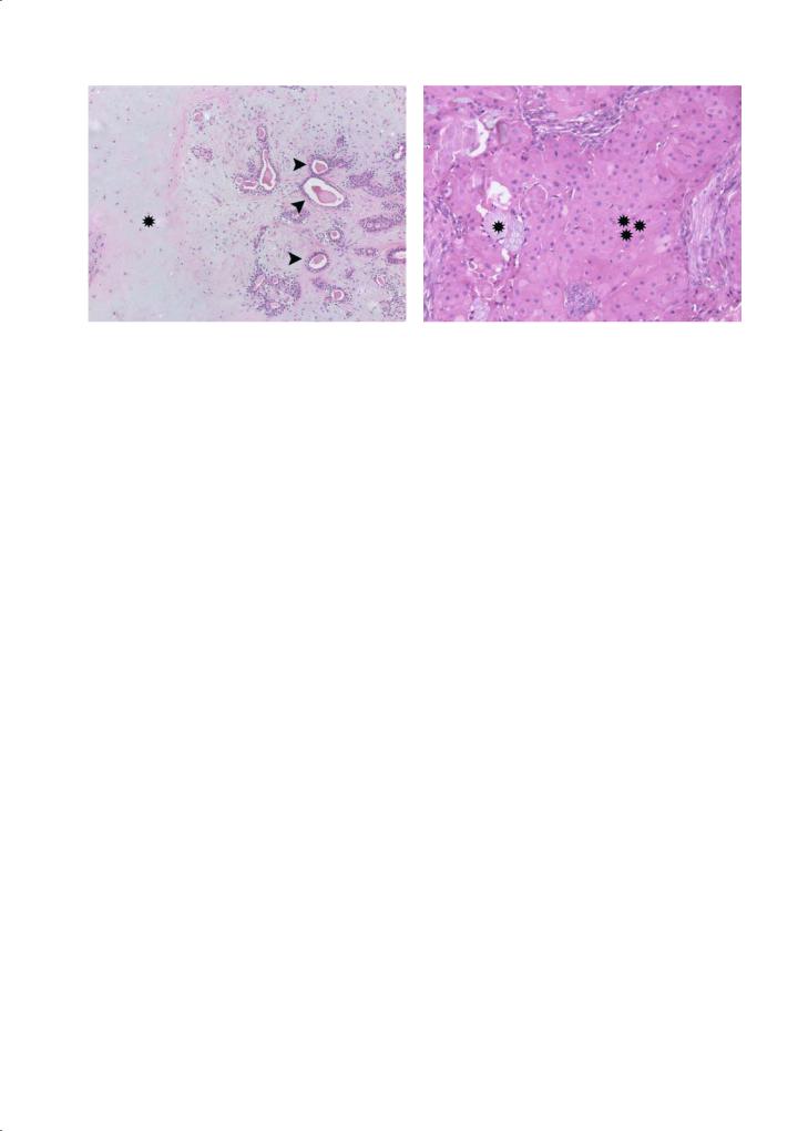
Fig. 1.15. Microscopical appearance of the same pleomorphic adenoma as in Fig. 1.14. Note the chondromyxoid matrix (asterisk), in which ductal structures (arrows) can be noted. (Courtesy Raf Sciot, MD, PhD)
V. Vander Poorten
Fig. 1.16. Intermediate grade mucoepidermoid carcinoma of the parotid gland. Epithelial solid tumour (three asterisks) with some islands of mucinous cells (asterisk). (Courtesy Raf Sciot, MD, PhD)
1.2.2.2.2.2 Warthin’s Tumour
Warthin’s tumour is the second most frequent benign salivary gland tumour. It occurs exclusively in the parotid gland and the immediately adjacent level II lymph nodes. Between 6%–10% of parotid tumours are Warthin’s tumours (Ellis and Auclair 1996a). There is a male to female preponderance of 5 to 1. Warthin’s tumours can occur bilaterally in about 10% of patients (Heller and Attie 1988). Microscopically there is typically a two-layered eosinophylic epithelium and a lymphoid stroma, hence the name adenolymphoma.
1.2.2.2.3 Malignant Tumours
1.2.2.2.3.1
Mucoepidermoid Carcinoma
About one in six (Vander Poorten et al. 2003) to one in three (Spiro 1986) malignant salivary gland tumours are mucoepidermoid carcinomas. Macroscopically the cut surface is solid but can contain cysts. Microscopically the tumour consists of a variable combination of glandular cells lining cystic spaces and epidermoid basaloid type cells forming solid areas (Fig. 1.16). A histological grading system is based on the relative proportion of mucinous versus epidermoid cells. Tumours containing 90% solid area made up of epidermoid cells are designated high grade (Seifert and Sobin 1992) and are associated with a 72% disease specific death rate versus only 6%–8% disease specific death rate in low grade, more mucus containing, low grade tumours (Healey et al. 1970).
1.2.2.2.3.2
Adenoid Cystic Carcinoma
Adenoid cystic carcinoma accounts for about one out of six parotid carcinomas (Vander Poorten et al. 2003). It occurs more frequently in other salivary gland sites with about 45% of submandibular and minor salivary gland carcinomas being of this type (Vander Poorten et al. 1999a) (Vander Poorten et al. 2000). Macroscopically it is a frequently infiltrating, rather hard tumour with an irregular extension pattern. The tumour is notorious for extension via major cranial nerves and in this respect MRI imaging is often an essential imaging modality to determine the real anatomical extent. It is also a tumour with a well known capacity for distant metastasis in about 40% of patients (Spiro and Huvos 1992), mostly to the lungs (Fig. 1.17), and in this case a protracted clinical course can result in disease related deaths even after more than 10 years following the initial diagnosis (Vander Poorten et al. 1999b; Spiro and Huvos 1992). Microscopically the tumour is often composed of cylindrical cystic spaces separated by solid septae of tumour cells, and this is called the “cribriform pattern” of appearance (Fig. 1.18).
1.2.2.2.3.3
Acinic Cell Carcinoma
About one in five parotid carcinomas is diagnosed as acinic cell carcinoma (Vander Poorten et al. 2003). The majority of these tumours have a clinically low grade course, and following adequate resection, low stage tumours are not considered to need additional radiotherapy (Armstrong et al. 1990). Macroscopically acinic cell carcinomas are