


Protein Domains and Signal Transduction
Similar processes occur in clathrin-mediated endocytosis. At the plasma membrane, the components that lead to the formation of endocytic vesicles are recruited to PI(4,5)P2. At an early stage, one of the key proteins that promotes membrane invagination is Epsin,34 which binds to PI(4,5)P2 through its ENTH domain (Epsin N-terminal homology domain). This interaction causes its N-terminal residues to fold as an amphipathic helix that inserts into the inner leaflet of the bilayer, causing the membrane to bend. (This property is not restricted to Epsin. Other proteins involved in trafficking, including the GTPase ARF and some BAR domain-containing proteins, can also form amphipathic helices.) Epsin also associates with the adaptor AP2 that recruits clathrin monomers to form the coat that supports vesicle budding and mediates
cargo selection. A further essential protein involved in the tethering of clathrin is AP180/CALM. This also binds PI(4,5)P2 through its ANTH domain (AP180/ CALM N-terminal homology). These domains are about 50% larger than ENTH domains and both are arranged as solenoids of -helices. Although there is a region that is structurally similar in both, they bind PI(4,5)P2 differently.
Yet another phosphoinositide-binding domain is the C2 domain, discussed in the next section.
For more detail see www. endocytosis.org.
Polypeptide modules that bind Ca2
Calcium-binding motifs and domains
Many enzymes are sensitive to changes in the local Ca2 concentration. For instance, when the cytosolic [Ca2 ] rises above the resting level (40–100 nmol L 1), signalling proteins with Ca2 -binding motifs or domains
become activated (see Chapter 8). In addition, cytosolic enzymes that possess Ca2 -binding C2 domains may be recruited to membrane sites through a Ca2 -dependent interaction with membrane phospholipids.
The EF-hand motif
Many Ca2 -binding regions on proteins possess helix–loop–helix structures known as EF-hand motifs. The letters E and F denote helical regions of the muscle protein parvalbumin, in which the motif was first identified. The Ca2 binding site is formed by the loop of 12 amino acids that links the two -helices
(Figure 24.6). EF-hands generally occur as adjacent pairs. This arrangement allows them to fold compactly as an EF-hand unit. In general, the core of the EF-hand structure is reasonably conserved, but the outer regions vary and the KD for Ca2 can range from 10 5 to 10 7 M. The widely expressed, intracellular Ca2 -sensing protein calmodulin possesses two EF-hand units (i.e. four EF-hands). In each motif, the chemical ligands that coordinate Ca2 are oxygen atoms provided
by aspartate and glutamate side chains, a peptide bond carbonyl group, and a water molecule.35,36 Conformational changes in calmodulin that result from Ca2 binding are described in Chapter 8 (see page 225).
FIG 24.6 EF-hand structure.
One of the EF-hands of calmodulin showing a bound Ca2 ion (green sphere, almost hidden). The seven oxygen atoms that form the coordination shell are shown as red spheres (1cll.pdb37).
779
Signal Transduction
The binding of one ligand alters the affinity of
other site(s). In this case, positive cooperativity denotes an enhancement of the affinity at other sites.
Synaptotagmin is a transmembrane protein in secretory vesicles that is important in exocytosis.
C2 domains
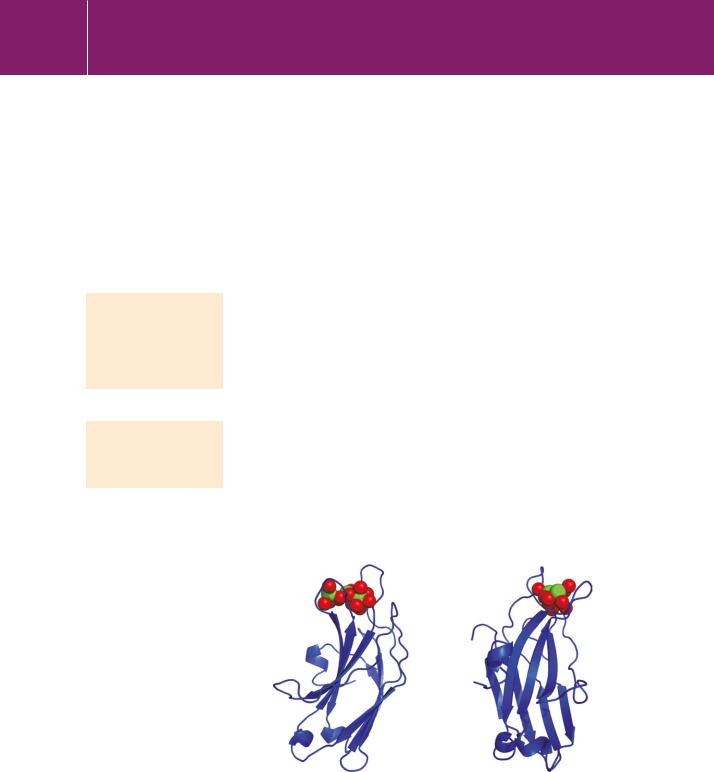
Unlike the small EF-hand motifs, C2 domains are substantial structures ( 130 residues), named after the‘second conserved’regulatory domain of protein kinase C (see Figure 9.8, page 254). They exist in a wide range of intracellular proteins and were originally known as Ca2 and lipid-binding domains, but like the EF-hands their affinity for Ca2 is variable. A typical C2 domain is arranged as a rigid, eight-stranded, antiparallel sandwich. Ca2 -binding is confined to a region defined by three loops on one edge of the structure. In the C2 domain of protein kinase C (Figure 24.7), five aspartate residues within two of the loops present oxygen atoms that form a binding pocket which can accommodate up to three Ca2 ions. These bind in a cooperative manner. Note, however, that the coordination sphere around each Ca2 ion is not quite complete. Apart from the anionic residues that bind the Ca2 ions, the side chains of other residues in the loops are mostly positively charged. Thus, when calcium ions enter the pocket, the negative charges are neutralized and the protein can then bind electrostatically to the anionic head group of the membrane lipid phosphatidylserine. This completes the coordination shell around the Ca2 ion and brings about membrane attachment. In a similar fashion, phospholipid
binding by the C2 domain of synaptotagmin requires the binding of Ca2 , which acts like an electrostatic switch, changing the surface potential of the Ca2 - binding loops and enabling them to bind to acidic phospholipid head groups.38
The tethering of signalling molecules to membranes through C2 domains can be strengthened by interactions involving regions of the domain away from its Ca2 -binding loops. For example the C2 domain of PKCpossesses additional basic residues in the loop between -strands 3 and 4, a region on
FIG 24.7 C2 domain structure.
Bound Ca2 ions are shown (green spheres) with the coordinating oxygen atoms (red spheres). The view on the right shows the structure rotated 90° about a vertical axis (1a25.pdb41).
780