

Chapter 2
First Messengers
The natural extracellular ligands that bind and activate receptors have been called first messengers, although they may be subdivided as hormones, neurotransmitters, cytokines, lymphokines, growth factors, chemoattractants, etc. Each of these terms attempts to define a class of agents that take effect
in a particular setting. However, there can be much overlap, as none of them adheres to a strict definition. This is illustrated by Table 2.1, which provides examples of extracellular first messengers that function as hormones, growth factors, and inflammatory mediators, and Table 2.2, which lists a selection of substances that can act as neurotransmitters.
Particular examples of multiple functions are:
•
•
The coenzyme ATP and the cellular metabolite glutamate are neurotransmitters when they are secreted at synapses.
The gut hormones gastrin, cholecystokinin, and secretin are also present in the central nervous system (CNS), where they have diverse functions as neuromodulators (influencing the release of other neurotransmitters).
21

Signal Transduction
Table 2.1 A selection of first messengers found in the circulation
Class |
Messenger |
Origin |
Target |
Major effects |
|
|
|
|
|
Messengers |
adrenaline |
adrenal |
heart smooth |
increase in pulse rate and |
derived |
(epinephrine) |
medulla |
muscle liver and |
blood pressure contraction |
from amino |
|
|
muscle adipose |
or dilatation glycogenolysis |
acids |
|
|
tissue |
lipolysis |
|
noradrenaline |
adrenal |
arteriolar smooth |
vasoconstriction |
|
(norepinephrine) |
medulla |
muscle |
|
|
serotonin (5-hydroxy |
platelets |
arterioles and |
vasodilatation and increased |
|
tryptamine) |
|
venules |
vascular permeability |
|
Histamine |
mast cells, |
arterioles and |
vasodilatation and increased |
|
|
basophils |
venules |
vascular permeability |
|
|
|
|
|
Peptide |
insulin |
pancreatic |
multiple tissues |
glucose uptake into cells |
hormones |
|
cells |
|
carbohydrate catabolism |
|
|
|
|
protein synthesis lipid |
|
|
|
|
synthesis (adipose tissue) |
|
|
|
|
Note: also a growth factor |
|
glucagon |
pancreatic |
liver adipose |
glycogenolysis lipolysis |
|
|
cells |
tissue |
|
|
gastrin |
intestine |
stomach |
secretion of HCl and pepsin |
|
secretin |
small |
pancreas |
secretion of digestive enzymes |
|
|
intestine |
|
|
|
cholecystokinin |
small |
pancreas |
secretion of digestive enzymes |
|
|
intestine |
|
|
|
atrial natriuretic |
heart |
kidney |
Na and water diuresis |
|
factor (ANF or ANP) |
|
|
|
|
adrenocorticotropic |
anterior |
adipose tissue |
lipolysis production of cortisol |
|
hormone (ACTH) |
pituitary |
adrenal cortex |
and aldosterone |
|
follicle stimulating |
anterior |
oocyte/ovarian |
growth oestrogen synthesis |
|
hormone (FSH) |
pituitary |
follicles ovarian |
|
|
|
|
follicles |
|
|
luteinizing hormone |
anterior |
oocyte ovarian |
maturation oestrogen and |
|
(LH) |
pituitary |
follicles |
progesterone synthesis |
|
thyroidstimulating |
anterior |
thyroid |
generation and release of |
|
hormone (thryotropin |
pituitary |
|
thyroid hormones |
|
or TSH) |
|
|
|
|
parathyroid hormone |
parathyroid |
bone kidney |
release of Ca2 and phosphate |
|
(PTH) |
|
|
Ca2 reabsorption |
|
|
|
|
Continued |
22

First messengers
Table 2.1 Continued
Class |
Messenger |
Origin |
Target |
Major effects |
|
|
|
|
|
|
vasopressin |
posterior |
kidney arteriolar |
water reabsorption from urine |
|
(antidiuretic hormone |
pituitary |
smooth muscle |
vasoconstriction to increase |
|
or ADH) |
|
|
blood pressure |
|
Oxytocin |
posterior |
uterine smooth |
dilation of the cervix milk |
|
|
pituitary |
muscle milk ducts |
ejection |
|
|
|
|
|
Growth |
epidermal growth |
multiple cell |
epidermal and |
growth |
factors |
factor (EGF) |
types |
other cells |
|
|
Somatotropin |
anterior |
liver |
production of somatomedins |
|
(growth hormone |
pituitary |
|
|
|
or GH) |
|
|
|
|
somatomedins 1 and |
liver |
bone, muscle and |
growth |
|
2 (insulin-like growth |
|
other cells |
|
|
factors, IGF1 and 2) |
|
|
|
|
Transforming growth |
multiple |
multiple |
growth control |
|
factor (TGF ) |
locations |
|
|
|
|
|
|
|
Eicosanoids |
Prostaglandins |
most body |
multiple |
inflammation, vasodilatation |
|
PGA1,PGA2, PGE2 |
cells |
|
|
Hormones |
Thyroid hormone (T3, |
thyroid |
multiple |
metabolic regulation |
with |
tri-iodothyronine) |
|
|
|
intracellular |
|
|
|
|
receptors |
|
|
|
|
|
progesterone |
corpus |
uterine |
preparation of endometrial |
|
|
luteum |
endometrium |
layer maintenance of |
|
|
placenta |
|
pregnancy |
|
|
|
|
|
•
•
•
•
•
Somatostatin, identified originally as a hypothalamic agent suppressing the secretion of growth hormone from the pituitary, also operates in the CNS as a neurotransmitter or neuromodulator. More than this, it is a paracrine agent in the pancreas and a hormone for the liver.
The transforming growth factor TGFβalso acts as a chemoattractant and as a growth inhibitor.
Insulin acts not only to regulate glucose metabolism, but also as a growth factor and a regulator of food intake.
Noradrenaline (norepinephrine), depending on whether it is released at a synapse or from the adrenal medulla, can be considered as a neurotransmitter or as a hormone.
Thrombin is a growth factor but is also involved in blood clotting as an activator of platelet function.
23
Signal Transduction
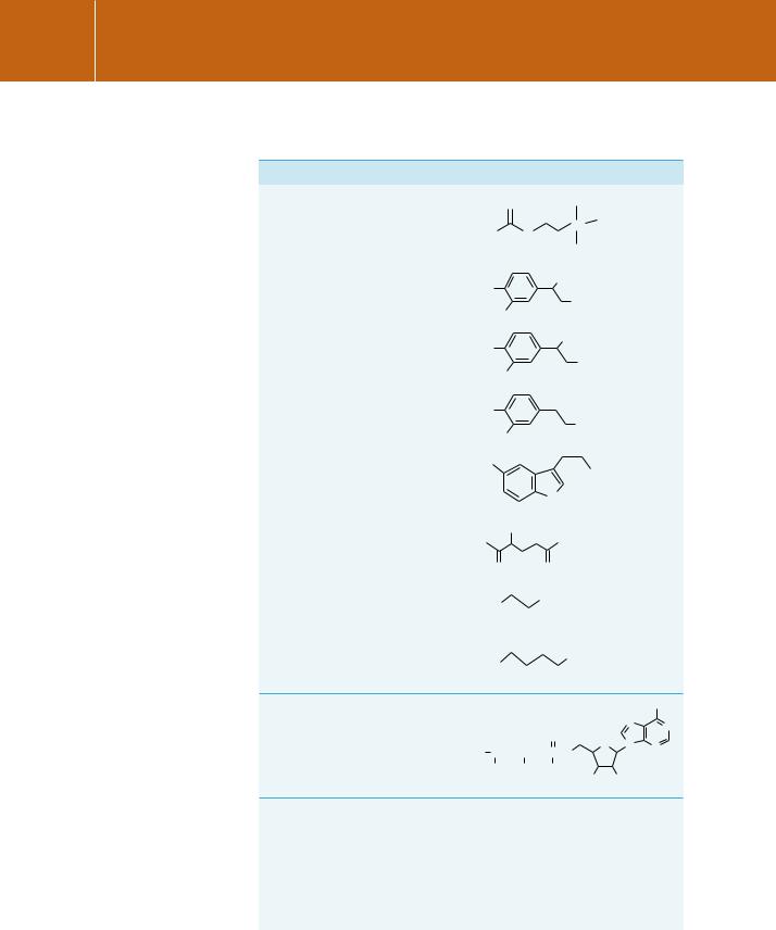
Table 2.2 A selection of compounds that act as neurotransmitters
Type |
Transmitter |
Structure |
|
|||||
|
|
|
|
|
|
|
|
|
|
|
O |
|
|
|
H3C |
|
|
|
acetylcholine |
H3C |
|
|
|
N+ |
CH3 |
|
|
|
O |
|
|||||
|
|
|
|
|
|
CH3 |
|
|
|
|
|
|
|
|
|
|
|
Tyrosine or |
adrenaline |
HO |
|
|
|
OH |
|
|
tryptophan- |
+ |
|
CH3 |
|||||
|
|
|
||||||
derived |
(epinephrine) |
HO |
|
|
|
NH2 |
|
|
|
||||||||
|
|
|
|
|
|
|
||
|
noradrenaline |
HO |
|
|
|
OH |
|
|
|
|
|
|
NH+3 |
|
|||
|
(norepinephrine) |
HO |
|
|
|
|
||
|
|
|
|
|
|
|
|
|
|
dopamine |
HO |
+ |
|
||||
|
|
|
||||||
|
|
|
|
|
|
NH3 |
|
|
|
|
HO |
|
|
|
|
|
|
|
|
HO |
|
|
|
|
NH+3 |
|
|
serotonin |
|
|
|
|
|
||
|
|
|
|
|
|
|
|
|
|
|
|
|
|
NH |
|
||
|
|
|
|
|
|
|
|
|
|
|
NH+3 |
|
|||||
Amino acids |
glutamate |
O− |
|
|
|
O− |
|
|
|
|
|
|
|
|
|
||
|
|
O |
|
|
O |
|
||
|
|
+ |
|
|
O− |
|
||
|
glycine |
H3N |
|
|
|
|
|
|
|
|
|
|
|
|
|
||
|
|
|
O |
|
||||
|
GABA |
+ |
|
|
|
O− |
|
|
|
|
|
|
|
|
|
||
|
|
H3N |
|
|
|
|
|
|
|
|
|
|
O |
|
|||
|
|
|
|
|
|
|||
Purine based |
ATP |
|
|
|
|
|
|
|
|
|
|
|
|
|
|
NH2 |
|
|
|
|
|
|
|
|
|
|
|
|
|
N |
N |
|
|
|
|
|
|
|
|
|
|
|
|
|
|
|
O |
|
|
O |
|
O |
|
|
|||||||
O− |
|
|
|
|
|
|
|
|
|
|
|
O |
N |
N |
|
|
|
|
|
|
|
|
|||||||
P |
|
O |
|
P |
|
O |
|
P |
|
O |
|
|||
|
|
|
|
|
|
|
||||||||
O− |
|
|
O− |
|
O− |
|
|
|||||||
|
|
|
|
|
|
|
|
|
|
|
|
OH |
OH |
|
Neuropeptides |
enkephalins |
Tyr-Gly-Gly-Phe-Leu |
|
|
Tyr-Gly-Gly-Phe-Met |
|
substance P |
Arg-Pro-Lys-Pro-Gln-Gln-Phe-Phe-Gly- |
|
|
Leu-Met |
|
angiotensin II |
Asp-Arg-Val-Tyr-Ile-His-Pro-Phe |
|
somatostatin |
Ala-Gly-Cys-Lys-Asn-Phe-Phe-Trp-Lys- |
|
|
Thr-Phe-Thr-Ser-Cys |
|
|
|
24

First messengers
Hormones
Hormones are commonly released in small amounts at sites remote from the organs they target. On entering the circulation, they are diluted enormously and are subject to enzymes that break them down. Many of them circulate as complexes with specific binding proteins, reducing their free concentration still further. The result of all this is that their level in the vicinity of a target cell is likely to be extremely low and accordingly, the cell receptors must possess high affinity. Another consequence is that although a target cell may react in milliseconds to hormone binding, overall response times are in the range of seconds to hours.
Examples of water-soluble hormones are included in the list of first messengers shown in Table 2.1. Adrenaline and noradrenaline (epinephrine and norepinephrine) are released from the adrenal gland and can act within seconds. The peptide hormones vary considerably in size, ranging from a few amino acids to full-scale proteins. For example, the hypothalamic factor TRH (TSHreleasing hormone) that induces the release of thyroid stimulating hormone (TSH), consists of only 3 amino acid residues, whereas FSH (follicle stimulating hormone) and TSH are heterodimeric glycoproteins each of about 200 residues.
Growth factors
The first reports that fragments of biological tissue could be maintained in a living state in vitro appeared nearly 100 years ago,1 but routine culturing of dispersed cells did not become established until the 1950s. The successful maintenance of dividing mammalian cells depends on the composition of the culture medium. This is traditionally a concoction of nutrients and vitamins in a buffered salt solution. A key ingredient is an animal serum, such as fetal bovine serum. Without such a supplement, most cultured cells will not replicate their DNA and therefore will not proliferate. An important early discovery was that the best mitogenic stimulus (i.e. inducing mitosis or cell division) is provided by natural clot serum and not plasma. A 30 kDa polypeptide released by platelets was later isolated and shown to have mitogenic properties. It was named platelet-derived growth factor, PDGF (see page 303).
PDGF is a member of a class of messengers, the growth factors, comprising over 40 polypeptides, ranging in size from insulin (5.7 kDa) and EGF (6 kDa) to transferrin (78 kDa). As with the hormones, cells have specific high-affinity receptors for growth factors, though binding may elicit multiple effects. Apart from stimulating (or inhibiting) growth, they may also initiate programmed cell death (apoptosis), differentiation, and gene expression. The effects of growth factors, unlike those for hormones, may last for days. Also, unlike the hormones their effects are short range, generally influencing the growth and function of neighbouring cells.
Endocrine (Greek
ενδoν within, κρiνεi to separate) denotes the ‘action at a distance’ of hormones secreted from the ductless glands that may pervade the whole organism, searching out specific target tissues.
Paracrine denotes the action of an extracellular messenger that takes effect only locally.
When a substance affects the same cell from which it has been released, the activity (perhaps part
of a negative feedback pathway) is termed autocrine.
When a cell affects another cell with which it is in contact, usually through membranebound proteins, the mode of communication is termed juxtacrine.
25

Signal Transduction
Cytokines
In parallel with the discovery of growth factors, several extracellular signalling proteins were identified that interact with cells of the immune system. Because they activate or modulate the proliferative properties of this class of cells,
they were initially termed ‘immunocytokines’. As it became apparent that they also act upon cells outside the immune or inflammatory systems, the name became shortened to cytokine. The functions of growth factors and cytokines are so diverse that a clear distinction cannot be made. Thus cytokines include the growth factors already mentioned, along with such molecules as the interferons, tumour necrosis factor (TNF), numerous interleukins, granulocyte– monocyte colony stimulating factor (GM-CSF) and many others. Chemokines are cytokines that bring about local inflammation by recruiting inflammatory cells by chemotaxis and subsequently activating them.
Vasoactive agents
Physical damage to tissues or damage caused by infection generates an inflammatory response. This is a defence mechanism in which specialized cells (mostly leukocytes), recruited to the site, act in a concerted fashion to remove, sequester or dilute the cause of the injury. The process is complex, involving many interactions between cells and numerous extracellular messengers. Among these are cytokines that induce inflammation (pro-inflammatory mediators), or reduce it (anti-inflammatory mediators). To enable the recruitment of leukocytes and to retain a local accumulation of (diluting) fluid, there is vasodilatation and a regional increase in vascular permeability. Agents that bring this about include histamine (released by mast cells and basophils), serotonin (released by platelets), and pro-inflammatory mediators such as bradykinin.
The eicosanoids are another important family of vasoactive compounds. They are derived from the polyunsaturated fatty acid, arachidonic acid (5,8,11,14-eicosatetraenoic acid) (Figure 2.1). Because arachidonic acid and many of its derivatives possess 20 carbon atoms, they are termed eicosanoids (from the Greek word for 20). They include the prostaglandins, thromboxanes, and leukotrienes and they operate over short distances as potent paracrine or autocrine agents, controlling many physiological and pathological cell functions. In the context of inflammation, they cause plasma leakage, skin
Fig 2.1
26

First messengers
reddening, and the sensation of pain. The drug aspirin possesses antiinflammatory and pain-relieving properties because it inhibits a key enzyme in the pathway generating prostaglandins, such as the vasoconstrictor prostaglandin E2 (Figure 2.1).
Neurotransmitters and neuropeptides
Neurotransmitters are also first messengers, but their release and detection at chemical synapses contrasts strongly with that of endocrine signals. In the presynaptic cell, neurotransmitter-containing vesicles are organized to
release their contents locally into the tiny volume defined by the synaptic cleft. Individual neurons contain only very small quantities of transmitter and this arrangement ensures that an effective concentration is quickly attained within the cleft following the secretion (by exocytosis) of the contents of relatively few vesicles. The released transmitter then diffuses across the cleft and binds to receptors on the postsynaptic cell. On the macroscopic scale, diffusion is a slow process, but across the short distance that separates the preand postsynaptic neurons ( 0.1 m or less), it is fast enough to allow rapid communication between nerves or between nerve and muscle at a neuromuscular junction. Table 2.2 shows the structures of some important neurotransmitters. In the central nervous system glutamate is the major excitatory transmitter, whereas GABA and glycine are inhibitory. Acetylcholine has its most prominent role at the neuromuscular junction, where it is excitatory. Whether an excitatory or inhibitory effect is evoked depends on the nature of the ion channel that is regulated by the neurotransmitter receptor (see page 54).
First messengers with intracellular receptors
There is a class of hormones that differs in several major respects from the first messengers that we have examined so far. These are the steroid and thyroid hormones (see Figure 10.1, page 275). Firstly, since many are hydrophobic they are able to cross the plasma membrane of cells and then interact with receptors that are intracellular. The hormone-bound receptors then bind to DNA and regulate the expression of particular proteins or sets of proteins. For example, the steroid cortisol can penetrate cells and bind to internal glucocorticoid receptors. These, in turn, bring about transcription of DNA sequences and thus the expression of enzymes that lead to glycogen synthesis. Such a process is slow, requiring hours to take effect. It forms a paradigm for the action of steroid and other hormones that bind to nuclear receptors (thyroid hormone, retinoic acid, vitamin D) and many other non-hormonal lipophilic messengers. This form of signalling is discussed in detail in Chapter 10.
In addition to the transcriptional actions of hydrophobic messengers, there is increasing evidence for non-transcriptional effects, which take place rapidly and involve interactions with cell surface receptors. A recently described
Almost 40 years ago there were already indications that oestrogens can cause a rapid (within 15 s) elevation of cAMP in rat uterus.4 Indeed,
as early as 1941, Hans Selye reported that steroids introduced into the peritoneum of rats induce deep anaesthesia within 15 min.5,6
Steroid derivatives as anaesthetics in surgery have been used since the 1950s (hydroxydione sodium or Viadril, 21- hydroxypregnane-3,20- dione sodium succinate).
27

Signal Transduction
example of steroid interaction with a well-established cell surface receptor is that of progesterone, essential for establishing and then maintaining
pregnancy in mammals. At the cell surface it interferes with the binding of the peptide hormone oxytocin to its receptor in rat uterine tissue.2 It is possible that it is through inhibition of oxytocin binding that progesterone maintains uterine quiescence during pregnancy. The oxytocin receptor is a member of the seven transmembrane (7TM) protein superfamily of G-protein-coupled receptors (see Chapter 4) and acts to elevate the concentration of intracellular Ca2 . Progesterone can also act as an inhibitor of the nicotinic acetylcholine receptor.3 The rapid, non-genomic effects of lipophilic messengers are also discussed in Chapter 10.
Oxytocin (from the Greek meaning swift birth) is generated in the brain of both males and females. It can be added to the examples already cited which have the actions of both hormones and neuromodulators. Not only does it act as a regulator of uterine smooth muscle contraction and milk ejection but it also acts as a regulator of emotional states, enhancing trust and bonding between pairs of individuals. Sometimes called the ‘cuddle hormone’, it plays a role in the expression of maternal, sexual, social, stress, and feeding behaviours, as well as in learning and memory. When the natural release of oxytocin is blocked, for instance, mothers – from sheep to rats – reject their own young, while virgin female rats injected with oxytocin nuzzle the pups of other females protecting them as if they were their own. Oxytocin in females and the closely related vasopressin in males are keys to pair bonding. These actions of the hormone, which must have their origins far back in mammalian evolution, prove to be readily adaptable in today’s hectic world dominated by the god Mammon.7,8
In a money game, a double-blind trial involving the investment of serious money, volunteers playing the roles of investor and trustee were offered a sniff of oxytocin or placebo. It was found that the hormone had the effect of
increasing the trust of the investors while having no effect on the trustworthiness of the trustees. The possibility of abuse of these substances with the aim of manipulating trust in business and other (e.g. love) situations must be self evident.
Common aspects
Although we have described differences in the nature of selected first messengers, our aim in this book is to accentuate their common aspects. This will become more apparent in later chapters, where it will be seen that similar intracellular mechanisms are used to promulgate an extracellular message. The ultimate effects of these first messengers are determined, not by their chemical natures, but by the anatomical context within which they operate and by the characteristics of the receptor molecules that sense their presence in different classes of cell.
28