

Chapter 16
Traffic of White Blood Cells
Inflammation and leukocytes
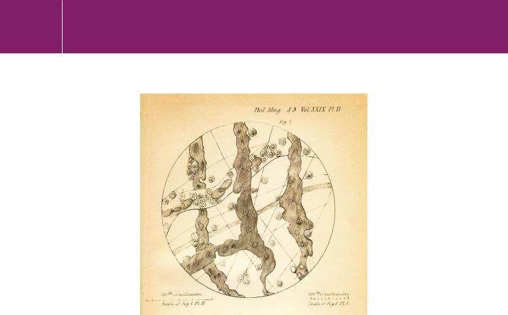
The first sightings of white cells adhering to the walls of the finer vessels and then emigrating into the tissues under conditions of inflammation were recorded more than 150 years ago (Figure 16.1).1–3 Cohnheim’s detailed
histological description of 18824,5 remained the basis of most textbook accounts of inflammation for a further 80 years. Later, the first electron microscopic investigation of leukocyte adherence and migration provided much-needed detail but failed to reveal how cells adhere to the inflamed endothelium.6 Nor were any ideas forthcoming about how the cells penetrate an apparently coherent endothelial cell layer.
Inflammatory mediators
It is essential that migratory cells such as leukocytes can tolerate changes in their environments. They must be able to move from the bone marrow (where they are attached) into the blood (where they float free) and then
483
Signal Transduction
Fig 16.1 Augustus Waller’s microscopic examination of cell adherence and extravasation from the vessels of an inflamed frog tongue.
‘The blood, as we are aware, consists of a transparent fluid holding in suspension numerous particles, most of which are red and of a flattened shape, while a few others are colourless, and spherical in form … The peculiar manner in which the lymph-globules, or corpuscles, conduct themselves when in the capillaries, when in an organ in a state of irritation, has of late engaged much attention. The experiments of Mr W. Addison of Malvern, have greatly contributed to show these important functions in inflammation. In the tongue of the frog and toad they may be frequently seen circulating with the red particles in the vessels, down to the minutest capillaries. As it has already been pointed out, these spherules are generally found, when they come into contact with the parieties of the vessels, to retain their adherence with greater force than is manifested by the red particles in the like circumstances; as in the figure, where the current was observed to continue for many minutes without displacing the globules near the sides of the vessel. Thus we frequently see a lymph-globule remain in the same place, notwithstanding the current of red particles sweeping and pushing by it. The appearance in the larger vessels of these spherules, adherent to their inner surface, has been very aptly compared to so many pebbles or marbles over which a stream runs without disturbing them … The corpuscles, which are transparent, are occasionally seen to be granulated … Let us now examine the admirable manner in which nature has solved the apparent paradox, of eliminating, from a fluid circulating in closed tubes, certain particles floating in it, without causing any rupture
or perforation in the tubes, or allowing the escape of the red particles, which are frequently the smaller of the two, or that of the fluid part of the blood itself … After the observation had continued for half an hour, numerous corpuscles were seen outside the vessels, together with a very few blood discs in the proportion of about one to ten of the former. No appearance of rupture could be seen in any of the vessels. The corpuscles were generally distant about
0mm 03 from their parieties. After the experiment had lasted about two hours, thousands of these corpuscles were seen scattered over the membrane, with scarcely any blood discs … No trace of the corpuscular extravasation could be seen, except the presence of the corpuscles themselves……. I consider therefore as established, 1st , the passage of these corpuscles ‘de toute pièce’ through the capillaries; 2ndly, the restorative power in the blood, which immediately closes the aperture thus formed … In endeavouring to account for the fact of the passage of the corpuscles through the vessels we find considerable difficulties. It cannot be referred to the influence of vitality, as it is observed likewise to take place after death. It may be surmised, either that the corpuscle, after remaining a certain time in contact with the vessel, gives off by exudation from within itself some substance possessing a solvent power over the vessel, or that the solution of the vessel takes place in virtue of some of those molecular actions which arise from the contact of two bodies; actions which are now known as exerting such extensive influence in digestion, and are referred to what is termed the catalytic power’. Paraphrased from Waller.2,3
484

Traffic of White Blood Cells
into the tissues, particularly the lymph nodes (where they become attached again). To do all this they must be able to switch their integrins off and on. If the integrins on leukocytes were permanently switched on, the cells could never leave the bone marrow. Once in the blood, the integrin function must be switched off. Leukocytes leave the circulation when they encounter an appropriate signal of inflammation or infection, or more generally in the continuous process of immune surveillance. For monocytes and neutrophils, departure from the circulation is a one-way journey to their destiny with death in the tissues. By contrast, lymphocytes move from the blood into the tissues, then through the lymphatic system and then back into blood. We focus here on the regulation of leukocyte adhesion and extravasation under inflammatory conditions (Figure 16.2).
The many abbreviations used in this chapter are collected together at the end of the chapter.
Fig 16.2 Generation of inflammatory mediators at sites of infection.
Tissue damage and bacterial infection induce the release of inflammatory mediators from various cell types. Bacterial protein A binds and activates the TNFreceptor. Bacterial lipopolysaccharide (LPS), which acts on local fibroblasts, dendritic cells, vascular endothelial cells, mast cells and resident leukocytes induces release of Gro, histamine, IFN- , IL-1 , IL-6, IL-8, MCP-1, RANTES, and TNF- . Collectively, these mediators are responsible for the up-regulation of
adhesion molecules on vascular endothelial cells and the activation of integrins on leukocytes. This results in their extravasation followed by migration to sites of damage/infection. Formylmethionyl peptides released from bacteria (as well as from the damaged mitochondria of host cells) facilitate the migratory response (chemotaxis). These processes allow the rapid accumulation of leukocytes, in particular neutrophils, at sites of infection as a first line of defence. It also initiates the process of tissue remodelling and repair (wound healing).
485

Signal Transduction
In bacteria, formylmethionine (fMet) is the initiating amino acid in protein synthesis; in eukaryotes, it is methionine. Synthetic peptides containing the sequence fMetXY (where X and Y are hydrophobic amino acids) act as powerful stimulants for neutrophil activation; among the most potent is fMetLeuPhe.7 fMet peptides isolated from bacterial cultures also activate neutrophils.8 Collectively, these have been called chemotactic peptides, although they act to stimulate all the multiple functions of these cells such as the respiratory burst and secretion of lysosomal enzymes in addition to chemotaxis.
At sites of inflammation (caused by injury or infection) mediators are released that affect the expression and affinity of adhesion molecules and affect
the release of chemokines from endothelial cells. LPS, acting through its receptor TLR4 (Toll-like receptor: see Chapter 15) induces the expression of inflammatory mediators including the cytokines and chemokines. Other bacterial and cellular components (listed in Figure 16.2 and Table 16.1), can be added to the list of ‘patterns’ that provoke the release of inflammatory mediators. One of these is protein A, a staphylococcal surface protein
that binds to the TNFreceptor. Another is the class of formylmethionyl peptides, generated as the initiation sequences of prokaryotic proteins that are released by microbes. Histamine, released from mast cells and basophils, plays an important role in locally widening the vasculature, therefore allowing accumulation of blood and thus the accumulation of leukocytes at the site of inflammation.
In the following sections we describe the signalling pathway that emanates from the TNFreceptor (TNFR1) in vascular endothelial cells. This results in elevated cell surface expression of adhesion molecules and the release of yet more inflammatory mediators. It also plays a crucial role in regulating the extravasation of circulating leukocytes (also known as transendothelial cell migration or diapedesis).
Tumour necrosis factor- , potential anti-tumour agent or inflammatory cytokine?
The observation that the size of a tumour occasionally diminishes after bacterial infection has a long history. As early as 1848, Legrand noted two cases of long-standing scrofulous lymphoma which appeared to regress after infection with erysipelas.13 In 1888, P. Bruns reported on the spontaneous regression of human tumours following infection with
S. erysipelas.14
Protein A. Derived from Staphylococcus aureus, protein A binds with very high affinity to both the constant (Fc ) and variable (Fab) regions of IgG and IgM antibodies.9 Protein A contributes to the pathology of Staphylococcus aureus by directly binding to TNFreceptors on the respiratory epithelium. This plays an important role in the onset of inflammation of the lower respiratory tract, leading to severe pneumonia associated with tissue damage and sometimes sepsis.10 Protein A can also bind some membrane-associated B cell antigen receptors and promote antibody production.11 This plays a role in the defence against the pathogens but is also associated with autoimmune disorders such as rheumatoid arthritis.
486

Traffic of White Blood Cells
‘Die öfters beobachtete Thatsache, dass Neubildungen, namentlich malingner Natur, durch ein interkurrentes Erysipel zur Verkleinerung oder zum Verschwinden gebracht werden, ist von hervorragendem theoretischem und praktischem Interesse. In ersterer Hinsicht verdient diese Thatsache gerade gegenwärtig besondere Beachtung, wo die Untersuchung der Aetiologie der malignen Neubildungen auf der Tagesordnung steht, deren Ergebnisse vielleicht geeignet sind, auch über die Art und Weise der salutären Wirkung des Erysipels Licht zu verbreiten. In praktischer Beziehung haben jene Beobachtungen zu Versuchen mit der kunstlichen Erzeugung eines kurativen Erysipels Veranlassung gegeben, um inoperable bösartige Neubildungen zur Heilung zu bringen.
Allein die Zulässichkeit.solcher Versuche ist noch eine offene Frage …’:
‘The frequently observable fact that new formations, particularly those of a malign nature may be caused to regress or to disappear by a concurrent
erysipelas is of very great interest both in theory and practice. In respect of the former this fact merits particular attention, especially at the present time, given the current preoccupation with the study of the aetiology of new formations, the results of which may well be capable of shedding new light upon the beneficial effects of erysipelas. On the practical front these observations have given rise to experiments in which curative erysipelas is artificially induced in order to bring about the healing of inoperable new formations. However the admissibility of such experiments remains an open question …’
Table 16.1 Sources of inflammatory mediators
Source |
Examples |
|
|
Release from |
Lipopolysaccharide (LPS or endotoxin), shed from |
invading |
the surface. Also N-formylmethionyl peptides (fMet- |
microorganisms |
peptides) derived from the initiator sequence of |
|
bacterial protein synthesis and protein A, a cell surface |
|
protein of Staphylococcus aureus that binds to and |
|
activates the TNF receptor-1 |
|
|
Release from |
TNF-αand IL-1βfrom activated macrophages or |
resident cells |
fibroblasts, histamine from mast cells and chemokines |
|
(MCP-1, IL-8, Gro) from endothelial cells or resident |
|
leukocytes |
|
|
Release from the |
Peptides derived from the N-terminus of proteins (such |
mitochondria of |
as NAD dehydrogenase) coded by the mitochondrial |
damaged cells12 |
genome. These share with eubacteria the characteristic |
|
N-formylmethionyl initiator sequence |
|
|
Components of |
C5a and thrombin respectively |
complement and of |
|
fibrinolysis |
|
|
|
487

Signal Transduction
Erysipelas: an acute superficial form of cellulitis involving the dermal lymphatics, usually caused by infection with group A streptococci. Formerly called St Anthony’s fire.
The principle of bacterial infection as a component in the armory of antitumour medicine persists today. Lovaxin C and other Listeria-based vaccines currently under development, at the stage of phase I/II clinical trials, target cervical, breast, ovarian, and
lung cancers. In animal models of breast cancer, the Lovaxin B vaccine, composed of Listeria with the Her2/neu antigen, appears to stop tumour growth.28
W. B. Coley15 reported several cases of tumour regression and even disappearance following repeated inoculation of erysipelas. Speculating on possible mechanisms, he considered
1) that the erysipelas coccus has a direct destructive action upon the cell elements of the new growth; 2) that the high temperature (induced by the infection) alone is sufficient; 3) that sarcoma and carcinoma are both of bacterial origin and the erysipelas germ has a direct antagonistic effect upon the cancer bacillus … it seems in the light of present knowledge not improbable that all of the three theories may contain an element of
truth, and that a larger theory combining all these elements is necessary to explain the curative action of erysipelas.
In the firm belief that the cure of cancer was at hand, Coley and others applied cell-free filtrates of streptococci as ‘Coley’s toxins’ over a period of about
40 years, apparently with some success.15–17 The effect of these bacterial products is associated with severe haemorrhagic reaction mainly confined to the core of the tumour which rapidly sloughs off. However, it generally leaves a ring of viable tissue which, unfortunately, continues to grow.
Not surprisingly, such treatments also tend to cause widely disseminated systemic effects and all too frequently, these can lead to circulatory collapse and death. Early attempts to separate the haemorrhagic from the cytotoxic components in filtrates of cultured Serratia marcescens led to the isolation of LPS, lipopolysaccharide (or endotoxin, see page 457).18,19 Among its many biological activities, this could elicit haemorrhagic necrosis of both experimental and primary subcutaneous tumours.20
All this provoked the hunt for immune mediators, cytotoxic for transformed cells but not causing septic shock.25 The first of these, the tumour necrosis factors TNFand TNF- , attracted immense interest.26,27 However, TNF was also found to possess general pro-inflammatory properties and to provoke severe systemic toxicity. Thus the focus of interest shifted from tumour necrosis to mechanisms of inflammation.
TNFas target for anti-inflammatory drugs. An excess of TNFis associated with chronic inflammatory disorders. The best studied in this respect are Crohn’s disease (destructive inflammation of the bowel) and rheumatoid arthritis (destructive inflammation of the synovial joints). TNFis the key cytokine regulating expression of all other inflammatory cytokines. It was concluded that by blocking its action it would be possible to reduce the general state of inflammation.21 Two approaches to lower the TNFconcentration have been exploited successfully. One uses a humanized antibody against TNF- (infliximab), the other an extracellular fragment of the TNFR2 coupled to IgG (etanercept). Both these treatments provide considerable relief.22,23 More recently, the pre-ligand assembly domains of the TNFR1 and -R2 have been targeted by a soluble cysteine-rich region to prevent homophilic trimerization of the receptors. This too reduces the symptoms of arthritis in animal models.24
488