

Chapter 11
Growth factors: setting the framework
The trails that led to the discovery of the growth factors (and related messengers) are very different from those that revealed the first hormones
and neurotransmitters. For a start, people knew, more or less, what to look for and where to go looking, and in general, the tale is somewhat less romantic and less fraught with angst and vehement disagreements. However, what began with a simple search for factors that would sustain living cells in laboratory conditions has expanded into a plethora of subject areas which are subjects in their own right. These include inflammation, wound healing, immune surveillance, development, and carcinogenesis. To confront this
bewildering prospect we first set out some details of how it evolved initially so that the reader is aware of how the major questions developed and have been confronted. A difficulty arises from the convergence of different disciplines, each bringing with it the baggage of its favoured nomenclature. We return to this matter at the end of this chapter.
For the ultimate in aggressive rivalry and claims to primacy in hormone discovery, coupled to barely restrained personality conflict, see Nicholas Wade’s account of the discovery by Roger Guillemin and Andrew Schally of thyrotropin releasing factor and other hypothalamic peptides.1–3
297
Signal Transduction
Viruses and tumours
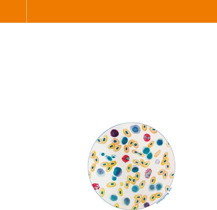
The first report of a tumour linked to a virus appeared in 1908, when Ellermann and Bang obtained a filterable agent from a chicken leukaemia and were able to make six passages of it, from fowl to fowl, producing the same disease each time.4 Their report was generally disregarded. Leukaemia was not considered to be a tumour, though as should have been evident from the work of Aldred Warthin (Figure 11.1), first reported in 1904,5, 6 a link between leukaemias and tumours, and hence cell growth and proliferation, had already been established:
These conditions are comparable to malignant tumors. The formation of metastases, the infiltrative and destructive growth, the failure of innoculations and transplantations etc., all favor the view that they are neoplasms, and present the same problems as do the malignant tumors.
More than 20 years were to pass before the leukaemias were eventually recognized as being tumours or neoplastic diseases.
FIG 11.1 Stained blood smear from a leukaemic fowl.
‘In December 1905, there came into my hands a Buff Cochin Bantam hen showing signs of illness in the way of indisposition to move about and a general weakness of a progressive character. No symptoms of ordinary fowl diseases were present . . . Examination of blood smears showed, however, a great increase of white cells of the large lymphocyte type . . . A diagnosis of leukemia was therefore made . . . A great variety of staining methods were used, including the most recent methods for the staining of spirochetes and protozoan parasites . . . No evidence of the existence of any infective agent could be obtained.6 (Note that avian red blood cells (yellow stain) are nucleated.)
298

Growth factors: setting the framework
In 1910, Francis Peyton Rous described a chicken tumour, identified as a sarcoma, that could be propagated by transplanting its cells, these then multiplying in their new hosts giving rise to tumours of the same sort.7 The cells yielded a virus, now known as Rous sarcoma virus (RSV), from which, in 1980, the first protein tyrosine kinase, v-Src, was isolated.8,9 Writing at the end of his career in 1967, Rous10 gives an apt description of how things were done:
Those were primitive times in the raising of chickens. They were sold in a New York market not far from the institute, and many individual breeders brought their stock there. Every week F. S. Jones, a gifted veterinarian attached to my laboratory, went to it, not only to buy living chickens with lumps which might be tumors, but any that seemed sickly and had a pale comb, as perhaps having leukemia. Thus within less than four years we got more than 60 spontaneous tumors of various sorts . . .
The discovery of NGF . . . and EGF
Among the fractions that I assayed in vitro the following day, there was one containing snake venom. Having not been told which of the fractions had been specially treated, I was completely stunned by the stupendous halo radiating from the ganglia. I called Stan in without telling him what I had seen. He looked through the microscope’s eyepieces, lifted his head, cleaned his glasses which had fogged up, and looked again. ‘Rita,’ he murmured, ‘I’m afraid we’ve just used up all the good luck we’re entitled to. From now on, we can only count on ourselves . . . .’ Events were to prove him wrong Rita Levi-Mpntaicini.
Nerve growth factor (NGF) may perhaps be regarded as the first identified growth factor, but there were many early clues hinting at their existence. The embryologist Hans Spemann (Nobel Prize winner in 1935, see page 618) had described the eponymous ‘organizer’ that directs the creation of the anteroposterior axis in the gastrula stage in the development of the amphibian embryo. This work had famously indicated that soluble factors made by the embryonic cells must be instrumental in regulating cell proliferation and differentiation.13
Rita Levi-Montalcini’s affair with growth factors had its origin at that time. Dismissed from her university post by the Nazi racial edicts, she determined to continue on alone. She had been alerted to the work of Spemann by an article written by one of his protégés, Viktor Hamburger. He described the concept of the inductive reaction of certain tissues on others during early development. In particular, he cited the effect of ablating the embryo limb buds of chicks upon the reduction in the volume of the motor column and the spinal ganglia responsible for the innervation of the limbs. The idea was that the failure of
Francis Peyton Rous
(1879–1970) shared the 1966 Nobel Prize for Physiology or Medicine for his discovery of tumour-inducing viruses.
Sarcoma is a form of cancer that arises in the supportive tissues such as bone, cartilage, fat or muscle. A carcinoma is defined as a malignant new growth that arises from epithelium, found in skin or, more commonly, the lining of body organs, for example: breast, prostate, lung, stomach, or bowel. Carcinomas tend to infiltrate into adjacent tissue and spread (metastasize) to distant organs, e.g. to bone, liver, lung, or brain.
v indicates a viral origin, commonly a transforming mutant. c indicates the cellular wild type. Src, sarcoma.
The quotations in this section are taken from:
In Praise of Imperfection, My life and work by
Rita Levi-Montalcini.11 A facsimile collection of her major papers, with annotations by the author, is presented in Levi-Montalcini.12
299

Signal Transduction
Stanley Cohen and Rita Levi-Montalcini were awarded the Nobel Prize in 1986 ‘for their discoveries of growth factors’.
the cells to differentiate and to develop is due to the absence of an inductive factor normally released by the innervated tissues. She aimed to understand how the excision of non-innervated tissue could affect differentiation and subsequent development.
Confined to working in her bedroom she made use of just the most basic materials and instruments needed for histological investigation. In her examination of this problem, Levi-Montalcini found that nerve cell
differentiation proceeds quite normally in the embryos with excised limbs, but that a degenerative process (what we would now call apoptosis) commences as soon as the cells emerge from the cord and ganglia appear at the stump
of the amputated limb.14,15 It appeared to her that the failure to develop was best explained by the absence of a trophic factor.
In 1946, she sailed for the USA in the company of Renato Dulbecco (see below), a friend from her student days. She was destined for St. Louis, he for Bloomington. Intending to pay a brief visit of a few weeks to the laboratory of Viktor Hamburger, she remained there for 30 years. The work that led eventually to the discovery of NGF had its origins in the observation by a late student of Hamburger’s, Elmer Bueker. He had had reported that fragments of an actively growing mouse tumour, grafted on to chick embryos, caused a great ramification of nerve fibres into the mass of tumour cells.16 Seeking advice and encouragement, he suggested that the tumour generated conditions favourable for the differentiation of the nerve cells which was reflected in the increased volume of the ganglia. Repeating the experiment, Levi-Montalcini describes the extraordinary spectacle of seeing bundles of nerve fibres passing between the tumour cells like rivulets of water flowing steadily over a bed of stones. In no case did they make any connection with the cells, as is the rule when fibres innervate normal embryonic or adult
tissue. Later she describes how the sympathetic fibres invaded the embryonal viscera, even entering into lumen of the venous, but not the arterial blood vessels so that the smaller veins were quite obstructed.
The penetration of the nerve fibers into the veins, furthermore, suggested to me that this still unknown humoral substance might be exerting a neurotropic effect, or what is known as a chemotactic directing force, one that causes nerve fibers to grow in a particular direction . . . . Among these, I guessed, was undoubtedly also the humoral growth factor that the cells produced. This hypothesis would explain this most atypical finding of sympathetic fibers gaining access inside the veins . . . Now, with the hindsight of the nearly forty years gone by since those moments of keenest excitement – it appears that the new field of research that
was opening up before my eyes was, in reality, much vaster than I could possibly have imagined.
300
Growth factors: setting the framework
It was clearly necessary to develop an in vitro assay. These were early days in the field of cell culture and it took the best part of 6 months to develop a
practical method that could be used as the basis for measuring the biological activity of fractions in protein purification. Only then was the point reached when a biochemical approach could usefully be applied in the pursuit of NGF, the name by which the factor became known shortly after.
‘Rita,’ Stan said one day, ‘you and I are good, but together, we are wonderful’.
After a year’s intense work, they had narrowed down the factor as a nucleoprotein, though Stanley Cohen suspected that the nucleic acid component was likely to be a contaminant. On the advice of Arthur Kornberg, he applied an extract of snake venom as a source of nuclease activity with the aim of removing nucleic acids, present as an impurity in their material. To their great surprise this yielded a preparation that enhanced neuronal growth still further. It emerged that the snake venom alone was active.17 Making the connection between venom and saliva, they tested mouse salivary glands and found that this too is an excellent source of NGF activity. (If they had been able to purchase purified nuclease enzyme, the course of the discovery would surely have been prolonged.)
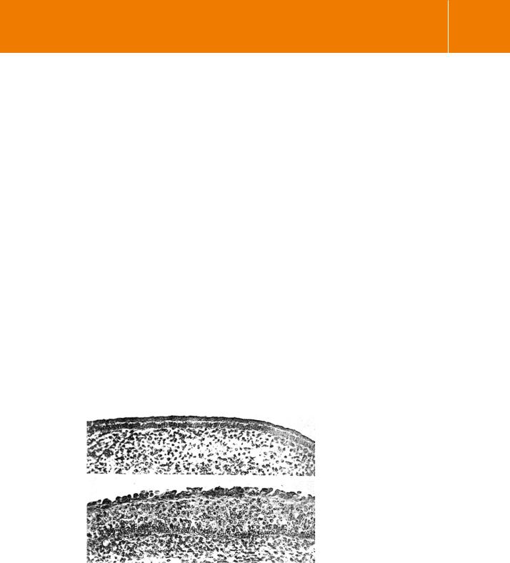
Later, Cohen discovered a new phenomenon that ‘was destined to become a magic wand that opened a whole new horizon to biological studies.’ A
contaminating factor was present that caused precocious growth in epidermis (Figure 11.2). It was also found that the factor has a powerful proliferative effect on connective tissues and it became clear that there is a link between the mechanisms that control normal proliferation and neoplastic growth.
FIG 11.2 An early demonstration of the effects of EGF. Thickening of the epidermal layer in skin explants from chick embryos after 3 days in culture.18
301

Signal Transduction
Since, under culture conditions, the stimulus to proliferate could not involve systemic or hormonal influences, Cohen called the new protein epidermal growth factor (EGF).18,19. Later, it was shown that mouse EGF enhances
DNA synthesis in cultured human fibroblasts. It was also found that EGF is similar to uragastrone, a peptide that had been isolated from human urine and recognized by pharmacologists because of its ability to inhibit gastric acid secretion.20 Out of 53 residues in the amino acid sequence, 37 share a common location and the two polypeptides have similar effects on both gastric acid secretion and the growth of epidermal cells.
All this now paved the way for a more molecular approach using isolated cells. It was found that rat kidney cells, transformed with the Kirsten sarcoma virus (see Chapter 4) fail to bind EGF. This down-regulation, which is due
to internalization of the receptor, is caused by the elevated expression and release of EGF by the cells themselves (an autocrine mechanism of feedback regulation). The possibility that internalization might be a necessary step initially found many adherents, but it became apparent that the transduction mechanism emanates from events at the plasma membrane. In particular, ligand binding directly induces phosphorylation (on tyrosine residues) of a membrane protein, later shown to be the EGF receptor (EGFR) itself.21 This was an important breakthrough because tyrosine kinase activity had already been associated with virus-induced sarcomas (the v-src gene product). Thus, a firm link was established between neoplasia and the physiological regulation of cellular growth.
Further evidence for the role of growth factors in tumour generation came with the revelation that the avian erythroblastosis virus oncogene, v-erb-B, codes a product having similarities with the EGF receptor. Indeed, it became apparent that the transformation derives from inappropriate acquisition from the (cellular) c-erb-B gene, of a truncated receptor, lacking the binding site for EGF and which is constitutively activated.22
The development of this field has generated much excitement but also frustration. It has been exciting, because it has yielded a good understanding of cell transformation and growth factors, and frustrating because it has become clear that cancer cells do not readily lend themselves as specific targets for drugs. The main impetus behind these studies was that nonmammalian genes might be the cause of disease. The hope was to discover targets which might be exploited to kill tumour cells selectively, for example the products of the viral genes. We now realize that these are initially hijacked from the mammalian genome itself, then inaccurately transcribed by sloppy DNA or RNA polymerases in the virus which then offers them back to the host upon infection. Apart from this, the large proportion of human tumours is
of non-viral origin, arising as a consequence of ageing, tumour-promoting substances, radiation, etc.
302