

Signal Transduction
proliferation40 and that it is a very potent chemotactic factor for neutrophils41 and fibroblasts.42 It has been proposed that a common name for these factors should be cytokines.43 While offering no clues to their various actions, this definition represents a move towards coordinating our understanding of their roles as first messengers. The unity of the cytokines is a concept as important as that of the hormones, defined by Bayliss and Starling nearly 90 years ago (see Chapter 1). However, while the pharmacology of the hormones has been so extensively (some might say exhaustively) investigated, for the protein (growth) factors, this area remains relatively unexplored. Attention here is firmly concentrated on intracellular events.
Essay: Cancer and transformation
Definitions
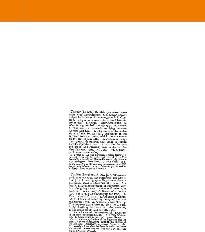
FIG 11.4 Definitions of cancer, from the Shorter Oxford English Dictionary (3rd ed. 1944, corrected 1977).
306
Growth factors: setting the framework
The essence of cancer
The essence of cancer is that cells run out of control. They no longer respond correctly to the demands and influences of the environment in which they exist. Cancer in a particular tissue manifests itself by an increased cell mass which
we describe as a tumour, meaning a swelling. The acquisition of cell mass is a consequence of new growth (neoplasm). In some cancers the primary tumour gives rise to the dissemination of cells that invade other tissues to form secondary tumours (metastasis). In addition, cancer cells tend to be poorly differentiated. As a consequence, they can lose their capacity to carry out the normal functions of the tissue from which they are derived, or they may take on an entirely inappropriate function (ectopic tumour). This means they may either fail to produce important factors or produce wrong factors in overwhelming excess. Under the microscope, cancerous cells are typically characterized by an enlarged nucleus with a very large nucleolus, less cytoplasm, and generally altered morphology. Cancer cells are often more rounded than their normal counterparts and there is generally less contact between them and their neighbours.
Most tumours develop in stages, going from benign to malignant. This is the result of a sequence of mutations that gradually enhance the sensitivity of the cells to growth factor signals, reduce the requirement of cell–cell and cell–matrix contact and render cells insensitive to the signals that determine programmed cell death. This change in phenotype is also called transformation, and so cancerous cells are often called transformed cells. The classification benign tumour means that there is an increase in the number of cells (hyperplasia, there may be a lump), but that the normal functions and morphology are retained. Importantly, there is no infiltration of other tissues, particularly the lymph nodes draining the areas in the vicinity of the tumour. By contrast, m alignancy refers to the loss or perversion of normal physiological function, altered morphology, and infiltration of other tissues, including metastasis.
Alterations dictating malignancy
Malignancy is a consequence of interplay between the transformed cell and its environment. In order for cells to proliferate in excess and to disseminate, they acquire new functions and they also induce the collaboration of the surrounding tissue.44 For instance, transformed cells release angiogenic factors that induce the formation of new vasculature.45 Surrounding
tissue also provides activators of metalloproteinases that cause the matrix degradation necessary for tissue invasion.46
Fully transformed cells possess one or more of the following characteristics:
•They may not require an exogenous growth signal. In culture conditions, this means that the provision of serum is no longer required. It also means that the cells no longer have to adhere to an extracellular matrix; they can grow in soft agar. They also grow well in athymic (nude) mice.
307

Signal Transduction
•
•
•
•
•
They may be insensitive to growth-inhibitory signals. For cells in culture, this means that proliferation of cultured cells continues beyond the point of formation of a tight monolayer. It also means that cells have become insensitive to the cell cycle checkpoints in G1, G2, and mitosis.
They can evade programmed cell death (apoptosis). This means that these cells have an inbuilt rescue mechanism.
They have limitless replicative potential. Normal cells can undergo 50 divisions. This is because progressive erosion of the telomeres (several thousand repeats of 6 bp sequence elements) results in exposure of chromosomal ends. When the telomeres are exhausted, chromosome fusion ensues causing nuclear chaos, cell senescence and cell death. By contrast, tumour cells can continue to divide without limit.
They can induce angiogenesis. During their development, tumours undergo a so-called angiogenic switch enabling them to induce the formation of new vasculature. This provides them with nutrients and oxygen and allows them to disseminate. Of course, dissemination is only possible when they have also acquired the capacity to survive when detached from the extracellular matrix (see Chapter 14).
They may invade other tissues (metastasis). This is the ultimate consequence of the preceding alterations. The cells have acquired the capacity to detach without dying, to destroy extracellular matrix in order to migrate through tissue, to be carried by the lymph or blood stream, to attach and then colonize sites where they would not normally survive due to the absence of specific adhesion and growth factors.
Genetic alterations at the basis of malignancy
Cell transformation in cancer is the consequence of mutations of several different types, some inherited, some due to environmental factors, others due to ageing. Loss-of-function or gain-of-function mutations have been found in genes (some 380 in total)53 that code for the following:
•
•
•
•
Molecules involved in the signalling of growth factors. These are either growth factors themselves (c-Sis, PDGF), their receptors (ErbB, the EGF receptor), or their downstream signalling components (Ras) (see Chapter 12). Molecules involved in the control of the cell cycle (so-called tumour suppressors). Most important are the mutations in the retinoblastoma protein (Rb) , the p53 protein or the failure to express p15INK4A and p15INK4B, inhibitors of the cyclin-dependent protein kinases.47
Adhesion molecules. These are mainly implicated in cadherin-mediated cell-cell adhesion (cadherin, -catenin, APC) and integrins (see Chapter 14). Molecules involved in the rescue from apoptosis. Important here are Bcl2 or Bcl-Xl, intracellular inhibitors of apoptosis, and the survival factors IGF-1 or IL-3.48
308

Growth factors: setting the framework
•Molecules involved in the signal transduction pathway that regulates cell survival (PTEN) (see Chapter 18).
•Molecules implicated in telomere maintenance. Transformed cells upregulate the expression of the catalytic subunit of the telomerase reverse transcriptase (hTERT).49
•Angiogenic factors or their inhibitors. For instance, VEGF, FGF, or thrombospondin-1.45
Cells will normally die rather than allow somatic (non-inherited) alterations to their genetic code to persist. The natural rate of somatic mutation (due, for instance, to natural environmental carcinogens or bombardment by cosmic rays) is low, and it is unlikely that a tumour would ever develop over the decades of a human life. Yet, in western Europe and North America, one in
three people will develop a cancerous growth. Certain mutations create a form of genomic instability that allows an enhanced and more generalized mutation frequency. The loss of function of the tumour suppressors p53 and Rb or other molecules involved in cell cycle control may be crucial here, the cells becoming too eager to replicate, failing to halt at the appropriate check points and failing to repair their DNA.50 From here on things go from bad to worse.
Constructing cancer in a dish
Cells possess mechanisms which protect the organism against the deleterious effects of their possible transformation. The responses that form this line of defence are set out in Table 11.1.
To understand the mechanism of transformation, it is necessary to establish its minimal requirements. How may this be achieved? Occasionally it has been possible to select rare, spontaneously arising immortalized cells. Alternatively, cells can be transformed by applying chemical and physical agents or a virus.
Table 11.1 Defences against transformation
Contingency |
Consequence |
|
|
Loss of contact with the extracellular matrix |
apoptosis |
|
|
Damage to DNA |
cell cycle arrest (G1) |
|
|
Defects in replication |
cell cycle arrest (G1) |
|
|
Failure of DNA repair |
apoptosis |
|
|
Excessive proliferation signals |
senescence or apoptosis |
|
|
Loss of telomeres (after 50 cell divisions) |
senescence and apoptosis |
|
(after 50 cell divisions) |
|
|
Unlike human cells, rodent cells express telomerase activity and, as a result, there is no erosion of the telomeres during ageing. Because of this, the loss of telomeres does not represent a barrier to transformation in rodents and only two oncogenic mutations
are necessary to cause transformation.
309
Signal Transduction
These can be said to represent shotgun approaches from which little can be learned. However, in certain human cells the imposition of just three mutations is sufficient to generate a transformed phenotype.51
•
•
•
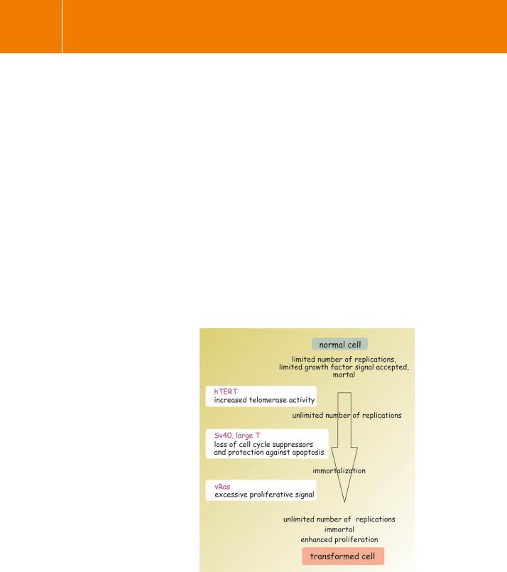
High telomerase activity to maintain telomere size and allow unlimited replication. Achieved by inserting the gene (hTERT) that codes for the catalytic subunit of the enzyme.
An antitumour suppressor activity in the form of a viral oncoprotein (SV40 large-transforming antigen, Large T). This disables the function of p53 and Rb, preventing arrest of the cell cycle in G1, and enables the cells to evade apoptosis.
A proliferative signal provided by a constitutively active form of Ras (V12G).
The sequence here is important.52 Enhanced telomerase activity is rather easily tolerated by the cells, whereas an excessive growth factor signal is not. The construction of a transformed human cell line was achieved by inducing telomerase activity first, then large-transforming antigen, and lastly Ras (see Figure 11.5). With just these three oncogenes, cells continue to proliferate
and grow well in soft agar or in immunodeficient nude mice. In human cancer six oncogenic mutations seem to be required for full transformation.
FIG 11.5 Construction of a transformed cell line.
310