

Chapter 14
Adhesion molecules in the regulation of cell differentiation: Mainly about Wnt
Here we consider the adhesion molecules as mediators of differentiation with regard to epithelial tissues. We discuss how cytokines affect
adhesion, with special emphasis on Wnt signalling. Errors in these pathways cause a transition towards a loosely attached mesenchymal phenotype that gives rise to cancer. We also discuss the importance of Wnt family proteins in the maintenance of the self renewal capacity of stem cells in the crypts of the intestinal tract.
Destabilization of adherens junctions causes cellular de-differentiation
The surfaces that separate interior from exterior, such as the skin, the linings of the body cavities, and the gut are formed from tight sheets of epithelial cells. Similarly, the linings of the vasculature, including the heart, blood vessels and lymphatics, are formed from endothelial cells which are
417
Signal Transduction
The many abbreviations used in this chapter are collected together at the end of the chapter.
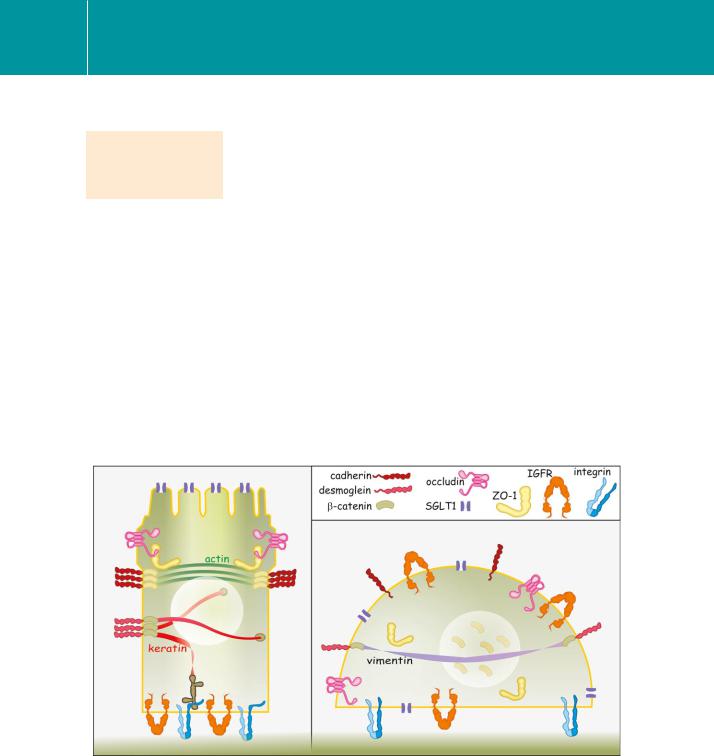
specialized epithelial cells. Unlike bone or interstitium, the extracellular matrix is minimal, comprising a thin sheet, the basal lamina. It is the cells themselves that bear the brunt of physical stresses such as the peristalsis of the gut and the pulsatile movement of the vessels. Tissue structure is maintained by an elaborate series of cellular attachments, tight junctions (also known as zonula occludens), adherens junctions and desmosomes (Latin occludo, to block up; desmos, meaning band or ligament) (see Figures 13.5 and 13.11).
If the adherens junctions stabilizing a cell layer are artificially destabilized, for instance by antibodies against cadherin, there is a loss of cell polarity and the spatial distribution of he membrane proteins becomes randomized.
Insulin-like growth factor receptor type-I (IGFR, normally present on the basal surface) and the glucose transporters (SGLT, on the apical surface), are now distributed all over the surface of the cell. The tight junctions disassemble through detachment of the zonula adherens-associated proteins (ZO-1) and the link between the cadherins and the catenins is broken; ZO-proteins are scattered into the cytosol1 and -catenin is present in the nucleus2 (Figure 14.1). The cells produce more fibronectin (extracellular matrix) and switch from the production of keratin to vimentin (both forming intermediate filaments). In
FIG 14.1 Loss of cell–cell contact and the dissipation of cell polarity.
The polarized state is well illustrated by the segregation of the components of the tight and adherent junctions on lateral surfaces: IGFRs at the basal surface and the Na /glucose symporter (SGLT1) at the apical surface. With the dissipation of the adherent junctions, this functional polarization is lost. The Na /glucose symporters and IGF receptors become randomly distributed and keratin is replaced by vimentin. The cells become de-differentiated.
418