


Chapter 6 |
263 |
|
|
Labour and delivery
Labour: overview 264 Labour: 1st stage 266 Labour: 2nd stage 268 Labour: 3rd stage 270
Induction of labour: indications 272 Cervical ripening 274
Induction of labour: methods 276
Induction of labour: special circumstances 278 Fetal surveillance in labour: overview 280 Fetal surveillance: cardiotocography 282
Fetal surveillance: cardiotocography abnormalities 284 Fetal surveillance: cardiotocography classification 288 Meconium-stained liquor 290
Operative vaginal delivery: overview 292 Operative vaginal delivery: instruments 294 Operative vaginal delivery: criteria 296 Operative vaginal delivery: trial 298 Episiotomy 300
Perineal tears 302
Thirdand fourth-degree tears 304 Caesarean section: overview 306 Caesarean section: indications 308 Caesarean section: types 309 Caesarean section: complications 310
Prelabour rupture of membranes at term 312 Prelabour rupture of membranes: management 314 Abnormal lie: transverse and oblique 315 Malpresentations in labour: overview 316 Malpresentations: brow and face 318
Retained placenta and placenta accreta 320 Post-partum haemorrhage 322
Home birth: overview 324
Home birth: risks and GP involvement 325 Home birth: the evidence 326

264 CHAPTER 6 Labour and delivery
Labour: overview
Labour is the process by which the fetus is delivered after the 24th week of gestation. The onset of labour is defined as the point when uterine contractions become regular and cervical effacement and dilatation becomes progressive. Hence, it is difficult to define the precise time of the onset. For clinical management the duration of observed labour is considered and not the duration the mother had painful contractions at home. Show and rupture of membranes may or may not be associated with labour, and these characteristics in themselves do not suggest onset of labour. In most cases, labour is characterized by:
•Onset of uterine contractions, which iin frequency, duration, and strength over time.
•Cervical effacement and dilatation.
•Rupture of membranes with leakage of amniotic fluid.
•Descent of the presenting part through the birth canal.
•Birth of the baby.
•Delivery of the placenta and membranes.
The mechanism of labour
The head usually engages in the transverse position and the passage of the head and body follows a well-defined pattern through the pelvis (Fig. 6.1). Not all the diameters of the fetal head can pass through a normal pelvis (see b Diameters of the female pelvis, p. 12). The process of labour therefore involves the adaptation of the fetal head to the various segments of the pelvis.
Sequence for the passage through the pelvis for a normal vertex delivery
•Engagement and descent: the head enters the pelvis in the occipitotransverse position with flexion ias it descends.
•Internal rotation to occipitoanterior: occurs at the level of the ischial spines due to the forward and downward sloping of the levator ani muscles.
•Crowning: the head extends, distending the perineum until it is delivered.
•Restitution: the head rotates so that the occiput is in line with the fetal spine.
•External rotation: the shoulders rotate when they reach the levator muscles until the biacromial diameter is anteroposterior (the head externally rotates by the same amount).
•Delivery of the anterior shoulder: occurs by lateral flexion of the trunk posteriorly.
•Delivery of the posterior shoulder: occurs by lateral flexion of the trunk anteriorly and the rest of the body follows.
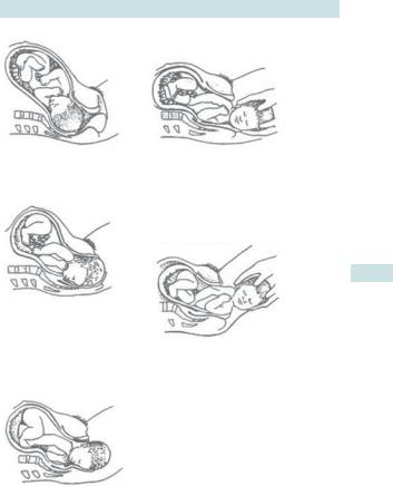
(1)
First stage of labour. The cervix dilates. After full dilation
the head flexes futher and descends further into the pelvis.
(2)
During the early second stage the head rotates at the levels of the ischial spine so the occiput lies in the anterior part of the pelvis. In late second stage the
head broaches the vulval ring (crowning) and the perineum stretches over the head.
(3)
LABOUR: OVERVIEW 265
(4)
Birth of the anterior shoulder. The shoulders rotate to lie in the anteroposterior diameter of the pelvic outlet. The head rotates externally, ‘restitutes’, to its direction at onset of labour. Downward and backward traction of the head by the birth attendant
aids delivery of the anterior shoulder.
(5)
Birth of the posterior shoulder is aided by lifting the head upwards whilst maintaining traction.
The head is born. The shoulders still lie transversely in the midpelvis.
Fig. 6.1 Mechanism of labour and delivery. Reproduced from Collier J, Longmore M, Brinsden M. (2003). Oxford Handbook of Clinical Specialties, 6th edn. Oxford: OUP. By permission of Oxford University Press.
266 CHAPTER 6 Labour and delivery
Labour: 1st stage
The 1st stage is divided into two phases:
•Latent phase: the period taken for the cervix to completely efface and dilate up to 3cm.
•Active phase: from 3cm to full dilatation (10cm).
Braxton Hicks contractions are mild, often irregular, non-progressive contractions that may occur from 30wks gestation (more common after 36wks) and may often be confused with labour. However, contractions in labour are painful, with a gradual increase in frequency, amplitude, and duration.
Failure to progress is suspected if:
•There is <2cm dilatation in 4h (on a 4hr action line partogram the plotted progress falls to the right).
•Slowing in progress in parous women.
2Consideration should also be given to effacement of cervix and descent of the head.
•If labour is slow from onset, it is pdysfunctional labour.
•If there was previous adequate progress then it is sarrest.
Some causes of poor progress in the 1st stage
•Inefficient uterine activity (power—commonest cause).
•Malpositions, malpresentation, or large baby (passenger).
•Inadequate pelvis (passage).
•A combination of two or more of the above.
Poor progress in the 1st stage
Assessment
•Review the history.
•Abdominal palpation, frequency, and duration of contractions.
•Review fetal condition; fetal heart rate and colour/quantity of amniotic fluid.
•Review maternal condition including hydration and analgesia.
•Vaginal assessment; cervical effacement, dilatation, caput, moulding, position, and station of the head.
Management
•Amniotomy (artifical rupture of membranes (ARM)) and reassess in 2h.
•Amniotomy + oxytocin infusion and reassess in 2h: this should always be considered in nulliparous women.
•Lower segment CS (if there is fetal distress).
For multiparous women and those with a previous CS an experienced obstetrician should review before starting oxytocin.
NICE. (2007). Intrapartum care: management and delivery of care to women in labour. NICE guideline CG55. Mhttp://guidance.nice.org.uk/CG55
LABOUR: 1ST STAGE 267
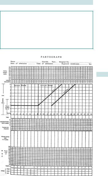
Monitoring in labour (recorded on the partogram (Fig. 6.2))
•The FHR should be monitored every 15min (or continuously with a CTG).
•The contractions should be assessed every 30min.
•Maternal pulse should be checked hourly.
•BP and temperature should be checked 4-hourly.
•VE should be offered every 4h to assess progress.
•Maternal urine is tested 4-hourly or when passed for ketones and protein.
Fig. 6.2 Partogram.

268 CHAPTER 6 Labour and delivery
Labour: 2nd stage
The 2nd stage is the time from full cervical dilatation until the baby is born.
2 If the woman has an epidural and the CTG is reassuring, 1h is usually allowed for passive descent before active pushing is commenced.
During this hour it is important to ensure that good contractions are maintained and oxytocin may be commenced.
2Birth should take place within 3h of the start of 2nd stage for nulliparous women and within 2h for multiparous women.
Description of a normal 2nd stage
•The active 2nd stage commences when the mother starts expulsive efforts using her abdominal muscles with the Valsalva manoeuvre to ‘bear down’.
•Women may choose many different positions to deliver in: squatting, standing, on all fours, or supine: lithotomy is required for instrumental deliveries.
•As the head comes down, it distends the perineum and anus: a pad may be used to support the perineum and cover the anus, while the other hand is used to maintain flexion and prevent sudden deflexion and to control the rate of delivery of the head (this attempts to slow perineal distension, minimizing tears by preventing rapid delivery).
•An episiotomy may be performed if there is concern that the perineum is tearing towards the anal sphincter: episiotomy should not be used routinely.
•With the next contraction gentle traction guides the head towards the perineum until the anterior shoulder is delivered under the subpubic arch.
•Gentle traction upwards and anteriorly helps to deliver the posterior shoulder and the remainder of the trunk.
•The cord is double-clamped and cut: delaying clamping for 2–3min results in higher haematocrit levels in the neonate.
•The condition of the baby is assessed at 1, 5, and 10min using the Apgar scoring system, and if all is well, baby is handed to the mother as soon as possible.

LABOUR: 2ND STAGE 269
Delay in the 2nd stage of labour
Nulliparous women
•Suspected if delivery is not imminent after 1h of active pushing: VE should be offered and amniotomy recommended.
•If not delivered in 2h: requires review by obstetrician to consider instrumental delivery or CS.
Multiparous women
•If delivery is not imminent after 1h of active pushing: requires review by obstetrician to consider instrumental delivery or CS.
Delay in the 2nd stage in a multiparous woman must always raise suspicions of malposition or disproportion.

270 CHAPTER 6 Labour and delivery
Labour: 3rd stage
The 3rd stage is the duration from delivery of the baby to delivery of the placenta and membranes.
Active management of the 3rd stage
Consists of:
•Use of uterotonics.
•Clamping and cutting of the cord.
•Controlled cord traction.
Benefits
•dRates of PPH >1000mL.
•dMean blood loss and postnatal anaemia.
•dLength of the 3rd stage.
•dThe need for blood transfusions.
Adverse effects
•Nausea and vomiting.
•Headache.
Physiological management of the 3rd stage
Consists of:
•No Syntometrine® or oxytocin is given.
•Cord is allowed to stop pulsating before it is clamped and cut.
•The placenta is delivered by maternal effort alone.
The cord must not be pulled and the uterus not pushed on in any direction to help to expel the placenta.
A planned physiological 3rd stage should be changed to active management in the event of:
•Haemorrhage.
•Failure to deliver the placenta within 1h.
•Maternal desire to shorten the 3rd stage.

LABOUR: 3RD STAGE 271
Description of an actively managed 3rd stage
•Syntometrine® IM (ergometrine 0.5mg + oxytocin 5IU) or oxytocin 10IU IM is given as the anterior shoulder of the baby is born.
•A dish is placed at the introitus to collect the placenta and any blood loss, and the left hand is placed on the abdomen over the uterine fundus.
•As the uterus contracts to 20-wk size, the placenta separates from the uterus through the spongy layer of the decidua basalis.
•The uterus will then feel firmer, the cord will lengthen, and there is often a trickle of fresh blood (separation bleeding).
•Controlled cord traction (CCT) is applied with the right hand, whilst supporting the fundus with the left hand (Brandt–Andrew’s technique).
Multiple pregnancy must be excluded before uterotonics are given.
2NICE recommends the use of oxytocin 10IU rather than Syntometrine® as it appears to have similar efficacy but with fewer side effects.
Care immediately after delivery
•Most complications occur in the first 2h after delivery, including;
•PPH
•uterine inversion
•haematoma formation.
•Usually women are kept in the delivery unit during this time to observe: pulse, BP, temperature, uterine size and contractions, fresh bleeding per vaginum, or painful swelling of the vulva, vagina, or perineum.
•Where there is an increased risk of PPH (e.g. in multiple pregnancy), an oxytocin infusion (40U in 500mL saline) should be given prophylactically for 3–4h.
•Encouragement should be given for skin-to-skin contact as soon as possible and the mother and baby should not be separated for the 1st hour.
•Support should be provided for breast-feeding, which should be initiated in the 1st hour.
•If there are no complications during these 2h, the mother may then be transferred to the postnatal ward: some women may then go home after a further 3–4h of observation.

272 CHAPTER 6 Labour and delivery
Induction of labour: indications
•10–20% of all pregnancies are induced.
•Overall success rate is about 60–80% at term.
•Chance of achieving vaginal birth after IOL <34wks is <35%.
•The indication may be obstetric or medical.
•IOL on maternal request should be avoided as it is associated with risks for both mother and fetus.
Obstetric indications
•Uteroplacental insufficiency (one of the most common indications).
•Prolonged pregnancy (41–42wks).
•IUGR.
•Oligoor anhydramnios.
•Abnormal uterine or umbilical artery Dopplers.
•Non-reassuring CTG.
•PROM.
•Severe pre-eclampsia or eclampsia after maternal stabilization.
•Intrauterine death of the fetus (IUD).
•Unexplained antepartum haemorrhage at term.
•Chorioamnionitis.
XThere is inadequate evidence for induction for suspected fetal macrosomia. Some advocate IOL around 40–41wks with an aim of preventing further intrauterine growth and associated risks like shoulder dystocia and birth trauma.
Medical indications
With underlying maternal medical conditions, planned early IOL may potentially limit the maternal risks associated with pregnancy. Careful timing is required to balance the best interests of the mother with any potential risks of prematurity.
Such situations may include:
•Severe hypertension.
•Uncontrolled diabetes mellitus.
•Renal disease with deteriorating renal function.
•Malignancies (to facilitate definitive therapy).
This page intentionally left blank

274 CHAPTER 6 Labour and delivery
Cervical ripening
Predictors for successful induction of labour
Strong predictors for a successful IOL are:
•Gestational age at induction.
•Parity.
•Modified Bishop’s score of the cervix (Table 6.1): overview of the ‘ripeness’ of the cervix (iscore, isuccess).
Mechanical methods of cervical ripening
Separation of the membranes from the cervix leads to the local release of prostaglandins.
•A common method is artificial separation (‘stretch and sweep’).
•This requires that the cervical os admits a finger, and involves digitally separating the membranes from the cervix.
•It is uncomfortable and may lead to some bleeding.
•30% will go into spontaneous labour in <7 days.
•In the majority it results in a more favourable cervix.
Pharmacological methods
Prostaglandins (PGE2 = dinoprostone)
•Preferred agents for cervical ripening.
•Usually given intravaginally into the posterior fornix.
•The gel form is absorbed well.
•Tablet forms are easier to remove if hyperstimulation occurs (5–7%).
•Vaginal prostaglandins (3mg tablets, 2mg gel) increase vaginal delivery rates within 24h with no increase in operative delivery rates.
Oxytocin infusion
•Has been shown to increase cervical prostaglandin levels.
•As most receptors are located in the myometrium, it is more suitable for initiating uterine contractions.
•Best used where membranes have ruptured, whether spontaneously or after amniotomy.
2 Other agents have been tried but there is limited evidence for their safety and efficacy.
Other methods
There is no evidence to suggest that the following are effective in cervical ripening or IOL:
•Sexual intercourse.
•Herbal remedies (raspberry leaf tea).
•Nipple stimulation.
•Acupuncture.
•Castor oil.

CERVICAL RIPENING 275
Table 6.1 Modified Bishop’s score: to assess the favourability for induction of labour. A total score of >8 indicates a favourable cervix
Score |
0 |
1 |
2 |
Position of cervix |
Posterior |
Axial |
Anterior |
Length of cervix |
2cm |
1cm |
<0.5cm |
Consistency of cervix |
Firm |
Soft |
Soft and |
|
|
|
stretchy |
Dilatation of cervix |
0 |
1cm |
>2cm |
Station of the presenting part |
−2 |
−1 |
0 |
(distance in cm in relation to the |
|
|
|
ischial spines) |
|
|
|
Data from Kennedy et al., 1982.
276 CHAPTER 6 Labour and delivery
Induction of labour: methods
Amniotomy
•ARM or amniotomy releases local prostaglandins causing cervical ripening and myometrial contractions.
•If regular, painful uterine contractions are not initiated or there are no cervical changes after 2h, then oxytocin infusion should be commenced.
•Starting oxytocin at the time of amniotomy has been shown to decrease the induction–delivery interval, thereby decreasing both the fetal and maternal risk of sepsis.
•ARM alone is not recommended for IOL.
Prostaglandins for induction of labour
• A CTG should be performed 30min before, as well as after insertion of prostaglandins to confirm fetal well-being and to detect possible hyperstimulation.
• VE after 6h: if the cervix is not favourable, another dose may be administered (>2 doses need to be reviewed by a consultant; multiparous women seldom require more than 1 dose).
•Oxytocin should not be started for 6h to avoid the risk of uterine hyperstimulation.
Synthetic oxytocin for induction or augmentation of labour
•It should be started on a low dose (1–4mU/min).
•It is increased (usually doubled) every 30min to achieve optimal contractions (3–4 every 10min, each lasting 40–60s).
•Continuous CTG monitoring should be used:
•the sensitivity of the myometrium to oxytocin iduring labour and it may be necessary to dthe rate of infusion as labour advances
•infusion pumps should be used to carefully control the amount given and avoid the risk of uterine hyperstimulation.
X Women should be advised that the use of oxytocin will reduce the length of labour and may reduce the operative births due to dystocia without any major impact on neonatal outcomes (Cochrane review).

INDUCTION OF LABOUR: METHODS 277
Risks and complications of IOL
•Prematurity:
•may be iatrogenic (severe pre-eclampsia)
•unintentional (failure to correctly assess the gestational age).
•Cord prolapse with rupture of membranes if the presenting part is not engaged.
•Side effects of pharmacological agents used:
•pain or discomfort
•uterine hyperstimulation
•fetal distress
•uterine rupture (rare but iin grand multipara or a scarred uterus).
•Prostaglandins rarely cause non-selective stimulation of other smooth muscle leading to:
•nausea and vomiting
•diarrhoea
•bronchoconstriction (caution in asthmatics)
•maternal pyrexia may result due to the effect on thermoregulation in the hypothalamus.
•CS due to failed induction.
•Atonic post-partum haemorrhage.
•Intrauterine infection with prolonged induction.
Oxytocin has the properties of ADH, and U&E should be checked if it has been used for >12h as it may very rarely cause dilutional hyponatraemia.

278 CHAPTER 6 Labour and delivery
Induction of labour: special circumstances
Prelabour rupture of membranes
Prostaglandins should be used before starting syntocinon for IOL if the cervix is unfavourable.
Stabilizing induction
This is carried out when the presenting part is not engaged or when there is an unstable lie, to avoid the risk of cord prolapse.
•The head is ‘stabilized’ by an assistant holding it suprapubically and if possible by pushing the head into the pelvic brim.
•Amniotomy is performed.
•Once cord prolapse is excluded, oxytocin infusion is started.
2This is usually performed in the delivery unit with the theatre and team available should an emergency of cord prolapse occur. It is good to have an epidural that could be topped up sufficiently to allow an emergency CS.
Grand multipara (≥ para 5)
The risk of uterine rupture is higher and hence caution should be exercised.
XProstaglandin gel should only be used in exceptional circumstances.
•Onset of labour is awaited for up to 4h after ARM.
•In the absence of contractions oxytocin infusion can be started and titrated to get 3–4 every 10min, each lasting >40s.
•Once contractions are established it should be possible to stop the oxytocin as most will continue to labour and deliver normally.
Malpresentation (obstructed labour) must be excluded before starting oxytocin.
Induction for intrauterine death at term
The WHO and RCOG Guidelines recommend misoprostol 25 micrograms every 2–4h.
•As this strength is not available in the UK 200 microgram tablets can be dissolved in 40mL water and 5mL aliquots administered.
•Although uniformity of strength cannot be guaranteed it is potentially safer than administering a higher dose.

INDUCTION OF LABOUR: SPECIAL CIRCUMSTANCES 279
IOL with previous CS
The risk of scar dehiscence with previous uterine surgery is:
•5:1000 with spontaneous labour.
•8:1000 with use of oxytocin.
•24:1000 with prostaglandins.
2 Women should be counselled regarding these risks and have continuous CTG monitoring throughout the whole of the induction process when contractions are present.
2Facilities should be available for immediate CS should there be a scar rupture and fetal bradycardia.
Further reading
Kennedy JH, Stewart P, Barlow DH, et al. (1982). Induction of labour: a comparison of a single prostaglandin E2 vaginal tablet with amniotomy and intravenous oxytocin. Br J Obstet Gynaecol 89:704–7.
NICE. (2011). Induction of labour, NICE guideline CG70. Mhttp://www.nice.org.uk/CG70
RCOG. (2011). Green-top guidelines. Induction of labour for intrauterine death at term. M www. rcog.org.uk
WHO. (2011). Recommendations on induction of labour. M http://whqlibdoc.who.int/ publications/2011/9789241501156_Eng.pdf

280 CHAPTER 6 Labour and delivery
Fetal surveillance in labour: overview
•It is estimated that 10% of CP is due to intrapartum hypoxia (the rest may be attributed to antenatal).
•Blood supply to the placental pool is restricted, with contractions (especially in the 2nd stage) placing a physiological strain on the fetus.
•Ability to withstand the stress is dependent on fetal reserve.
•A fetus that was coping in the antenatal period but has no extra reserve may decompensate in labour.
Intrapartum surveillance
•The options for intrapartum surveillance are:
•intermittent auscultation (IA)
•continuous CTG, also known as electronic fetal monitoring (EFM).
•On admission in labour, an assessment should be made to identify fetal and maternal risk factors (see Boxes 6.1 and 6.2).
•If the woman has no risk factors she should be offered intermittent auscultation performed for a full minute after a contraction:
•at least every 15min in the 1st stage
•every 5min or after every other contraction in the 2nd stage.
Electronic fetal monitoring
•Results in:
•iintervention and operative delivery rates
•no marked din CP.
•Most likely because:
•CTG is not specific enough in detecting fetal hypoxia
•failure to consider the clinical situation
•poor interpretation
•delay in taking action
•intrapartum hypoxia as a cause of CP is rare.
•Additional tests, such as fetal scalp blood sampling in labour, are required to ispecificity.
2Some centres use fetal ECG ST waveform analysis (STAN) to improve the positive predictive value of the CTG.
2 Cochrane review suggested that there is reduction of fetal scalp blood sampling and total operative delivery, but not CS and there is no reduction in poor outcome of the neonate.

FETAL SURVEILLANCE IN LABOUR: OVERVIEW 281
Box 6.1 Antenatal risk factors that should prompt recommendation of EFM in labour
Maternal
•Previous CS.
•Cardiac problems.
•Pre-eclampsia.
•Prolonged pregnancy (>42wks).
•Prelabour rupture of membranes (>24h).
•Induction of labour.
•Diabetes.
•Antepartum haemorrhage.
•Other significant maternal medical conditions.
Fetal
•IUGR.
•Prematurity.
•Oligohydramnios.
•Abnormal Doppler velocimetry.
•Multiple pregnancy.
•Meconium-stained liquor.
•Breech presentation.
Box 6.2 Intrapartum risks requiring EFM
•Oxytocin augmentation.
•Epidural analgesia.
•Intrapartum vaginal bleeding.
•Pyrexia >37.5ºC.
•Fresh meconium staining of liquor.
•Abnormal FHR on intermittent auscultation.
•Prolonged labour.
Further reading
Cochrane review of ST waveform analysis for intrapartum surveillance. Mwww.cochrane.org
NICE. (2007). Intrapartum care guidelines. M publications.nice.org.uk/intrapartum-care-cg55/ guidance#complicated-labour-monitoring-babies-in-labour
282 CHAPTER 6 Labour and delivery
Fetal surveillance: cardiotocography
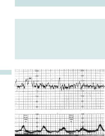
Definitions of terms used in EFM
•Baseline rate: mean level of the FHR when this is stable, and after exclusion of accelerations and decelerations.
•Baseline variability: degree to which the baseline varies, i.e. bandwidth of baseline after exclusion of accelerations and decelerations. Variability of 5–25 beats/min is defined as normal, 0–5 beats/min as reduced, and >25 beats/min as saltatory.
•Acceleration: a transient rise in FHR by at least 15 beats over the baseline lasting for 15s or more (Fig. 6.3).
•Deceleration: a reduction in the baseline of 15 beats or more for more than 15s.
Fig. 6.3 Cardiotocographic trace.
2 The most useful features in assessing fetal well-being are normal variability and presence of accelerations.
Always be concerned about a CTG if you cannot identify the baseline rate.

FETAL SURVEILLANCE: CARDIOTOCOGRAPHY 283
Causes of decreased baseline variability
•Fetal hypoxia.
•Fetal sleep cycle (should be for <40 and maximally 90min).
•Fetal malformation (CNS or cardiac) or arrhythmias.
•Administration of drugs including:
•methyldopa
•magnesium sulphate
•narcotic analgesics
•tranquillizers
•barbiturates
•general anaesthesia.
•Severe prematurity.
•Fetal heart block.
•Fetal anomalies.

284 CHAPTER 6 Labour and delivery
Fetal surveillance: cardiotocography abnormalities
Abnormalities in baseline rate
A bradycardia is a baseline FHR of less than 110 beats/min.
•100–110 beats/min is moderate baseline bradycardia and on its own is not considered to be associated with fetal compromise if the baseline variability is normal and accelerations are present.
•A baseline below 100 beats/min should raise the possibility of hypoxia or other pathology.
Beware of maternal heart rate being recorded as the FHR.
A tachycardia is a baseline FHR >160 beats/min and is associated with maternal pyrexia and tachycardia, prematurity, and fetal acidosis.
•160–180 beats/min is moderate baseline tachycardia and on its own is probably not indicative of hypoxia if the baseline variability is normal and accelerations are present.
•A baseline >180 beats/min should always raise suspicion of underlying pathology.
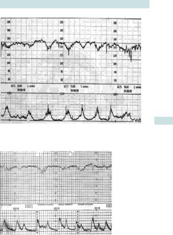
Decelerations
•Early decelerations: the peak of the deceleration coincides with the peak of the contraction (Fig. 6.4). This is related to head compression and, therefore, should only be seen in active second stage of labour.
•Late decelerations: have at least a 15s time lag between the peak of the contraction and the nadir of the deceleration (Fig. 6.5).
They may be suggestive of acidosis, especially if accompanied with tachycardia and reduced baseline variability.
Shallow, late decelerations in the presence of reduced baseline variability on a non-reactive trace should be of particular concern and may even be preterminal, especially if there are associated clinical risks including IUGR, absent FM, bleeding, infection, prolonged pregnancy, or severe pre-eclampsia.
•Variable decelerations: have variable pattern in timing, size, and shape and are associated with cord compression (Fig. 6.6):
•typical variables are U or V shaped, quick to drop and to recover, and often have ‘shouldering’ (not usually associated with hypoxia)
•atypical variables have a duration of >60s, a loss >60 beats from the baseline, slow recovery, a combined variable, and a late deceleration component
•with progressive hypoxia the decelerations become deeper and wider with rising baseline rate. Subsequent reduction of baseline variability suggests possible fetal acidosis.
Other abnormalities Sinusoidal pattern: a rare undulating pattern (sine wave) with little, or no, variability. Can indicate significant fetal anaemia, but in short spells (<10min) may be a result of fetal behaviour (thumb-sucking).
A sinusoidal pattern should always be taken seriously. Blood group antibodies, Kleihauer test, and a scan for middle cerebral artery velocity to detect fetal anaemia may be indicated.
FETAL SURVEILLANCE: CARDIOTOCOGRAPHY ABNORMALITIES 285
Fig. 6.4 Cardiotocograph trace with early decelerations.
Fig. 6.5 Cardiotocograph trace with late decelerations.
286 CHAPTER 6 Labour and delivery
Fig. 6.6 Cardiotocograph trace with variable decelerations.
This page intentionally left blank

288 CHAPTER 6 Labour and delivery
Fetal surveillance: cardiotocography classification
In order to help with the difficulties encountered when assessing a CTG a classification scheme was introduced that can be used to define a CTG as normal, suspicious, or pathological (Table 6.2).
|
|
Table 6.2 |
Fetal heart-rate feature classification |
|
||
|
|
|
Baseline |
Variability |
Decelerations |
Accelerations |
|
|
|
(beats/min) |
(beats/min) |
|
|
|
|
|
|
|
|
|
|
|
Reassuring |
110–160 |
≥5 |
None |
Present |
|
|
Non- |
100–109 |
<5 for ≥40 |
Early decelerations |
The absence of |
|
|
reassuring |
161–180 |
but <90min |
Variable |
accelerations |
|
|
|
|
|
decelerations |
in an otherwise |
|
|
|
|
|
being present |
normal CTG |
|
|
|
|
|
for 50% of |
is of uncertain |
|
|
|
|
|
contractions |
significance |
|
|
|
|
|
for ≥90min |
|
|
|
|
|
|
|
|
|
|
|
|
|
Single prolonged |
|
|
|
|
|
|
deceleration up to |
|
|
|
|
|
|
3min |
|
|
|
Abnormal |
<100 >180 |
<5 for |
Atypical variable |
|
|
|
|
Sinusoidal |
≥90min |
decelerations |
|
|
|
|
|
Late decelerations |
|
|
|
|
|
pattern for |
|
being present |
|
|
|
|
≥10min |
|
for >50% of |
|
contractions for ≥30min
Single prolonged deceleration >3min
NICE. (2011). Intrapartum care: management and delivery of care to women in labour. CG55.
Mhttp://www.nice.org.uk

FETAL SURVEILLANCE: CARDIOTOCOGRAPHY CLASSIFICATION 289
CTG classification using Table 6.2
•Normal: all four features are in the reassuring category.
•Suspicious: no more than one non-reassuring feature when analysing the CTG.
•Pathological: two or more non-reassuring features or one or more abnormal features.
Maternal factors that may contribute to an abnormal CTG
•The woman’s position: advise her to adopt left lateral.
•Hypotension.
•Vaginal examination.
•Emptying bladder or bowels.
•Vomiting.
•Vasovagal episodes.
•Siting and topping-up of regional anaesthesia.
Fetal blood sampling
•This is used to improve the specificity of CTG in the detection of fetal hypoxia.
•It should be obtained if the trace is pathological, unless obvious immediate delivery may be required (e.g. bradycardia of <80 beats/ min for >3min).
•The woman should be in left lateral.
Interpretation of the FBS results
•Normal (pH ≥7.25): repeat FBS within 1h if CTG remains pathological.
•Borderline (pH 7.21–7.24): repeat FBS within 30min if CTG remains pathological.
•Abnormal (pH 7.20): immediate delivery.

290 CHAPTER 6 Labour and delivery
Meconium-stained liquor
Meconium is made up of water, bile pigment, mucus, and amniotic fluid debris. Detection in amniotic fluid causes anxiety as it is associated with i perinatal morbidity and mortality; it may be aspirated by fetus.
•Meconium-stained amniotic fluid (MSAF) is rare in preterm infants (<5%) and is associated with infection and chorioamnionitis.
•Incidence of MSAF gradually increases from 36 to 42wks.
•Passage of meconium signifies the maturation of central nervous and gastrointestinal systems. Sometimes hypoxia causes peristalsis of the bowel and relaxation of anal sphincters resulting in MSAF.
Meconium aspiration syndrome
•Occurs in 1:1000 births in Europe.
•May happen in utero when fetal breathing movements draw amniotic fluid into the airway. Fetal gasping in utero is thought to be associated with prolonged decelerations that cause transient hypoxia, as 50% of mecorium aspiration syndrome (MAS) occur in fetuses that were not acidotic in labour.
•Meconium:
•causes mechanical blockage of the airway
•acts as a chemical irritant, causing pneumonitis and alveolar collapse
•predisposes to secondary bacterial infection.
Suction of the mouth and upper airway immediately after delivery is not currently recommended if the baby is active and crying.
If the newborn has respiratory difficulty, meconium should be cleared from the oroand nasopharynx and, if needed, from the trachea by using a laryngoscopy.
2This will not help with pre-existing in utero aspiration.
Classification of MSAF
•Grade 1 (light): meconium lightly stains the amniotic fluid that is usually copious.
•Grade 2 (moderate): dark green staining of amniotic fluid that appears opalescent.
•Grade 3 (thick): thick, opaque meconium in scanty amniotic fluid (‘pea soup meconium’).
Management of meconium-stained liquor
•Recommend immediate induction of labour if prelabour rupture of membranes.
•Advise continuous fetal monitoring.
•Advise delivery in a unit able to provide fetal blood sampling and advanced neonatal life support at birth:
•if the baby is born with depressed vital signs it will require laryngoscopy and suction by a healthcare professional trained in advanced neonatal life support
•if the baby is born in good condition it will still require close monitoring for 12h.
This page intentionally left blank

292 CHAPTER 6 Labour and delivery
Operative vaginal delivery: overview
CS in the 2nd stage of labour is associated with increased morbidity to the mother. Instrumental vaginal delivery helps to avoid maternal and perinatal morbidity and mortality and an emergency CS.
In the UK, the operative vaginal delivery rate is stable at between 10 and 15%. Important to appreciate that forceps and ventouse are complementary to each other and that operator’s skill and experience, as well as clinical findings, should decide which one to use.
2If in doubt, senior help must be called.
Indications for instrumental delivery
Maternal
•Exhaustion.
•Prolonged 2nd stage:
•>1h of active pushing in multiparous women
•>2h in primiparous women.
•Medical indications for avoiding Valsalva manouevre, such as:
•severe cardiac disease
•hypertensive crisis
•uncorrected cerebral vascular malformations.
•Pushing is not possible (paraplegia or tetraplegia).
Fetal
•Fetal compromise.
•To control the after-coming head of breech (forceps).
2 It is important to discuss with the woman why an operative delivery is indicated, the instrument chosen, the likelihood of success, and the alternatives available (emergency CS).
Consent (verbal or written) must be obtained by explaining the indication and it should be recorded.

OPERATIVE VAGINAL DELIVERY: OVERVIEW 293
Complications of operative vaginal delivery
•Forceps are associated with increased maternal trauma (including anal sphincter trauma).
•Rotational forceps may cause spiral tears of the vagina.
•Fetal injuries with forceps are rare but may occur (mostly due to incorrect application of the blades) including:
•facial nerve palsy
•skull fractures
•orbital injury
•intracranial haemorrhage.
•Ventouse is associated with fetal injuries including:
•scalp lacerations and avulsions (rarely, alopecia in the long term)
•cephalohaematoma
•retinal haemorrhage
•rarely, subgaleal haemorrhage and/or intracranial haemorrhage.
The use of sequential instruments (usually forceps after a failed ventouse) is associated with an increased risk of fetal trauma when attempted with no significant descent.
2 It is not uncommon for ventouse to slip when the head is at the introitus, then delivery is completed by a lift-out forceps—hence, the discrepancy in the Cochrane review of more failed ventouse deliveries, but a lower CS rate compared with forceps deliveries.

294 CHAPTER 6 Labour and delivery
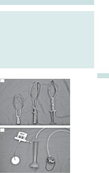
Operative vaginal delivery: instruments
Forceps
These consist of curved blades that sit around the fetal head and allow traction to be applied along the ‘flexion point’ of the head (3cm in front of the occiput). This is usually to speed up delivery, but may be used to slow rate of the head in a breech delivery (see Fig. 6.7).
Low cavity forceps (Wrigley’s)
•Short and light.
•These are also the forceps used at CS.
Mid-cavity non-rotational forceps (Neville-Barnes’, Haig Ferguson, Simpson’s)
•Used when the sagittal suture is in the direct anteroposterior position (usually direct occipito-anterior (DOA)).
•Malposition (direct occipito-posterior (DOP) or direct occipito-lateral (DOL)) can be corrected manually between contractions and the blades applied once the head is in the DOA position.
Mid-cavity rotational forceps (Keilland’s)
•Reduced pelvic curve on the blades of the forceps allows rotation about the axis of the handle.
•Helps to correct asynclitism and malposition.
•Must only be attempted by an experienced operator.
Vacuum extraction (ventouse)
Works on the principle of creating ‘negative pressure’ to allow scalp tissues to be sucked into the cup. This creates artificial caput called a ‘chignon’. The cup is held in place by the atmospheric pressure on the cup against the negative pressure created.
It should not be used at <34wks gestation (see Fig. 6.7).
Metal cup
•Available with 60, 50, or 40mm standard anterior (for positions) or posterior cup (for occipito-lateral (OL) or occipito-posterior (OP) positions).
•Pressure is created by a suction pump.
•Excessive traction is likely to cause fetal trauma.
Soft cup
•Soft and easier to apply (important in women without epidural) for OA positions.
•Moulds around the fetal head covering a greater surface area.
•Causes fewer scalp abrasions.
Kiwi Omni CupTM
•Single-use cup.
•Pressure created with hand pump (quick in an emergency).
•Allows application to flexion point in OL and OP position.
OPERATIVE VAGINAL DELIVERY: INSTRUMENTS 295
Comparison of forceps and vacuum extractor
•Ventouse is more likely to fail.
•Ventouse is more likely to cause fetal trauma such as:
•cephalohaematoma
•retinal haemorrhage.
•Ventouse is more likely to be associated with maternal concerns about the baby.
•Forceps are more likely to cause significant maternal genital tract trauma.
•There is:
•slightly less CS delivery with ventouse delivery
•no difference in low 5min Apgar scores
•no difference in need for neonatal phototherapy.
2Bottom line—ventouse appears safer for mother but forceps may be safer for baby.
(a)
(b)
Fig. 6.7 (a) Forceps (Wrigley’s, Neville-Barnes’, Keilland’s). (b) Ventouse cups (Kiwi Omni Cup™, silc cup, Bird’s cup). With permission of Clinical Innovations Europe Ltd, 2008, and Menox AB, Goteborg, Sweden, 2008.
296 CHAPTER 6 Labour and delivery
Operative vaginal delivery: criteria
2 The techniques involved with both ventouse and forceps can only be learned under direct supervision from an experienced operator and are therefore not described in the book.
The following criteria should be satisfied before attempting an operative vaginal delivery. This may be best remembered as ‘FORCEPS’.
•F Fully dilated cervix (i.e. confirm 2nd stage).
•O Obstruction should be excluded (head 1/5 palpable abdominally).
•R Ruptured membranes.
Review the procedure (if forceps blades don’t lock, or lack of rotation or descent despite three attempts at traction).
• C Consent.
Catheterize bladder (‘in and out’ technique, indwelling catheters must be removed).
Check instrument prior to application.
•E Explain the procedure to the patient. Epidural (or pudendal) analgesia.
Examine the genital tract to exclude genital tract trauma.
•P Check Presentation and Position of the head (must be sure before applying any instrument).
Power: are the contractions effective? Correct with syntoci- non—(propulsion is better than extraction).
Correct Placement of forceps blades or ventouse cup (ensure no maternal tissues are caught).
•S Station of the presenting part (not above ischial spines).
Senior help should be called if needed.
See Fig. 6.8.
Further reading
RCOG. (2011). Green-top guideline 26: Operative vaginal delivery. M http://www.rcog.org.uk/ womens-health/clinical-guidance/operative-vaginal-delivery-green-top-26
OPERATIVE VAGINAL DELIVERY: CRITERIA 297
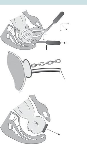
(a)
Line of pull
Push down with left hand
Pull on handle with right hand
(b)
Line |
of traction |
|
|
|
|
|
|
|
|
|
|
Tube to apply vacuum
Line of pull at this level of pelvis
Fig. 6.8 Assisted delivery techniques: (a) forceps delivery; (b) ventouse delivery. Reproduced from Chamberlain G, Steer P. (1999). ABC of labour care: operative delivery. Br Med J 318: 1260–4. With permission. © BMJ Publishing Group Ltd 1999.

298 CHAPTER 6 Labour and delivery
Operative vaginal delivery: trial
This term is used when it is not possible to determine with sufficient confidence that an instrumental delivery will be successful. It should, therefore, take place in theatre, where it is possible to move to an immediate CS, avoiding failed delivery in delivery room and subsequent delay in performing CS, which may compromise fetal well-being.
•The woman should be fully informed of the likely success and sign a consent form for ‘Trial of instrumental vaginal delivery +/– emergency CS’.
•If procedure is abandoned, assistance may be required to ‘push head up’ from vagina during CS as head may be impacted in pelvis.
Senior obstetric input is recommended in 2nd stage CS, especially after a failed trial of instrumental delivery.
When to abandon and deliver by emergency Caesarean
• No evidence of progressive descent with each pull (care must be taken with the ventouse to not interpret increasing caput as descent of the head).
•Where delivery is not imminent following three pulls of a correctly applied instrument by an experienced operator.
Risk factors for failed operative vaginal delivery
•BMI >30.
•EFW >4000g or clinically big baby.
•OP position.
•Mid-cavity delivery or if head is >1/5 palpable abdominally.
This page intentionally left blank

300 CHAPTER 6 Labour and delivery
Episiotomy
More than 85% of women delivering vaginally in the UK will sustain some degree of perineal trauma. Episiotomy is a surgical incision to enlarge the vaginal introitus. The decision to perform an episiotomy is made by the birth attendant. The worldwide rates of episiotomy vary dramatically (14% in England, 8% in the Netherlands, 50% in the USA). There is clear evidence to recommend a restricted use of episiotomy.
WHO recommends that episiotomy should be considered in the following circumstances:
•Complicated vaginal delivery:
•breech
•shoulder dystocia
•forceps
•ventouse.
•If there is extensive lower genital tract scarring:
•female genital mutilation
•poorly healed 3rd or 4th degree tears.
•When there is fetal distress.
2 It is also often recommended if there is an indication that there may be extensive perineal trauma such as the appearance of multiple vaginal/ perineal tears or perineal button-holing.
Types of episiotomy
•Mediolateral episiotomy extends from the fourchette laterally (thus reducing the risk of anal sphincter injury).
•Midline episiotomy extends from the fourchette towards the anus (common in the USA, but not recommended in the UK).
How to perform an episiotomy (see Fig. 6.9)
•If the woman does not have a working regional block (epidural) then the perineum should be infiltrated with lidocaine (lignocaine).
•Two fingers should be placed between the baby’s head and the perineum (to protect the baby).
•Sharp scissors are used to make a single cut in the perineum about 3–4cm long (ideally this should be at the height of the contraction when the perineum is at its thinnest).
2 Every effort should be made to anaesthetize the perineum early to provide sufficient time for effect.
2 It will cause bleeding so must not be done too early and should be repaired as soon as possible.
Always check for any extension or other tears (including a PR examination to ensure no trauma to the anal sphincter).

EPISIOTOMY 301
General complications of perineal trauma including episiotomy
•Bleeding.
•Haematoma.
•Pain.
•Infection.
•Scarring, with potential disruption to the anatomy.
•Dyspareunia.
•Very rarely, fistula formation.
Women who have undergone female genital mutilation should be seen antenatally and de-infibulation discussed. However, it they present in labour the episiotomy should be anterior and upwards. (See bFemale genital mutiliation, p. 470).
Line of incision of mediolateral episiotomy
Anal sphincter
Fig. 6.9 Performing an episiotomy. Adapted from Wyatt JP, Illingworth RN, Graham CA, et al. (eds) (2006). Oxford Handbook of Emergency Medicine. Oxford: OUP. By permission of Oxford University Press.

302 CHAPTER 6 Labour and delivery
Perineal tears
Classification of perineal tears
•1st-degree: injury to the skin only.
•2nd-degree: injury to the perineum involving perineal muscles (includes episiotomy).
•3rd-degree: injury to the perineum involving the anal sphincter complex:
•3a: <50% of the external anal sphincter (EAS) thickness torn
•3b: >50% of the EAS thickness torn
•3c: internal anal sphincter (IAS) torn.
•4th-degree: injury to perineum involving the anal sphincter complex (EAS and IAS) and the anal/rectal epithelium.
Principles of basic perineal repair (Fig. 6.10)
• Suture as soon as possible to reduce bleeding and infection risk.
• A rectal examination is recommended before starting, to ensure there is no trauma to the anal sphincter complex.
•The attendant should have adequate training for the type of tear: difficult trauma should be repaired in theatre under regional or general anaesthesia by an experienced operator.
•The woman should preferably be in lithotomy position.
•There should be a good light source and adequate analgesia.
•Use of rapid-absorption polyglactin suture material is associated with a significant reduction in pain.
•Apex of the cut should be identified and the suturing started from just above this point.
•A loose, continuous non-locking suturing technique used to appose each layer is associated with less short-term pain than the traditional interrupted method.
•Perineal skin should be sutured with a subcuticular suture as this is associated with less pain.
•Anatomical apposition should be as accurate as possible and consideration given to cosmetic results.
•Rectal examination after completion ensures that no suture has accidentally passed into the rectum or anal canal.
Needle and swabs must be counted afterwards (lost swabs are a recurring cause of litigation in obstetrics).
Further reading
RCOG. (2007). Green-top guideline 23: Perineal repair. Mhttp://www.rcog.or.uk
PERINEAL TEARS 303
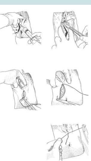
(1) |
(2) |
|
Swab the vulva towards the |
Place tampon with attached tape in |
|
perineum. Infiltrate with 1% |
upper vagina. Insert 1st suture above |
|
lignocaine → (arrows). |
apex of vaginal cut (not too deep as |
|
|
underlying rectal mucosa nearby). |
|
|
|
|
|
|
|
(3) |
(4) |
Bring together vaginal edges with |
Close perineal skin (subcuticular) |
continuous stitches placed 1cm apart. |
continuous stitch is shown here). |
Knot at introitus under the skin. Appose |
|
divided levator ani muscles with 2 or 3 |
|
interrupted sutures. |
|
(5) |
|
When stitching finished, remove |
|
tampon and examine vagina (to check |
|
for retained swabs). Do a PR to check |
|
that apical sutures have not penetrated |
|
rectum. |
|
Fig. 6.10 Episiotomy repair. Reproduced from Collier J, Longmore M, Brinsden M. (2006). Oxford Handbook of Clinical Specialties, 7th edn. Oxford: OUP. By permission of Oxford University Press.

304 CHAPTER 6 Labour and delivery
Thirdand fourth-degree tears
Approximately 1–3% of vaginal deliveries will result in injury to the anal sphinter. Prediction and prevention are both difficult.
Factors associated with increased risk of anal sphincter trauma
•Forceps delivery.
•Nulliparity.
•Shoulder dystocia.
•2nd stage >1h.
•Persistent OP position.
•Midline episiotomy.
•Birth weight >4kg.
•Epidural anaesthesia.
•Induction of labour.
Management of 3rdand 4th-degree tears
•All women sustaining genital tract injury should be carefully examined before suturing is started (including a rectal examination).
•Repair must be carried out by a trained senior clinician in theatre with adequate analgesia.
•The technique used can be end to end or overlapping for the EAS using either polydioxanone suture (PDS) or vicryl suture material.
•The IAS should be repaired with vicryl using interrupted sutures.
•Women must receive broad-spectrum antibiotics and stool softeners.
•They should receive physiotherapy input.
•Ideally, they should be reviewed 6wks later by an obstetrician or gynaecologist.
•Women must be warned of the risk of incontinence of faeces, fluid, and flatus: those experiencing symptoms at 6wks should be referred to a specialist gynaecologist or colorectal surgeon for investigation with endoanal ultra sonography.
•Around 60–80% will have a good result and be asymptomatic at 12mths.
•For future deliveries they should be advised that the result may not be so good from a 2nd repair: if symptomatic they should be given the option of delivery by CS.
Further reading
RCOG. (2007). Green-top guideline 29: Thirdand fourth-degree tears—management. Mhttp://www. rcog.org.uk/womens-health/clinical-guidance/management-third-and-fourth-degree-perineal- tears-green-top-29
This page intentionally left blank
306 CHAPTER 6 Labour and delivery
Caesarean section: overview
Background
•CS involves delivery of the fetus through a direct incision in the abdominal wall and the uterus.
•The rates vary in different countries and populations:
•the overall CS rate for nulliparous women in the UK has increased to about 24%
•for multiparous women who have not previously had a CS the rate is <5%
•for women who have had at least one previous CS the rate is increased to about 67%; most are elective.
Caesarean section
Associated with a higher incidence of:
•Abdominal pain.
•Venous thromboembolism.
•Bladder or ureteric injury.
•Hysterectomy.
•Very rarely maternal death.
Associated with a lower incidence of:
•Perineal pain.
•Urinary incontinence.
•Uterovaginal prolapse.
XLong-term effect of the last two are debated.
Some interventions to decrease morbidity from CS
•Preoperative haemoglobin check and correction of anaemia.
•Intraoperative prophylactic antibiotics given just before skin incision.
•Risk assessment and appropriate thrombo-prophylaxis (graduated stockings, hydration, early mobilization, and low molecular weight heparin).
•In-dwelling bladder catheterization during the procedure.
•Antacids and H2 receptor analogues before surgery.
•Antiemetics as appropriate.
•Regional rather than general anaesthesia.
•The risk of hypotension can be reduced using:
•intravenous ephedrine or phenylephrine infusion
•volume preloading with crystalloid or colloid
•lateral tilt of 15º.
•General anaesthesia for emergency CS should include preoxygenation and rapid sequence induction to reduce the risk of aspiration.

CAESAREAN SECTION: OVERVIEW 307
Vaginal birth after CS
•Uterine rupture is very rare but is increased with VBAC:
•50:10 000 with VBAC and spontaneous onset of labour
•1:10 000 with repeat CS.
•Intrapartum infant death is rare: 1:1000—the same as for primiparous women
•EFM is recommended during labour as FHR changes may be the earliest signs of scar rupture.
•Women should deliver in a unit where there is immediate access to CS and on-site blood transfusion.
•With induction of labour, there is increased risk of uterine rupture:
•8:1000 if oxytocin infusion is used
•24:1000 if prostaglandins are used.
•Women with both previous CS and a previous vaginal birth are more likely to give birth vaginally.

308 CHAPTER 6 Labour and delivery
Caesarean section: indications
The main indications for CS are:
•Repeat CS.
•Fetal compromise.
•‘Failure to progress’ in labour.
•Breech presentation.
X Maternal request accounts for 7% of CS, but is not, on its own, an indication for CS.
Categories to determine the timing of CS
•Immediate threat to the life of the woman or fetus (immediate, ‘crash CS’).
•Maternal or fetal compromise, which is not immediately lifethreatening (urgent).
•No maternal or fetal compromise but needs early delivery (scheduled).
• Delivery timed to suit woman and staff (elective).
In cases of suspected or confirmed acute fetal compromise, delivery should be as soon as possible. The accepted standard for category 1 (immediate) CS is within 30min.
Indications for category 1 CS
•Placental abruption with abnormal FHR or uterine irritability.
•Cord prolapse.
•Scar rupture.
•Prolonged bradycardia.
•Scalp pH <7.20.
Indications for category 2 CS
• Failure to progress with pathological CTG.
Indications for category 3 (scheduled) CS
•Severe pre-eclampsia.
•IUGR with poor fetal function tests.
•Failed induction of labour.
Indications for category 4 (elective) CS
•Term singleton breech (if ECV is contraindicated or has failed).
•Twin pregnancy with non-cephalic 1st twin.
•Maternal HIV.
•Primary genital herpes in the 3rd trimester.
•Placenta praevia.
•Previous hysterotomy or classical CS.
2 Elective CS is usually carried out after 39wks unless indicated, as the risk of respiratory morbidity (transient tachypnoea of the newborn) is increased at lower gestational ages.

CAESAREAN SECTION: TYPES 309
Caesarean section: types
Lower uterine segment incision
The two main types of skin incision are:
•Pfannenstiel incision: a straight horizontal incision 2cm above the symphysis pubis—superior cosmetic result.
•Joel–Cohen incision: also a straight horizontal incision, but higher, about 3cm below the level of the anterior superior iliac spines (ASIS)—allows quicker entry to the abdomen.
A transverse incision in the uterine lower segment is used in >90% of CS as it is associated with:
•Reduced adhesion formation.
•Decreased blood loss.
•Lower incidence of scar dehiscence in subsequent pregnancies.
However, if the lower segment is poorly developed, a low transverse incision carries a risk of lateral extension into the uterine vessels and haemorrhage.
Following delivery of the fetus and completion of the 3rd stage, the lower uterine segment is closed in one or two layers.
X Double-layer closure is usually practiced, but research comparing singlewith double-layer has no long-term results to compare scar integrity, although short-term morbidity showed no difference.
Classical CS
This involves a vertical incision into the upper uterine segment. It is rarely performed, but indications may include:
•Structural abnormality of the uterus.
•Difficult access to the lower uterine segment due to fibroids or severe adhesions over the lower segment.
•Postmortem CS delivery (if the fetus is viable).
•Anterior placenta praevia with abnormally vascular lower uterine segment.
•Contraction ring.
•Very preterm fetus (especially breech presentation) where the lower segment is poorly formed.
•Elective Caesarean hysterectomy.
•Transverse lie of the fetus with ruptured membranes.
This incision allows rapid delivery and has a lower risk of bladder injury. However, the closure is more complicated and time-consuming and there is a higher incidence of infection and adhesion formation.
There is a greater risk of uterine rupture in subsequent pregnancies with a greater risk of the fetus being expelled into the peritoneal cavity. For these reasons, a classical incision is an absolute contraindication to a trial of a vaginal delivery (VBAC).

310 CHAPTER 6 Labour and delivery
Caesarean section: complications
Intraoperative complications
Major complications are most common with an emergency CS and 82% of anaesthesia-related maternal deaths occurred in women undergoing CS, most frequently with general anaesthesia.
2Intraoperative complications occur in 12–15% of women and include:
•Uterine or uterocervical lacerations (5–10%).
•Blood loss >1L (7–9%).
•Bladder laceration (0.5–0.8%).
•Blood transfusion (2–3%).
•Hysterectomy (0.2%).
•Bowel lacerations (0.05%).
•Ureteral injury (0.03–0.09%.).
Risk factors predisposing to uterocervical lacerations include:
•Low station of the presenting part and full dilatation.
•Birth weight >4000g.
•iMaternal age.
•Category 1 CS.
Risk factors predisposing to intraoperative haemorrhage include:
•Placenta praevia or abruption.
•Extremes of fetal birth weight.
•BMI >25.
Postoperative complications
2Postoperative complications occur in up to 1/3 of women and include:
•Endometritis (5%).
•Wound infections (3–27%).
•Pulmonary atelectasis.
•Venous thromboembolism.
•Urinary tract infections.
Risk factors independently associated with infection are:
•Preoperative remote infection.
•Chorioamnionitis.
•Maternal severe systemic disease.
•Pre-eclampsia.
•High BMI.
•Nulliparity.
•iSurgical blood loss.

CAESAREAN SECTION: COMPLICATIONS 311
Long-term effects of CS
In subsequent pregnancies there is a higher risk of:
•Uterine rupture (1:200 with spontaneous labour).
•Placenta praevia (47% iof background risk).
•Placenta accreta.
•Antepartum stillbirth: risk doubles with a previous CS.
Women undergoing multiple CS (≥3) are at higher risk of:
•Excessive blood loss (8%).
•Difficult delivery of the neonate (5%).
•Dense adhesions (46%).
•The risk of any major complication is higher (9%).
•Complications are iwith inumber of CS:
•4% for 2nd
•8% for 3rd
•13% for 4th.

312 CHAPTER 6 Labour and delivery
Prelabour rupture of membranes at term
Definition
Prelabour rupture of membranes (PROM) at term is defined as leakage of amniotic fluid in the absence of uterine activity after 37 completed weeks of gestation.
Incidence
8% of term pregnancies (2–3% before 37wks).
Aetiology
•Unknown.
•Clinical or subclinical infection.
•Polyhydramnios.
•Multiple pregnancy.
•Malpresentations.
Clinical assessment
It is important to establish a correct diagnosis to plan further management. If unnecessary interventions are undertaken there is a risk of increased maternal and fetal morbidity.
History
Women give a history of a sudden gush of fluid leaking from the vagina, recurrent dampness, or constant leaking.
Examination
•There is no need to carry out a speculum examination with certain history of ruptured membranes at term with liquor seen on the pad or undergarments.
•If the history is uncertain, a speculum examination should be offered (liquor should be seen pooling in the upper vagina or trickling through the cervical os):
•coughing or straining (Valsalva manoeuvre) may help to demonstrate leaking fluid
•note the colour of the liquor (?blood or meconium stained).
•Temperature, pulse, and BP.
•Obstetric examination of abdomen (including lie and presentation).
•CTG.
2 If conservative management is planned, avoid digital examination as it increases the incidence of chorioamnionitis, post-partum endometritis, and neonatal infection.
Any concern regarding fetal well-being is an indication to deliver.
Signs of chorioamnionitis should prompt treatment with antibiotics and rapid delivery.

PRELABOUR RUPTURE OF MEMBRANES AT TERM 313
Clinical features of chorioamnionitis
•Fetal tachycardia.
•Maternal tachycardia.
•Maternal pyrexia.
•Rising leucocyte count.
•Rising CRP.
•Irritable or itender uterus.

314 CHAPTER 6 Labour and delivery
Prelabour rupture of membranes: management
If there are no contraindications to waiting, women should be offered the choice between immediate induction and expectant management.
Expectant management vs. immediate induction
•60% of women will labour spontaneously within 24h.
•No evidence of a difference in the mode of delivery for either.
•1% risk of serious neonatal infection (compared with 0.5% for women with intact membranes).
With expectant management
•Women are more likely to develop chorioamnionitis and endometritis with expectant management of >24h.
•Baby is more likely to be admitted to SCBU: no evidence of a difference in eventual neonatal outcome (morbidity/mortality) with expectant management of <24h.
2The NICE guidelines recommend induction after 24h.
Conservative management advice
If the woman opts for this she should be advised to:
•Record her temperature every 4h (during waking hours).
•Urgently report any change in colour or offensive smell.
•Avoid sexual intercourse (showering and bathing is okay).
•Report any din fetal movements.
•Deliver in a unit with neonatal services and remain in hospital for >12h after delivery to allow close observation of the baby.
•Consider induction if not in labour by 24h.
•Seek medical advice if any concerns regarding the baby’s well-being in the 1st 5 days of life (especially in the first 12h).
XUse of antibiotics is controversial. NICE does not recommend prophylactic antibiotics for either mother or baby in absence of symptoms, even if her membranes have been ruptured for >24h.
2In labour, regular maternal observations are essential to pick up signs of infection early. Fetal heart rate monitoring should be carried out as it may be tachycardic in the presence of infection.
If there is clinical evidence of infection a full course of broad spectrum IV antibiotic therapy should be started after blood cultures have been sent.
Known group B streptococcus carriers
See bGroup B streptococcus, p. 172.
•Immediate induction should be encouraged (dneonatal infection).
•Mothers should be offered benzylpenicillin in labour.
•Neonates should be screened soon after birth.
Further reading
NICE. (2007). Intrapartum care, guideline September 2007. Mhttp://www.nice.org.uk/Search.do?x= 17&y=18&searchText=Intrapartum+care&newsearch=true#/search/?reload

ABNORMAL LIE: TRANSVERSE AND OBLIQUE 315
Abnormal lie: transverse and oblique
Transverse and oblique lie occur in 1:300 pregnancies and result in a shoulder, limb, or cord presentation. If this persists, vaginal delivery is not feasible (Fig. 2.4).
Diagnosis
•The maternal abdomen is unusually wide and the fundus is lower than expected for the gestation.
•Neither fetal pole is palpable entering the pelvis.
•Fetal head is identifiable at one side.
•On vaginal examination the pelvis is empty.
•A limb or cord may prolapse through the cervix.
Management
When this presents in labour, fetal well-being should be established and a USS performed to try to identify the cause.
Exclude placenta praevia before attempting vaginal examination.
•CS is indicated in almost all cases.
•An unstable lie at term due to multiparity alone may warrant a gentle attempt at ECV if the following criteria are met:
•the membranes must be intact
•labour not advanced
•the fetus must have no signs of compromise.
•If ECV is successful cord presentation or prolapse should be excluded before labour is allowed to establish.
•CS for transverse lie, especially with placenta praevia or fibroids, requires an experienced obstetrician and cross-matched blood.
•Vertical uterine incision on the uterus or acute tocolysis with a transverse incision may be necessary for safe delivery of fetus.
316 CHAPTER 6 Labour and delivery
Malpresentations in labour: overview
•>95% of fetuses at term present with vertex (area subtended by two parietal eminences, anterior, and posterior fontanelle).
•Malpresentation describes any presentation other than vertex lying in close proximity to internal os of the cervix and includes:
•breech (most common malpresentation with an incidence of 3–4% at term; see bBreech presentation: delivery, p. 86)
•brow
•face
•shoulder
•arm
•cord.
Some causes of malpresentation
Maternal
•Multiparity.
•Pelvic tumours.
•Congenital uterine anomalies.
•Contracted pelvis.
Fetal
•Prematurity.
•Multiple pregnancy.
•Intrauterine death.
•Macrosomia.
•Fetal abnormality including:
•hydrocephalus
•anencephaly
•cystic hygroma.
Placental
•Placenta praevia.
•Polyhydramnios.
•Amniotic bands.
This page intentionally left blank

318 CHAPTER 6 Labour and delivery
Malpresentations: brow and face
Brow presentation
Incidence ranges between 1:1000 and 1:3500 deliveries. The head occupies a position midway between full flexion (vertex) and full extension (face). It can revert to a face or vertex presentation, but if it persists vaginal delivery is not usually possible (see Fig. 6.11a).
Diagnosis
•Often diagnosed in advanced labour (may be suspected on abdominal palpation when both occiput and chin are palpable).
•The head does not descend below the ischial spines.
•Vaginal examination is diagnostic as the frontal sutures, anterior fontanelle, orbital ridges, eyes, and the root of nose are palpable.
Management
•Watch and wait: may become a vertex or face presentation.
•If progress is slow or if the brow persists then CS is indicated.
Face presentation
Incidence of face presentation is between 1:600 and 1:1500 deliveries. It is due to hyperextension of the fetal neck (see Fig. 6.11b).
Diagnosis
•Face presentation is diagnosed in labour on vaginal examination.
•The orbital ridges, nose, malar eminences, mentum, gums, and mouth can be distinguished.
2It may be mistaken for a breech, but presence of gum margins will help to differentiate between a mouth and an anus.
Management
•90% are mentoanterior (MA) and head can flex to allow vaginal delivery.
•Expectant management should be considered with mentoposterior (MP) as about 20–30% will rotate on reaching the pelvic floor.
•Persistent MP face presentations cannot deliver vaginally as it would require the head to overextend.
•If there is poor progress or failure to rotate, CS is indicated.
•Fetal monitoring should be external and fetal blood sampling is contraindicated.
•The use of ventouse is absolutely contraindicated but forceps delivery is possible with an MA position well below spines.
Attempts to convert face presentations manually into vertex or use of forceps to rotate persistent MP positions can lead to complications of cord prolapse and fetal cervical cord injury.
bSee Figs 1.6 and 1.7.
MALPRESENTATIONS: BROW AND FACE 319
Cord presentation
This occurs when one or more loops of cord lie below the presenting part and the membranes are still intact. It is associated with malpresentation, abnormal lie, and a high head. The risk is of cord prolapse when the membranes rupture. This is an obstetric emergency (see b Cord prolapse, p. 401).
Diagnosis
•The diagnosis of cord presentation is often made on USS, but may be found on VE in labour.
•It can be suspected clinically when persistent variable fetal heart decelerations occur early in labour.
•ARM is contraindicated as it will cause cord prolapse.
(a) Brow presentation |
(b) Face presentation |
|
|
Fig. 6.11 Malpresentations: (a) brow presentation; (b) face presentation.

320 CHAPTER 6 Labour and delivery
Retained placenta and placenta accreta
Retained placenta should be suspected if it is not delivered within 30min of the baby in an actively managed 3rd stage and 1h in a physiological 3rd stage.
Care must be taken as blood can gather behind placenta leading to significant occult blood loss—beware of high uterus full of blood!
Management of retained placenta
•IV access, FBC, and cross-match.
•If it was physiological management, revert to active management:
•give Syntometrine® or oxytocin
•try controlled cord traction.
•If the oxytocin is not effective within 30min, transfer to theatre for regional block and manual removal of the placenta.
•Intraoperative prophylactic antibiotics should be given.
X The use of a 40IU IV oxytocin infusion to help deliver the placenta is controversial. The NICE guidelines do not recommend using it before the placenta is delivered.
Placenta accreta, increta, and percreta
Abnormal placentation occurs in about 1:7000 pregnancies, but is much more common if there have been prior Caesarean deliveries. The placenta is normally separated from the myometrium by the decidua basalis. However, if the decidua is abnormal the villi may invade further through the uterine wall. There are three types, but they are all often referred to as just accreta:
•Placenta accreta: placental villi are attached to the myometrium.
•Placenta increta: villi invaded into >50% of the myometrium.
•Placenta percreta: villi pass through the whole myometrium up to the serosa, potentially involving other viscera (bladder or bowel).
Risk factors for placenta accreta may include:
•Uterine surgery such as CS or myomectomy.
•Repeated surgical termination of pregnancy.
Management of placenta accreta post delivery
•With heavy bleeding:
•blood replacement
•tamponade with balloon (e.g. Rusch)
•hysterectomy.
•With minimal bleeding, leaving the placenta in situ is an option, with close monitoring.

RETAINED PLACENTA AND PLACENTA ACCRETA 321
How to perform manual removal of placenta (MROP)
•One hand is placed on abdomen to steady uterus (reduces the risk of perforation).
•Other hand is gently inserted through cervix into uterus.
•Fingers are used to identify plane between placenta and uterine wall, and gently separate it.
•Placenta should be removed in one piece and inspected to ensure it is complete.
•Uterine cavity is then explored again to make sure it is completely empty.
•Oxytocin infusion is continued for 4h prophylactically.
•IV antibiotics are given.
•Mother observed for bleeding or infection by observing vaginal bleeding, fundal height, change in pulse, BP, temperature, urinary output, and Hb%.

322 CHAPTER 6 Labour and delivery
Post-partum haemorrhage
•Primary PPH is defined as blood loss of 500mL or more from the genital tract occurring within 24h of delivery.
•Secondary PPH is defined as ‘excessive’ loss occurring between 24h and 6wks after delivery.
•Major cause of maternal morbidity and mortality: globally >125 000 women die of PPH each year.
•Major cause of maternal deaths in the UK (often after CS).
•Incidence is 2–11% in the UK.
•With a low BMI or low Hb, <500mL loss may cause haemodynamic disturbance requiring prompt and appropriate management.
Causes of primary PPH
•Uterine atony.
•Genital tract trauma.
•Coagulation disorders.
•Large placenta.
•Abnormal placental site.
•Retained placenta.
•Uterine inversion.
•Uterine rupture.
Uterine atony (90%)
Caused by failure of uterus to contract effectively after delivery. May be due to many factors—overdistended uterus with twins or polyhydramnios, prolonged labour, infection, retained tissue, failure to actively manage 3rd stage of labour, or, rarely, due to placental abruption (diffuse bleeding into uterine muscle preventing contraction).
Genital tract trauma (7%)
Tears, episiotomy, lacerations of the cervix, and rupture of uterus.
Coagulation disorders
Severe PET, placental abruption, and sepsis may contribute to PPH. Autoimmune diseases, liver disease, and inherited or acquired coagulation disorders are rare causes. Sometimes patients may be on heparin, which can lead to excessive bleeding.
Abnormal placental site
Placenta praevia, accreta, and percreta are associated with PPH. Appropriate preplanning is needed to avoid morbidity and mortality.
Uterine inversion and rupture are rare causes of excessive bleeding.
For management see b Massive obstetric haemorrhage: medical management, p. 386.

POST-PARTUM HAEMORRHAGE 323
Antenatal risk factors for PPH
•Previous PPH.
•Previously retained placenta.
•Maternal Hb ≤8.5g/dL at onset of labour.
•iBMI.
•Para 4 or more.
•Antepartum haemorrhage.
•Overdistention of uterus (multiple pregnancy or polyhydramnios).
•Uterine abnormalities.
•Low-lying placenta
•Maternal age >35yrs.
The presence of any risk factors for PPH should lead to the woman being advised to deliver in an obstetric unit (facilities for blood transfusion and surgical management of PPH).
Intrapartum risk factors for PPH
•Induction of labour.
•Prolonged 1st, 2nd, or 3rd stage.
•Use of oxytocin.
•Precipitate labour.
•Vaginal operative delivery.
•CS.

324 CHAPTER 6 Labour and delivery
Home birth: overview
Home birth can be safe for women screened as low risk, and emotionally satisfying for the mother and her family. For women identified as having risk factors, hospital delivery is safer. Debates about the safety of home births focus on risk of preventable perinatal morbidity and mortality, and on broader issues of appropriate screening and referral.
The numbers
•Proportion of births at home fell from 80% in 1930 to 1% in 1990.
•As a result of Government committee recommendation (HMSO 1993), stating that a full choice including home births should be offered, further enhanced by ‘Maternity matters’, a Government white paper (2007), the UK home birth rate is increasing and is now about 2–3%.
•Some studies suggest that 10–14% of women would choose home birth if given the opportunity.
•In some regions where there is difficulty in geographical accessibility to a hospital the home birth rate could be about 10%.
•In women booked for home births:
•change to hospital care is nearly 29%
•transfer in labour is up to 15% in multiparae and 30% in nulliparae.
•most of these transfers are for failure to progress or pain relief.
•The risk of intrapartum fetal death in appropriately selected low-risk women is 1:1000.
•It is difficult to compare directly the perinatal mortality rates for home and hospital, as more complex deliveries occur in hospital.
•Recent prospective study suggests a slight increase in perinatal mortality with home births.
Discussion points when considering home birth
•In the presence of obvious risk factors (hypertension, diabetes, placenta praevia) the advice must be to deliver in hospital.
•If mother is low-risk and wishes to have home birth, she should be counselled appropriately with full information about the very slight increase in perinatal mortality and possibility of transfer in labour.
•If a risk arises before birth, the booking should be changed.
•If risk is minimal, the lead professional in charge should offer the woman and her partner the opportunity to review their choice and respect their decision.
Reasons for women to choose home birth
•Wish for a familiar setting where they feel relaxed and in control.
•Fear of hospital setting.
•To have a continuing relationship with a known midwife.
•To be with more family members who provide support.
•Previous home birth.
•To avoid intervention.
Further reading
Maternity Matters: choice, access and continuity & care in a safe service. Mhttp://www.dh.gov.uk/ en/publicationsandstatistics/publications/publicationspolicyandguidance/DH_073312

HOME BIRTH: RISKS AND GP INVOLVEMENT 325
Home birth: risks and GP involvement
The potential risks of home birth are rare, but should be discussed with the woman as part of her decision. These include:
•Should a complication occur, transfer to hospital may be required.
•Should there be a delay in transfer, response to acute complications, such as intrapartum fetal hypoxia or post-partum haemorrhage, may be delayed, potentially leading to a worse outcome although such complications are rare.
•The facilities for neonatal resuscitation will be limited but the midwife should be well trained in basic neonatal resuscitation.
•Inadequate lighting and analgesia may make diagnosis of the extent of perineal tears difficult, necessitating transfer to hospital.
Discussion of the risks and other factors, including type of pain relief available, will help the woman to make an informed choice. Clear documentation of these discussions in the antenatal period is essential for the mother not to regret her choice and for medico-legal reasons.
The role of the GP
•The GP should be fully informed about the local options for place of birth, and will then be in a position to provide the options to the woman in a clear, understandable, and balanced manner.
•GPs who do not wish to provide care for home births should refer women to the community midwife, supervisor of community midwives at the district maternity unit, or a GP who provides this care.
•In case of any unfortunate event occurring with intrapartum care of a woman being looked after by her GP and if the case proceeds to a
litigation, the GP would not be judged by the standards of a consultant obstetrician, but by those of a GP with similar skills and standing (the Bolam test).
•The GP does not have to attend a home birth even when the woman has been accepted by the GP for full maternity care, unless asked to do so by the midwife.
•The GP should provide support to the woman and midwife, help identify any deviations from normal course of labour, and arrange for hospital care.
•Where the midwife feels that the GP is supportive, the likelihood of transfer to hospital is reduced.
•In current practice very few GPs offer care in labour and delivery services.
Further reading
The GP’s guide to home birth. Mhttp://www.medicine.ox.ac.uk/bandolier/band32/b32-8.html
326 CHAPTER 6 Labour and delivery
Home birth: the evidence
Meta-analysis of several methodologically sound observational studies comparing the outcomes of planned home births (irrespective of the eventual place of birth) with planned hospital births for women with similar characteristics showed that there was no increase in maternal mortality. The rate is unlikely to be different as the maternal mortality is generally low, and good midwifery and ambulance services help to avoid such deaths, although occasional cases have been described.
Recent home birth study in the UK showed:
•Slightly increased perinatal mortality, but when all studies in literature are considered there is little statistically significant difference. This is partly due to the complexity of such studies, some being retrospective or prospective descriptive.
•In the home births group there were significantly fewer medical interventions (including in women transferred to hospital).
•Fewer babies had low Apgar scores, neonatal respiratory problems, and instances of birth trauma with home births.
Further randomized controlled trials are needed to resolve this controversy over relative safety of home and hospital births. Because maternal and perinatal mortality and morbidity are so low in low-risk pregnancies, to observe differences in these primary outcome measures large numbers need to be studied.
Home birth: general points
GPs and midwives have the responsibility for creating the right circumstances for safe and satisfying home births.
This means:
•Selecting women without risk factors.
•Establishing an infrastructure for safe obstetric care including:
•hygiene during delivery
•keeping the baby warm
•care of the eyes.
•Providing support and care during labour, delivery, and in immediate postnatal period.
•Arrangements for transfer to hospital in the event of any unforeseen complication.
•Care should be provided based on prearranged protocol that provides guidance as to conduct of labour and what action needs to be taken should the woman need help.