


xxiii
Contributors
Editors
Miss Sally Collins
John Radcliffe Hospital, Oxford,
UK
Professor Sabaratnam
Arulkumaran
St George’s Hospital, London, UK
Contributors
Miss Karolina Afors
St George’s Hospital, London, UK
Dr Christian Becker
John Radcliffe Hospital, Oxford, UK
Dr Amy Bennett
Dept of Genitourinary Medicine,
Oxford University Hospitals
NHS Trust, Oxford, UK
Mrs Rebecca Black
John Radcliffe Hospital, Oxford, UK
Dr Shabana Bora
St George’s Hospital, London, UK
Dr Brian Brady
John Radcliffe Hospital, Oxford, UK
Mr Paul Bulmer
St George’s Hospital, London, UK
Mr Edwin Chandraharan
St George’s Hospital, London, UK
Dr Noan-Minh Chau
Specialist Registrar rotation in Medical Oncology, London Deanery, UK
Dr Mellisa Damodaram
Queen Charlotte’s and Chelsea
Hospital, London, UK
Mr Kevin Hayes
St George’s Hospital, London, UK
Mr Simon Jackson
John Radcliffe Hospital, Oxford, UK
Mr Lawrence Impey
John Radcliffe Hospital, Oxford, UK
Miss Claudine Domoney
Chelsea and Westminster
Hospital, London, UK
Dr Stergios K.
Doumouchtsis
St George’s Hospital, London, UK
Dr Suzy Elniel
Chelsea and Westminster
Hospital, London, UK
Dr Cleave W. J. Gass
St George’s Hospital, London, UK
Dr Ingrid Granne
John Radcliffe Hospital, Oxford,
UK
Miss Catherine Greenwood
John Radcliffe Hospital, Oxford, UK
Mr Manish Gupta
John Radcliffe Hospital, Oxford, UK
Miss Pauline Hurley
John Radcliffe Hospital, Oxford, UK
Dr Nia Jones
Queens Medical Centre,
Nottingham, UK
Miss Brenda Kelly
John Radcliffe Hospital, Oxford, UK

xxiv CONTRIBUTORS
Dr Nigel Kennea
St George’s Hospital, London, UK
Dr Andy Kent
St George’s Hospital, London, UK
Dr Su-Yen Khong
John Radcliffe Hospital, Oxford, UK
Dr Emma Kirk
St George’s Hospital, London, UK
Dr Samatha Low
Royal Berkshire Hospital,
Reading, UK
Dr Jo Morrison
Musgrove Park Hospital,
Taunton, UK
Dr Neelanjana
Mukhopadhaya
St George’s Hospital,
London, UK
Dr Faizah Mukri
Specialist Registrar rotation, London Deanery, UK
Dr Santosh Pattnayak
St George’s Hospital,
London, UK
Dr Natalia Price
John Radcliffe Hospital,
Oxford, UK
Dr Aysha Qureshi
Royal United Hospital, Bath, UK
Dr Devanna Rajeswari
St George’s Hospital, London, UK
Dr Gowri Ramanathan
St George’s Hospital, London, UK
Dr Margaret Rees
John Radcliffe Hospital, Oxford, UK
Dr Jackie Sherrard
Dept of Genitourinary Medicine,
Oxford University Hospitals NHS
Trust, Oxford, UK
Dr Lisa Story
John Radcliffe Hospital, Oxford, UK
Ms Louise Strawbridge
University College London,
London, UK
Mr Alex Swanton
Royal Berkshire Hospital,
Reading, UK
Dr Linda Tan
St George’s Hospital, London, UK
Dr Katy Vincent
John Radcliffe Hospital, Oxford, UK
Miss Cara Williams
Clinical Fellow in Paediatric and
Adolescent Gynaecology,
University College London
Hospital, UK
Dr Niraj Yanamandra
St Peter’s Hospital, Chertsey, UK

Chapter 1 |
1 |
|
|
Normal pregnancy
Obstetric history: current pregnancy 2 Obstetric history: other relevant features 4 Obstetric physical examination 6 Engagement of the fetal head 8
The female pelvis 10
Diameters of the female pelvis 12 Fetal head 14
Diameters and presenting parts of the fetal head 16 Placenta: early development 18
Placenta: later development 19 Placenta: circulation 20 Placenta: essential functions 22
Physiology of pregnancy: endocrine 24 Physiology of pregnancy: haemodynamics 26 Physiology of pregnancy: cardiorespiratory 27
Physiology of pregnancy: genital tract and breast 28 Physiology of pregnancy: other changes 29 Preparing for pregnancy 30
Supplements and lifestyle advice 32 General health check 34
Diagnosis of pregnancy 36 Dating of pregnancy 38
Ultrasound assessment of fetal growth 40 Booking visit 42
Antenatal care: planning 44
Antenatal care: routine blood tests 46 Antenatal care: specific blood tests 47 Antenatal care: preparing for delivery 48

2CHAPTER 1 Normal pregnancy
Obstetric history: current pregnancy
Obstetric history taking has many features in common with most other sections of medicine, along with certain areas specific to the specialty. The basic framework can be easily learned; however, competence requires good clinical knowledge and a lot of practice. As obstetrics often requires intimate examination and discussion of sensitive information, it is important to ensure privacy, and to demonstrate respect and confidentiality. It is important to offer a health professional as a chaperone. Translation may be required and it is best to have an official translator. The family, especially the husband, translating may not divulge or may distort certain information. It is also important to ask about domestic violence when the mother is alone and offer help if appropriate.
A carefully obtained history taken in a logical sequence avoids inadvertent omission of important details, and guides the examination to follow.
Current pregnancy
Much of this information will be contained in the patient’s ‘hand-held’ notes:
•Name.
•Age.
•Occupation.
•Relationship status.
•Gravidity (i.e. number of pregnancies, including the current one).
•Parity (i.e. number of births beyond 24wks gestation).
The expected date of delivery (EDD) can be calculated from the last menstrual period (LMP) using Naegele’s rule (add 1yr and 7 days to the LMP and subtract 3mths), most often done with an obstetric calendar (‘wheel’). Enquire about details that may affect the validity of the patient’s EDD as calculated from her LMP including:
•Long cycles.
•Irregular periods.
•Recent use of the combined oral contraceptive pill (COCP).
2 Dating scans between 8 and 13wks are more reliable than LMP and should be used to provide an EDD where possible.
Enquire about the current pregnancy, including:
•General health (tiredness, malaise, and other non-specific symptoms).
•If >20wks, enquire about fetal movements.
•General details of pregnancy to date (previous admissions and current problems).
•Results of all antenatal (AN) blood tests—routine and specific.
•Results of anomaly and other scans (details of results can be cross-checked with the notes).
•If she is postnatal:
•labour and delivery
•history of the postnatal period.

OBSTETRIC HISTORY: CURRENT PREGNANCY 3
An obstetric history
Should include:
•Current pregnancy details.
•Past obstetric history.
•Past gynaecological history.
•Past medical and surgical history.
•Drug history and allergies.
•Social history, including:
•recreational drug use
•domestic violence
•psychiatric illness especially in the postnatal period.
•Family history especially with regard to:
•multiple pregnancy
•diabetes
•hypertension
•chromosomal or congenital malformations.
Gravidity and parity explained
The terminology used is gravida x, para a + b:
•x is the total number of pregnancies (including this one).
•a is the number of births beyond 24wks gestation.
•b is the number of miscarriages or termination of pregnancies before 24wks gestation.
Example
A woman who is pregnant for the 4th time with 1 normal delivery at term, 1 termination at 9wks, and 1 miscarriage at 16wks would be gravida 4, para 1+2.

4CHAPTER 1 Normal pregnancy
Obstetric history: other relevant features
2 History often repeats itself, so previous AN, intrapartum, or postpartum complications should influence the management of this pregnancy.
Past obstetric history includes:
•Details of all previous pregnancies (including miscarriages and terminations).
•Length of gestation.
•Date and place of delivery.
•Onset of labour (including details of induction of labour).
•Mode of delivery.
•Sex and birth weight.
•Fetal and neonatal life.
Clear details of any complications or adverse outcomes (such as shoulder dystocia, postpartum haemorrhage, or stillbirth).
Past gynaecological/medical/surgical history
•Method of contraception before conception.
•Previous gynaecological procedures.
•Cervical smear history.
•Medical conditions, such as hypertension, epilepsy, or diabetes.
•Details of any consultations with other physicians (neurologist or endocrinologist, psychiatrists).
•Involvement of multidisciplinary teams (MDT).
•Details of any previous surgery.
Drug and allergy history
•Current medications.
•Medications taken at any time during the pregnancy.
•Any allergies and their severity (anaphylaxis or a rash?).
Family history
Any history of hereditary illnesses or congenital defects is important and is required to ensure adequate counselling and screening is offered.
•Familial disorders such as thrombophilias.
•Previously affected pregnancies with any chromosomal or genetic disorders, hypertensive disorders, early pregnancy loss, or preterm delivery.
•Consanguinity.
Social history
•Smoking.
•History of drug or alcohol abuse.
•Plans for breast-feeding.
•Social aspects, such as plans for childcare arrangements.
•Domestic violence screening.
This page intentionally left blank
6CHAPTER 1 Normal pregnancy
Obstetric physical examination
At initial visit, a complete physical examination should be undertaken.
Abdominal examination: inspection
•Note the apparent size of the abdominal distension.
•Note any asymmetry.
•Fetal movements.
•Cutaneous signs of pregnancy:
• linea nigra (dark pigmented line stretching from the xiphi sternum through the umbilicus to the suprapubic area)
•striae gravidarum (recent stretch marks are purplish in colour)
•striae albicans (old stretch marks are silvery-white)
•flattening/eversion of umbilicus (due to iintra-abdominal pressure).
•Superficial veins (alternative paths of venous drainage due to pressure on the inferior vena cava by a gravid uterus).
•Surgical scars (a low Pfannenstiel incision may be obscured by pubic hair, and laparoscopy scars hidden within the umbilicus).
Abdominal examination: palpation
•Symphysis fundal height (SFH):
•palpated <20wks
•measured in centimetres >20wks.
•Estimation of number of fetuses: ?multiple fetal poles.
•Fetal lie (relationship of longitudinal axis of fetus to that of the uterus):
•longitudinal—fetal head or breech palpable over pelvic inlet
•oblique—the head or breech is palpable in the iliac fossa and nothing felt in the lower uterus
•transverse—fetal poles felt in flanks and nothing above the brim.
•Presentation (part of the fetus overlying the pelvic brim):
•cephalic (this could be vertex, face, or brow presentations determined vaginally)
•breech
•other (shoulder, compound).
•Amniotic fluid volume:
•itense abdomen with fetal parts not easily palpated
•dcompact abdomen with fetal parts easily palpable.
Auscultation of the fetal heart
The fetal heart (FH) is best heard at the anterior shoulder of the fetus:
•A Doppler ultrasound device (Sonicaid) from about 12wks.
•A fetal stethoscope (Pinard) from about 24wks gestation.
•In a breech presentation it is often heard at, or above, the level of the maternal umbilicus.
•Rate and the rhythm of the FH should be determined over 1min.
•The recent NICE guidelines raise the need for routine fetal heart rate (FHR) auscultation in the presence of fetal movement (FM); but mothers enjoy listening to the fetal heart.

OBSTETRIC PHYSICAL EXAMINATION 7
General examination
•Body mass index (BMI) calculated [weight (kg)/height (m)2].
Pregnancy complications are increased with a BMI <18.5 and >25.
•Blood pressure (BP) measured in the semi-recumbent position (45° tilt).
Use an appropriate size cuff; too small a cuff gives a falsely high BP.
•Auscultation of the heart and lungs:
•flow murmurs are common in pregnancy and are not significant
•cardiac murmurs may be detected for the first time in pregnancy.
•Thyroid gland (exclude a goitre).
•Breasts (exclude any lumps).
•Varicose veins and skeletal abnormalities (kyphosis or scoliosis): normal pregnancy is associated with an increase in lumbar lordosis, which can lead to lower backache.
Normal uterine size
•The uterus normally becomes palpable at 12wks gestation.
•It reaches the level of the umbilicus at 20wks gestation.
•It is at the xiphi sternum at 36wks gestation.
Symphysis fundal height
2 The SFH detects approximately 40–60% of small-for-gestational age fetuses, but its predictive value in detecting large-for-dates fetuses is considerably less.
The uterine size is objectively measured with a tape measure from the highest point of the fundus to the upper margin of the symphysis pubis (see Fig. 1.1).
Appropriate growth is usually estimated to be the number of weeks gestation in centimetres (at 30wks the SFH should be 30 ± 2cm):
•± 2cm from 20 until 36wks gestation.
•± 3cm between 36 and 40wks.
•± 4cm at 40wks.
40 weeks
36 weeks
22 weeks
16 weeks
12 weeks
Fig. 1.1 Typical fundal heights at various stages of pregnancy. Reproduced from Wyatt JP, Illingworth RN, Graham CA, et al. (eds) (2006). Oxford Handbook of Emergency Medicine. Oxford: OUP. By permission of Oxford University Press.

8CHAPTER 1 Normal pregnancy
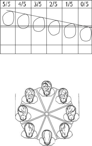
Engagement of the fetal head
Conventionally, engagement or the passage of the maximal diameter of the presenting part beyond the pelvic inlet is estimated using the palm width of the five fingers of the hand (Fig. 1.2). If five fingers are needed to cover the head above the pelvic brim, it is five-fifths palpable, and if no head is palpable, it is zero-fifths palpable.
•Normally, the fetus engages in an attitude of flexion in the larger transverse diameter of the pelvic inlet, unless the pelvis is very roomy where it may engage in any diameter (see Fig. 1.3).
•In nulliparous women, engagement usually (not in all) occurs beyond 37wks, but in multiparous women it may not occur until the onset of labour.
•Rare causes of non-engagement should always be considered and investigated with an ultrasound scan (USS) (including placenta praevia and fetal abnormality).
•In women of Afro-Caribbean origin, engagement may only occur at the onset or during the course of labour, even in nulliparous women due to the shape of the pelvic inlet.
Paulik’s grip
This is a one-handed technique that uses a cupped right hand to grasp and assess the lower pole of the uterus (usually the fetal head).
2This can be very uncomfortable and is not necessary if the head can be palpated using two hands.
Engagement
•A head that is only two-fifths palpable is usually considered to be engaged (and therefore fixed in the pelvis; see Fig. 1.2).
•Put simply, an easily palpable head is not engaged, whereas a head more difficult to palpate is more likely to be deeply engaged.
Care must be taken, as a breech presentation can sometimes be mistaken for a deeply engaged head.
|
|
ENGAGEMENT OF THE FETAL HEAD |
9 |
||||
s |
|
Abdomen |
|
|
|
|
|
|
s |
|
|
|
|
||
|
|
|
|
|
|
|
|
o |
o |
s |
s |
|
|
|
|
|
|
|
|
|
|
||
|
|
o |
|
|
|
|
|
|
|
o |
s |
|
|
|
|
|
|
|
|
|
|
||
|
|
|
|
s |
|||
|
|
|
|
|
|||
Pelvic brim |
|
|
|
o |
o |
||
|
|
|
|
|
|||
|
|
Pelvic cavity |
|
|
|
|
|
|
|
|
|
|
|
||
|
|
|
|
|
|
||
Completely |
Sinciput |
Sinciput |
Sinciput |
Sinciput |
None of |
||
high |
easily felt |
felt |
felt |
head |
|||
above |
Occiput |
Occiput |
Occiput |
Occiput |
palpable |
||
|
easily felt |
felt |
just felt |
not felt |
|||
Fig. 1.2 Clinical estimation of descent of the fetal head and engagement. Reproduced from Arulkumaran S, Symonds IM, Fowlie A. (2004). Oxford Handbook of Obstetrics and Gynaecology. Oxford: OUP. By permission of Oxford University Press.
|
Occipitoposterior (OP) |
ROP |
LOP |
Mother’s |
Mother’s |
right side |
left side |
Right occipito- |
Left occipito- |
transverse |
transverse |
(ROT) |
(ROT) |
ROA |
LOA |
Occipitoanterior (OA)
Front
Fig. 1.3 Fetal position. Reproduced from Collier J, Longmore M, et al. (2008).
Oxford Handbook of Clinical Specialties, 8th edn. Oxford: OUP. By permission of Oxford University Press.

10 CHAPTER 1 Normal pregnancy
The female pelvis
The bony ring of the pelvis is made up of two symmetrical innominate bones and the sacrum. Each innominate bone is made up of the ilium, ischium, and the pubis, which are joined anteriorly at the symphysis pubis and posteriorly to the sacrum at the sacroiliac joints.
The female pelvis has evolved for giving birth, and differs from the male pelvis in the following ways:
•The female pelvis is broader, and the bones more slender than those of the male.
•The male pelvic brim is heart-shaped and widest towards the back, whereas the female pelvic brim is oval-shaped transversely and widest further forwards; the sacral promontory is less prominent.
•The female pelvic cavity is more spacious and has a wider outlet than the male pelvis.
•The subpubic angle is rounded in a female pelvis (like a Roman arch) and more acute in the male pelvis (like a Gothic arch).
Pelvic muscles and ligaments
The pelvis gains its strength and stability through numerous muscles and ligaments. The inner aspect of the pelvic bones is covered by muscles. Above the pelvic brim are the iliacus and psoas muscles; the obturator internus and its fascia occupies the side walls; the posterior wall is covered by the pyriformis; and the levator ani and coccygeus, with their opposite counterparts, constitute the pelvic floor.
Pelvic ring stability is provided by the following ligaments:
•Sacrospinous ligament: extending from the lateral margin of the sacrum and coccyx to the ischial spine.
•Sacrotuberous ligament: extending from the sacrum to the ischial tuberosity.
•Iliolumbar ligament: extending from the spine to the iliac crest at the back of the pelvis.
•Dorsal sacroiliac ligament: a heavy band passing from the ilium to the sacrum posterior to the sacroiliac joint.
•Ventral sacroiliac ligament: bridging the sacroiliac joint anteriorly, and is an important stabilizing structure of the joint.
•Inferior and superior pubic ligament: a band across the lower and upper part of the symphysis respectively, providing further strength to the joint.
•Inguinal ligament: running from the anterior superior iliac spine of the ilium to the pubic tubercle of the pubic bone.
The remaining ligaments that surround the pelvis are ligaments that do not provide stabilization of the pelvis.

THE FEMALE PELVIS 11
Pelvic boundaries
The pelvis is divided by an oblique plane passing through the prominence of the sacrum, the arcuate, and pectineal lines, and the upper margin of the symphysis pubis, into the greater and the lesser pelvis. The circumference of this plane is termed the pelvic brim. This pelvic brim separates the false pelvis above from the true pelvis below. The plane of the pelvis is at an angle of 55° to the horizontal.
Pelvic shapes
There are four basic shapes of the female pelvis, as illustrated in Fig. 1.4.
•Gynaecoid: the classical female pelvis with the inlet transversely oval and a roomier pelvic cavity.
•Anthropoid: a long, narrow and oval-shaped pelvis due to the assimilation of the sacral body to the fifth lumbar vertebra.
•Android: the inlet is heart-shaped and the cavity is funnel-shaped with a contracted outlet.
•Platypolloid: a wide pelvis flattened at the brim with the sacral promontory pushed forward.
Name
Platypolloid Gynaecoid Anthropoid Android
%
women
20
25
50
5
Pelvic |
Pelvic |
Pelvic |
Pelvic |
shape |
inlet |
outlet |
arch |
Fig. 1.4 Basic shapes of the female pelvis. Reproduced from Abitbol M, Chervenak F, Ledger WJ. (1996). Birth and human evolution: anatomical and obstetrical mechanics in primates. New York: Bergin & Garvey.

12 CHAPTER 1 Normal pregnancy
Diameters of the female pelvis
The female bony pelvis is not distensible, and only very minor degrees of movement are possible at the symphysis pubis and the sacroiliac joints. Its dimensions are, hence, critical for normal childbirth.
2The diameters of the female pelvis vary at different parts of the pelvis:
•The true pelvis is bound anteriorly by the symphysis pubis (3.5cm long) and posteriorly by the sacrum (12cm long).
•The superior circumference of the true pelvis is the pelvic inlet and the
inferior circumference is the outlet (Fig. 1.5).
The true pelvis has four planes.
Plane of pelvic inlet
•This is bound anteriorly by the upper border of the pubis, laterally by the iliopectineal line, and posteriorly by the sacral promontory.
•The average transverse diameter is 13.5cm and the average anteroposterior diameter is 11cm (obstetric conjugate diameter) (transversely oblong).
•It is not possible to measure these diameters clinically, and the only diameter at the pelvic inlet amenable to clinical assessment is the distance from the inferior margin of the pubic symphysis to the midpoint of the sacral promontory (the diagonal conjugate), which is 71.5cm greater than the obstetric conjugate diameter.
Plane of greatest pelvic dimensions/cavity
•This is the roomiest part of the pelvis and has little clinical significance.
•It is almost round in shape with an average transverse diameter of 13.5cm and an average anteroposterior diameter of 12.5cm.
Plane of least pelvic dimensions/mid-pelvis (circular in shape)
•This is bound anteriorly by the apex of the pubic arch, laterally by the ischial spines, and posteriorly by the tip of the sacrum.
•The interspinous diameter is the narrowest space in the pelvis (10cm) and represents the level at which impaction of the fetal head is most likely to occur.
Plane of pelvic outlet
•This is bound anteriorly by the pubic arch, which should have a desired angle of >90°, posterolaterally by the sacrotuberous ligaments and ischial tuberosities leading to the coccyx posteriorly (anteroposteriorly oblong).
•The average intertuberous diameter is 11cm.

DIAMETERS OF THE FEMALE PELVIS 13
Assessment of ‘pelvic adequacy’
Examination of the pelvis before labour does not accurately discriminate between those who will achieve vaginal birth and those who will not. Even computed tomography (CT) or magnetic resonance imaging (MRI) scanning, together with ultrasound of the fetal head, is not helpful, unless there is a gross abnormality, which will be evident from the history or gait. This is because of the dynamic nature of labour, when the head ‘moulds’ (reducing the head circumference by a few centimetres) and the joints of the pelvis can move, increasing the pelvic dimensions slightly.
The ideal female pelvis has the following features:
•Oval brim.
•Shallow cavity.
•Non-prominent ischial spines.
•Curved sacrum with large sciatic notches (>90 ).
•Sacrospinous ligament >3.5cm long.
•Rounded subpubic arch >90 .
•Intertuberous distance of at least 10cm.
Diagonal conjugate diameter of at least 12cm.
Inlet
Mid |
pelvis |
|
Sacrum
Outlet Coccyx
Fig. 1.5 Median sagittal section of the female pelvis showing the pelvic inlet and outlet. Reproduced from Collier J, Longmore M, et al. (2008). Oxford Handbook of Clinical Specialties, 8th edn. Oxford: OUP. By permission of Oxford University Press.

14 CHAPTER 1 Normal pregnancy
Fetal head
Anatomy of the fetal skull
The fetal cranium is made up of five main bones, two parietal bones, two frontal bones, and the occipital bone. These are held together by membranous areas called sutures, which permit movement during birth (Fig. 1.6).
•The coronal suture separates the frontal bones from the parietal bones.
•The sagittal suture separates the two parietal bones.
•The lambdoid suture separates the occipital bone from the parietal bones.
•The frontal suture separates the two frontal bones.
When two or more sutures meet, there is an irregular membranous area between them called a fontanelle (Fig 1.6).
•The anterior fontanelle or bregma is a diamond-shaped space at the junction of the coronal and sagittal sutures; this measures about 3cm in anteroposterior and transverse diameters, and usually ossifies at 718mths after birth.
•The posterior fontanelle or the lambda is a smaller triangular area that lies at the junction of the sagittal and lambdoid sutures.
The positions of the sutures and fontanelles play a very important role in identifying the position of the fetal head in labour.
Regions of the fetal head
The fetal head has different regions assigned to help in the description of the presenting part felt during vaginal examination in labour.
•The occiput is the bony prominence that lies behind the posterior fontanelle.
•The vertex is the diamond-shaped area between the anterior and posterior fontanelles, and between the parietal eminences.
•The bregma is the area around the anterior fontanelle.
•The sinciput is the area in front of the anterior fontanelle, which is divided into the brow (between the bregma and the root of the nose) and the face (lying below the root of the nose and the supraorbital ridges).

FETAL HEAD 15
Caput and moulding of the fetal head
During labour, the dilating cervix may press firmly on the fetal scalp preventing venous blood and lymphatic fluid from flowing normally. This may result in a tissue swelling beneath the skin called caput succedaneum. It is soft and boggy to touch and usually disappears within 24h of birth.
There is usually some alteration in the shape of the fetal head and a reduction in the head circumference in labour by a process of overlapping of the cranial bones (a reduction of up to 4cm is possible). This moulding is physiological and disappears a few hours after birth. The frontal bones can slip under the parietal bones and, in addition, one parietal bone can override the other and in turn slip under the occipital bone.
The degree of moulding can be assessed vaginally:
•No moulding: when the suture lines are separate.
•1+ moulding: when the suture lines meet.
•2+ moulding: when the bones overlap but can be reduced with gentle digital pressure.
•3+ moulding: when the bones overlap and are irreducible with gentle digital pressure.
2 The presence of caput and moulding can play an important part in diagnosing obstructed labour.
Posterior fontanelle (λ)
Biparietal diameter 9.5cm
Sagittal suture
Anterior fontanelle (bregma)
Fig. 1.6 Fontanelles, sagittal suture, and biparietal diameter. Reproduced from Collier J, Longmore M, et al. (2008). Oxford Handbook of Clinical Specialties, 8th edn. Oxford: OUP. By permission of Oxford University Press.
16 CHAPTER 1 Normal pregnancy
Diameters and presenting parts of the fetal head
The region that presents in labour depends on the degree of flexion or deflexion of the fetal head on presentation to the maternal pelvis. The important diameters of the fetal head as well as the presenting parts are as described below (Fig. 1.7):
•Suboccipitobregmatic diameter (9.5cm): presentation of a well-flexed vertex. Diameter extends from the middle of the bregma to the undersurface of the occipital bone where it joins the neck. Fetal head circumference is smallest at this plane and measures 32cm.
•Suboccipitofrontal diameter (10.5cm): partially flexed vertex, with diameter extending from the prominent point of the mid-frontal bone to the undersurface of occipital bone where it joins the neck.
•Occipitofrontal diameter (11.5cm): presentation of a deflexed head. Diameter extends from the prominent point of the mid-frontal bone to the most prominent point on the occipital bone. Fetal head circumference at this plane measures 34.5cm.
•Mentovertical diameter (13cm): brow presentation, with the diameter extending from the chin to the most prominent point of the midvertex. Presents with the largest anteroposterior diameter.
•Submentobregmatic diameter (9.5cm): face presentation, with diameter extending from just behind chin to the middle of the bregma.
Other noteworthy diameters of the fetal head include:
•Biparietal diameter (BPD, 9.5cm): greatest transverse diameter of the head, extending from one parietal eminence to the other.
•Bitemporal diameter (8cm): greatest distance between two temporal eminences.
•Bimastoid diameter (7.5cm): distance between the tips of the two mastoid processes.
See bMalpresentations in labour: overview, p. 316.

DIAMETERS AND PRESENTING PARTS OF THE FETAL HEAD 17
 1
1
4
2
3
5
1 Suboccipitobregmatic 9.5cm flexed vertex presentation
2 Suboccipitofrontal 10.5cm partially deflexed vertex
3 Occipipitofrontal 11.5cm deflexed vertex
4 Mentovertical 13cm brow
5 Submentobregmatic 9.5cm face
Fig. 1.7 Different presenting diameters of the fetal head. Reproduced from Collier J, Longmore M, Turmezei T, et al. (2008) Oxford Handbook of Clinical Specialties, 8th edn. Oxford: OUP. By permission of Oxford University Press.
Useful definitions when discussing the presenting part
•Presentation is the lowermost part of the fetus presenting to the pelvis. In more than 95% of cases the vertex is the presenting part and is called normal presentation. Any other presentation (e.g. face, brow, breech, and shoulder) is called malpresentation.
•Denominator is the most definable peripheral landmark of the presenting part, i.e. occiput for the vertex, mentum for the face, and sacrum for the breech presentation.
•Position of the presenting part is the relationship of the denominator to the fixed points of the maternal pelvis, i.e. sacrum posteriorly, pubic symphysis anteriorly, sacro-iliac joints posterolaterally, and ileo-pectineal eminences anterolaterally.
•Station is the relationship of the most prominent leading part of the presenting part to the ischial spines expressed as ± 1,2,3cm.
2 In the vertex presentation more than 90% present in the occipitoanterior position, i.e. the occiput is in the anterior half of the pelvis and is called the normal position. If the occiput is pointing laterally or is in the posterior half of the pelvis, it is called malposition and is associated with deflexed head presenting a larger anteroposterior diameter of the vertex (11.5cm) and, hence, difficulties with progress of labour (Fig. 1.3).

18 CHAPTER 1 Normal pregnancy
Placenta: early development
The placenta is the organ responsible for providing endocrine secretions and selective transfer of substances to and from the fetus. It serves as an interface between the mother and developing fetus. Understanding the development of the placenta is important, as it is the placental trophoblasts that are critical for a successful pregnancy.
Embryological development
• After fertilization, the zygote enters the uterus in 3–5 days and continues to divide to become the blastocyst.
•Implantation of the blastocyst starts on day 7 and is finished by day 11:
•the inner cell mass of the blastocyst forms the embryo, yolk sac, and amniotic cavity
•the trophoblast forms the future placenta, chorion, and extraembryonic mesoderm.
•When the blastocyst embeds into the decidua, trophoblastic cells differentiate and the embryo becomes surrounded by two layers of trophoblasts:
•the inner mononuclear cytotrophoblast
•the outer multinucleated syncytiotrophoblast.
•The invading trophoblast penetrates endometrial blood vessels forming intertrophoblastic maternal blood-filled sinuses (lacunar spaces).
•Trophoblastic cells advance as early or primitive villi, each consisting of cytotrophoblast surrounded by the syncytium.
•These villi mature into sand 3° villi, and the mesodermal core develops to form fetal blood vessels (completed by day 21).
•On days 16–17, the surface of the blastocyst is covered by branching villi which are best developed at the embryonic pole: the chorion here is known as chorionic frondosum; the future placenta develops from this area.
•Simultaneously, the lacunar spaces become confluent with one another and, by weeks 3–4, form a multilocular receptacle lined by syncytium and filled with maternal blood: this becomes the future intervillous space.
•With further growth of the embryo the decidua capsularis becomes thinner, and both villi and the lacunar spaces in the decidua are obliterated, converting the chorion into chorionic levae.
•The villi in the chorionic frondosum show exuberant division and subdivision, and with the accompanying proliferation of the decidua basalis, the future placenta is formed.
•This process starts at 6wks and the definitive numbers of stem villi are established by 12wks.

PLACENTA: LATER DEVELOPMENT 19
Placenta: later development
Placental growth continues to term.
•Until week 16, the placenta grows both in thickness and circumference due to growth of the chorionic villi with accompanying expansion of the intervillous space.
•After 16wks growth occurs mainly circumferentially.
Placental villi
•Functional units of the placenta.
•There are 760 stem villi in human placenta with each cotyledon containing 3–4 major stem villi.
•Despite their close proximity (0.025mm), there is no mixing of maternal and fetal blood.
•Placental barrier is made of outer syncytiotrophoblast, which is in direct contact with maternal blood, the cytotrophoblast layer,
basement membrane, stroma containing mesenchymal cells, and the endothelium and basement membrane of fetal blood vessels
The placenta at term
•Circular, diameter 15–20cm, thickness 72.5cm at the centre.
•Weight 7500g (ratio of fetal:placental weight at term is about 6:1).
•Occupies 730% of the uterine wall at term and has two surfaces.
Fetal surface
•Covered by a smooth, glistening amnion with the umbilical cord usually attached at or near its centre.
•Branches of the umbilical blood vessels are visible beneath amnion as they radiate from the insertion of the cord.
•Amnion can be peeled off from underlying chorion, except at insertion of cord.
Maternal surface
•Rough and spongy appearance, divided into several velvety bumps called cotyledons (15–20) by septa arising from the maternal tissues.
•Each cotyledon may be supplied by its own spiral artery.
•Numerous small greyish spots may be visible on the maternal surface representing calcium deposition in degenerated areas.
Umbilical cord
•Vascular cable that connects the fetus to the placenta.
•Varies from 30 to 90cm long, covered by amniotic epithelium.
•Contains two umbilical arteries and one umbilical vein embedded into the Wharton’s jelly.
•Arteries carry deoxygenated blood from fetus to placenta and the oxygenated blood returns to fetus via the umbilical vein.
•In a full-term fetus, blood flow in the cord is 7350mL/min.

20 CHAPTER 1 Normal pregnancy
Placenta: circulation
The placental circulation consists of two distinctly different systems—the uteroplacental circulation and the fetoplacental circulation.
Uteroplacental circulation
•Uteroplacental circulation is the maternal blood circulating through the intervillous space (Table 1.1).
•Intervillous blood flow at term is estimated to be 500–600mL/min, and blood in the intervillous space is replaced 3–4 times/min.
• Pressure and concentration gradients between fetal capillaries and intervillous space favours placental transfer of oxygen and other nutrients to the fetus.
Arterial system
•Spiral arteries respond to the idemand of blood supply to the placental bed by becoming low-pressure, high-flow vessels.
•They become tortuous, dilated, and less elastic by trophoblastic invasion, which starts early in pregnancy and occurs in two stages:
•in first trimester, the decidual segments of the spiral arterioles are structurally modified
•in second trimester, second wave of trophoblastic invasion occurs, resulting in invasion of myometrial segments of spiral arteries.
2 Failure of this physiological change, particularly second wave of trophoblastic invasion, is implicated in development of pre-eclampsia and intrauterine growth restriction.
Venous system
•Blood entering the intervillous space from the spiral artery becomes dispersed to reach the chorionic plate and gradually the basal plate, being facilitated by mild movements of villi and uterine contractions.
•From basal plate, uterine veins drain the deoxygenated blood.
•Venous drainage only occurs during uterine relaxation.
•Spiral arteries are perpendicular and veins are parallel to uterine wall, making large volumes of blood available for exchange at the intervillous space even though the rate of flow is decreased during contraction, i.e. the veins are blocked for a longer time to allow pooling of blood in the retroplacental area.
Fetoplacental circulation (see Table 1.2)
•Two umbilical arteries carry deoxygenated blood from the fetus and enter the chorionic plate underneath the amnion.
•Arteries divide into small branches and enter the stem of the chorionic villi, where further division to arterioles and capillaries occurs.
•The blood then flows to the corresponding venous channel and subsequently to the umbilical vein.
•Maternal and fetal bloodstreams flow side by side, in opposite directions, facilitating exchange between mother and fetus.

|
|
PLACENTA: CIRCULATION |
21 |
||
|
|
|
|||
|
Table 1.1 Haemodynamics of uteroplacental circulation |
|
|||
|
Volume of blood in the intervillous space |
150mL |
|
|
|
|
Blood flow in the intervillous space |
500–600mL/min |
|
||
|
Pressure changes in the intervillous space |
|
|
|
|
|
Height of uterine contraction |
30–50mmHg |
|
||
|
Uterine relaxation |
10–15mmHg |
|
||
|
Pressure in the spiral artery |
70–80mmHg |
|
||
|
Pressure in the uterine veins |
8–10mmHg |
|
||
|
|
|
|||
|
|
|
|
|
|
Table 1.2 Haemodynamics in the fetoplacental circulation
Fetal blood flow through placenta |
400mL/min |
Pressure |
|
In the umbilical artery |
60–70mmHg |
In the umbilical vein |
10mmHg |
Oxygen saturation and partial pressure |
|
of oxygen |
|
In the umbilical artery |
60%; 20–25mmHg |
In the umbilical vein |
70–80%; 30–40mmHg |
|
|

22 CHAPTER 1 Normal pregnancy
Placenta: essential functions
The placenta is directly responsible for mediating and/or modulating the maternal environment necessary for normal fetal development.
Principal functions of the placenta
•To anchor the fetus and establish the fetoplacental unit.
•To act as an organ for gaseous exchange.
•Endocrine organ to bring the needed changes in pregnancy.
•Transfer of substances to and from the fetus.
•Barrier against infection.
The placenta as an endocrine organ
As an active endocrine organ, the placenta produces a number of hormones, growth factors, and cytokines. The production of human chorionic gonadotrophin (hCG), oestrogens, and progesterone by the placenta is vital for the maintenance of pregnancy.
hCG
•Primarily produced by syncytiotrophoblasts.
•Detected from 6 days after fertilization; forms basis of modern pregnancy testing.
•Concentrations reach a peak at 10–12wks gestation, then plateau for remainder of the pregnancy.
The placenta as a barrier
The placenta acts as a barrier for the fetus against pathogens and the maternal immune system.
Infection
The placenta forms an effective barrier against most maternal blood-borne bacterial infections. However, some important organisms, such as syphilis, parvovirus, hepatitis B and C, rubella, human immune deficiency virus (HIV), and cytomegalovirus (CMV) are able to cross it and infect the fetus during pregnancy.
Drugs
Many drugs administered to the mother will pass across the placenta into the fetus; exceptions include low-molecular-weight heparin (LMWH).
Some drugs may have little effect on the fetus and be considered ‘safe’ (e.g. paracetamol), but others (e.g. warfarin) may significantly affect development, structure, and function of the fetus—a process known as teratogenesis.
Before prescribing any drug to a pregnant woman it is the prescriber’s obligation to ensure it is considered safe for stage of pregnancy.
See bDrugs in pregnancy, p. 261.

PLACENTA: ESSENTIAL FUNCTIONS 23
Placental transfer
Although the placenta acts as a barrier to most substances, it allows exchange of gases, transfer of fetal nutrition, and removal of waste products in a highly effective manner. Speed of exchange and concentration of substance exchanged depends upon:
•Concentration of the substance on each side of the placenta.
•Molecular size.
•Lipid solubility.
•Ionization.
•Placental surface area.
•Maternofetal blood flow.
A low-molecular-weight lipid-soluble substance with a high concentration gradient across the placenta, for example, will be transferred quickly to the fetus. Actual transfer occurs by simple diffusion, facilitated diffusion, active transport, and/or endocytosis (Table 1.3).
Table 1.3 Transfer mechanisms across the placenta for common anabolites and catabolites
Substance |
Transfer mechanism(s) |
Direction of transfer |
Oxygen |
Simple diffusion |
To fetus |
Carbon dioxide |
Simple diffusion |
From fetus |
Glucose |
Simple and facilitated diffusion |
To fetus |
Amino acids |
Facilitated diffusion |
To fetus |
Iron |
Endocytosis |
To fetus |
Fatty acids |
Facilitated diffusion |
To fetus |
Water |
Simple diffusion |
To and from fetus |
Electrolytes |
Counter-transport mechanism |
To and from fetus |
Urea and |
Simple diffusion |
From fetus |
creatinine |
|
|
|
|
|

24 CHAPTER 1 Normal pregnancy
Physiology of pregnancy: endocrine
Physiological and anatomical changes occur during the course of pregnancy to provide a suitable environment for the growth and development of the fetus. Early changes are due, in part, to metabolic demands brought on by the fetus, placenta, and uterus, and, in part, to increasing levels of pregnancy hormones, particularly those of progesterone and oestrogen. Later changes are more anatomical in nature and are caused by mechanical pressure from the expanding uterus.
Endocrine changes
Progesterone ithroughout pregnancy
•Synthesized by the corpus luteum until 35 days and by the placenta thereafter.
•Progesterone promotes smooth muscle relaxation (gut, ureters, uterus) and raises body temperature.
•It is the principal hormone that prevents preterm labour and is now increasingly administered to prevent preterm labour.
Oestrogens, mainly oestradiol (90%)
•iBreast and nipple growth, and pigmentation of the areola.
•Promote uterine blood flow, myometrial growth, cervical softening.
•iSensitivity and expression of myometrial oxytocin receptors.
•iWater retention and protein synthesis.
Human placental lactogen (hPL)
•Has a structure and function similar to growth hormone.
•Modifies maternal metabolism to ithe energy supply to the fetus.
•iInsulin secretion, but dinsulin’s peripheral effect (liberating maternal fatty acids and sparing glucose enabling it to be diverted to the fetus).
The pituitary gland in pregnancy
•Enlarges mainly due to changes in the anterior lobe.
•Prolactin levels increase substantially, probably due to oestrogen stimulation of the lactotrophes.
•Gonadotrophin secretion is inhibited, whilst plasma adrenocorticotrophic hormone (ACTH) levels i.
•Maternal plasma cortisone output i, but the unbound levels remain constant.
The posterior pituitary releases oxytocin principally during the first stage of labour and during suckling.

PHYSIOLOGY OF PREGNANCY: ENDOCRINE 25
Effect of pregnancy on the thyroid
•The maternal thyroid gland enlarges due to idemand in pregnancy.
•iRenal clearance of iodine results in a relative iodide deficiency.
•The thyroid responds by tripling its iodide uptake from the blood, which results in follicular enlargement.
•Thyroid-binding globulin (TBG) is doubled by the end of the first trimester due to high oestrogen levels.
•As a result, total T3 (triiodothryonine) and T4 (thyroxine) levels rise early in pregnancy, then fall to remain within normal non-pregnant range.
•Thyroid-stimulating hormone (TSH) may decrease slightly in early pregnancy, but tends to remain within the normal range.
•T3 and T4 cross the placental barrier in very small amounts.
Iodine, antithyroid drugs, and long-acting thyroid stimulator (LATS) or antibodies associated with Graves’ disease can cross the placenta and affect the fetal thyroid function, which starts as early as 12wks.
26 CHAPTER 1 Normal pregnancy
Physiology of pregnancy: haemodynamics
Plasma volume
•iBy 10–15% at 6–12wks of gestation.
•Expands rapidly until 30–34wks.
•Total gain at term 71100–1600mL (total plasma volume of 4700–5200mL, a 30–50% ifrom the non-pregnant state).
• Acute excessive weight gain is commonly due to oedema.
Red cell volume (or red cell mass)
•Rises from 1400 to 1640mL at term (i18%).
•With iron and folate supplements, an iof 30% has been reported.
2 The discrepancy between the rate of i of plasma volume and that of red cell mass results in a relative haemodilution or ‘physiological anaemia’ with the haemoglobin (Hb) concentration, haematocrit, and red cell counts all d(particularly in the second trimester).
2Mean corpuscular Hb concentration remains constant.
Total white cell count
•iMainly due to the iin neutrophil polymorphonuclear leucocytes, which peaks at 32wks.
•A further massive neutrophilia occurs during labour.
•Eosinophils, basophils, and monocytes remain relatively constant, but there is a profound din eosinophils during labour, being virtually absent at delivery.
•Although lymphocyte count and the number of B and T cells remain constant, lymphocyte function and cell-mediated immunity are profoundly depressed, giving rise to lowered resistance to viral infections.
Platelets
•dSlightly during pregnancy.
•Platelet function is unchanged.
Clotting factors
•Pregnancy is a hypercoagulable state.
•Most clotting factors i, especially fibrinogen.
2Erythrocyte sedimentation rate (ESR) levels can also be elevated up to 4-fold in pregnancy.

PHYSIOLOGY OF PREGNANCY: CARDIO-RESPIRATORY 27
Physiology of pregnancy: cardio-respiratory
Cardiovascular changes
Major changes occur in the cardiovascular system in pregnancy; the most significant of these changes occur within the first 12wks.
•Cardiac output ifrom 5 to 6.5L/min by istroke volume (10%) and pulse rate (715 beats/min).
•During labour, contractions may icardiac output by 2L/min, probably due to injection of blood from the distended intervillous space.
•With progressive enlargement of the uterus, the heart and diaphragm are displaced upwards.
•The heart enlarges and iin volume by 70–80mL due to idiastolic filling and muscle hypertrophy.
2 Pregnancy may proceed normally even when the mother has an artificial cardiac pacemaker, compensation occurring mainly from increased stroke volume.
Blood pressure in pregnancy
•Peripheral resistance dby nearly 50% (probably due to the iproduction of vasodilator prostaglandins).
•BP (most noticeably diastolic) dmid-pregnancy by 10–20mmHg and ito non-pregnant levels by term.
•Profound dcan occur late in pregnancy when lying supine, due to compression of the inferior vena cava leading to dvenous return and dcardiac output (supine hypotension syndrome).
•Aortic compression may also occur causing a conspicuous difference between brachial and femoral pressures giving a pressure difference of 10–15% from the supine to the lateral position.
•The balance of vasoconstrictor and vasodilator factors regulating peripheral resistance may be the basis of BP regulation in pregnancy and implicated in development of pregnancy-induced hypertension.
•Vasodilatation and hypotension also stimulate renin–angiotensin release, which plays a part in BP regulation.
Respiratory system changes
•The level of the diaphragm rises in pregnancy and the intercostal angle ifrom 68° in early pregnancy to 103° in late pregnancy: breathing becomes more diaphragmatic than costal.
•Tidal volume i740% (500–700mL) due to effect of progesterone.
•Inspiratory capacity (tidal volume plus inspiratory reserve volume) iprogressively in late pregnancy.
•Respiratory rate changes slightly, hence the resting pregnant woman iventilation by breathing more deeply and not more frequently.
•Breathlessness is common in pregnancy as maternal partial pressure of carbon dioxide (pCO2) is set lower to allow the fetus to offload CO2.

28 CHAPTER 1 Normal pregnancy
Physiology of pregnancy: genital tract and breast
Uterus
•Undergoes a 10-fold iin weight to 1000g at term.
•Muscle hypertrophy occurs up to 20wks, after which stretching of the muscle fibres occurs.
•Uterine blood flow has been shown to ifrom 750mL/min at 10wks to 500–700mL/min at term.
•The uterine and ovarian arteries and branches of the superior vesical arteries undergo massive hypertrophy.
The uterus is divided functionally and morphologically into three sections:
•Cervix.
•Isthmus (which later develops into the lower segment).
•Body of the uterus (corpus uteri).
Cervix
•Reduction in cervical collagen towards term enables its dilatation.
•Hypertrophy of cervical glands leads to the production of profuse cervical mucus, and the formation of a thick mucus plug or operculum that acts as a barrier to infection.
•Vaginal discharge idue to cervical ectopy (proliferation of columnar epithelium into vaginal portion of the cervix) and cell desquamation.
Uterine body
•iIn size, shape, position, and consistency.
•Uterine cavity expands from 4 to 4000mL.
Vagina
•A rich venous vascular network in connective tissue surrounds vaginal walls with blood and gives rise to slightly bluish appearance.
•High oestrogen levels stimulate glycogen synthesis and deposition:
•action of lactobacilli on glycogen in vaginal cells produces lactic acid
•lactic acid lowers the vaginal pH to keep the vagina relatively free from any bacterial pathogens.
Breast
•The lactiferous ducts and alveoli develop and grow under the stimulus of oestrogen, progesterone, and prolactin.
•From 3–4mths, colostrum (thick, glossy, protein-rich fluid) can be expressed from the breast.
•Prolactin stimulates the cells of the alveoli to secrete milk:
•effect is blocked during pregnancy by the peripheral action of oestrogen and progesterone
•shortly after delivery the sudden din these hormones enables prolactin to act uninhibited on the breast, and lactation begins.
•Suckling further stimulates prolactin and oxytocin release: oxytocin stimulates contraction of the myoepithelial cells to cause ejection of milk.

PHYSIOLOGY OF PREGNANCY: OTHER CHANGES 29
Physiology of pregnancy: other changes
Urinary tract
Various anatomical and physiological changes occur in pregnancy:
•Kidney size iby about 1cm in length.
•Marked dilatation of the calyces, renal pelvis, and ureter from first trimester.
•Vesicoureteric reflux occurs sporadically: a combination of reflux and ureteric dilatation leads to urinary stasis and iinfection.
•Although bladder muscle relaxes in pregnancy, residual urine is not normally present after micturition.
•Uric acid clearance increases from 12 to 20mmol/mL, causing reduction in plasma uric acid levels: as pregnancy progresses, the filtered load of uric acid i, while the excretion remains constant, resulting in plasma levels returning to non-pregnant values.
•Renal blood flow iby 30–50% in the first trimester, in line with the iin cardiac output that occurs, and remains elevated:
•results in iglomerular filtration rate (GFR) and effective renal plasma flow, causing a din plasma levels of urea and creatinine
•plays an important role in the variable glycosuria (due to exceeding the tubular maximum of absorption caused by more volume filtered) and urinary frequency that occurs in pregnancy.
Creatinine within the normal range for non-pregnant women may indicate renal impairment in pregnancy.
Alimentary system
•dTone of oesophageal sphincter and displacement through the diaphragm due to iabdominal pressure causes reflux oesophagitis (heartburn).
•Gastric mobility is low and gastric secretion is reduced, resulting in delayed gastric emptying.
•Gut motility is generally d, and with possible isodium and water absorption in the large bowel, there is a tendency to constipation.
Skin
•Pigmentation in linear nigra, nipple, and areola or chloasma (brown patches of pigmentation seen especially on the face).
•Palmar erythema and spider naevi are also common.
•Incidence of striae varies in different populations:
•represents the effect of disruption of collagen fibres in the subcuticular zone
•probably related to the effect of iproduction of adrenocortical hormones, as well as to the actual stress in the skin associated with relatively rapid expansion of the abdomen.

30 CHAPTER 1 Normal pregnancy
Preparing for pregnancy
A woman’s body undergoes significant changes in pregnancy, with the developing fetus making increasing demands. Preparation for pregnancy should begin before conception, as fetal development begins from the third week after the last menstrual period. Damaging effects (e.g. exposure to drugs) may occur before the woman is even aware she is pregnant. Being as fit and healthy as possible before conception maximizes chances of a healthy pregnancy, but not all poor obstetric outcomes can be avoided. Pre-pregnancy counselling by a specialist team is recommended where specific risks and diseases are identified.
Specific risks for older mothers
•Advanced maternal age is a risk factor for adverse outcome.
•A woman >35yrs old has a reduced chance of conceiving. This rate of decline drops very quickly by 40yrs.
•Age also carries an increased risk of chromosomal abnormalities in the baby (most common abnormality being Down’s syndrome).
•Older mothers are more likely to develop complications in pregnancy, e.g. pre-eclampsia and diabetes mellitus.
Exercise and stress
•Moderate exercise should be encouraged, as it improves a woman’s cardiovascular and muscular fitness.
•Women should be reassured that beginning or continuing a moderate course of exercise during pregnancy is not associated with adverse outcome. Best exercises are low-impact aerobics, swimming, brisk walking, and jogging.
•Contact and high-impact and vigorous racquet sports that may involve the risk of abdominal trauma should be avoided.
•Exercise is also associated with higher self-esteem and confidence.
•Relaxation and avoiding stress should be encouraged when planning for pregnancy.
Scuba diving may result in fetal birth defects and fetal decompression disease and, therefore, is not recommended.
Stopping contraception
•There is no delay in return to fertility after stopping the pill or having the coil removed.
•Women using contraceptive injection may experience a delay of several months.
•Often recommended that women wait 3mths after stopping the pill before trying to conceive.
This page intentionally left blank

32 CHAPTER 1 Normal pregnancy
Supplements and lifestyle advice
Folic acid and other vitamins
Folic acid is the only vitamin supplement that is recommended for use before pregnancy and up to 12wks gestation for women who are otherwise eating a healthy balanced diet.
Recommended doses of folic acid
•400micrograms/day folic acid has been shown to reduce the occurrence of neural tube defects.
•For women at higher risk (e.g. previous affected child, women with epilepsy, diabetes, and obesity), a dose of 5mg/day is recommended.
Iron
•Routine supplementation is not necessary and should be only prescribed when medically indicated. However, it may be considered routine in areas where incidence of iron-deficiency anaemia is high.
•The amount of elemental iron in an adult female is 5g. She will need 1mg/day before menstrual age, 2mg/day during reproductive age, and 3mg/day during pregnancy.
Calcium
Supplementation may be necessary if intake of calcium is low; however, the ideal is increased calcium by dietary intake.
Iodine
Deficiency is endemic in some parts of the world, and can cause cretinism and neonatal hypothyroidism. Supplementation with iodinized salt or oil should be considered.
Zinc
Low serum levels have been associated with an increased risk of preterm labour and growth restriction, but increased intake from dietary sources, such as milk and dairy products, should be sufficient.
Vitamin A supplementation (intake >700micrograms/day) might be teratogenic and should be avoided, as should consumption of products high in vitamin A, such as liver and pate.
Alcohol, smoking, and recreational drugs
Excessive alcohol intake has been conclusively shown to cause fetal malformations. The exact threshold of alcohol that will cause malformation in the fetus has not been established.
Avoid alcohol or limit consumption to one standard unit per day.
Smoking during pregnancy has an adverse effect on the developing fetus (e.g. preterm labour, low birth weight). Women should be encouraged to stop and supported through smoking cessation. If they cannot stop, reduction should be promoted.

SUPPLEMENTS AND LIFESTYLE ADVICE 33
2Stopping smoking at any stage has a beneficial effect.
Recreational and illegal drugs cause significant problems including miscarriage, preterm birth, poor fetal development, and intrauterine death. Help and support for dealing with any addiction should be sought from the appropriate agencies.
Weight and diet
•Fertility may be reduced in women who are significantly overweight (BMI >30) or underweight (BMI <18.5).
•Obesity is the most common nutritional disorder in the industrialized world, with increased risks including gestational diabetes and hypertension; also, monitoring and assessment may be difficult during pregnancy and labour (see bObesity in pregnancy: maternal risks, p. 258).
•Obesity has been recognized by the Confidential Enquiry into Maternal Deaths (CEMD) as carrying a greater risk of maternal death (see b The Confidential Enquiry into Maternal Deaths, p. 408).
•Malnutrition, on the other hand, is a major life hazard in the developing world and is a cause of other problems such as anaemia that has its own inherent risk for both mother and fetus.
•Poor nutrition in pregnant women is associated with the delivery of low birth weight (<2500g) babies, and improving the nutritional status and maternal weight can have a positive effect on the birth outcome.
•Weight gain should be around 11–16kg during pregnancy, and women should consume an additional 350kcal (1500kJ) a day.
•A nutritious, well-balanced diet includes foods rich in protein, dairy foods (which supply calcium), starchy foods, and plenty of fruit and vegetables that supply vitamins and fibre.
•It is best to avoid a lot of sugary, salty, or fatty foods.
•Food delicacies such as undercooked meats and eggs, pates, soft cheeses, shellfish and raw fish, and underpasteurized milk should be avoided as they are potential sources of Listeria and Salmonella that could lead to adverse perinatal outcome.
Listeriosis in pregnancy is a known but rare cause of poor obstetric outcome and fetal death.

34 CHAPTER 1 Normal pregnancy
General health check
Planning a pregnancy provides a good opportunity for a general health check by the GP and may identify any potential obstetric risk factors well in advance.
Pre-pregnancy general health check
May include:
•A general examination including BP, heart, and lungs.
•Family history of inherited disorders or congenital abnormalities.
•Urine dipstick.
•Blood tests such as thalassaemia and sickle cell disease may be offered if at risk.
•Rubella (and hepatitis) status should be ascertained and vaccination given if not immune (women should be advised to avoid pregnancy for 3mths after immunization).
•HIV screening if at risk.
•Dental examination.
Pre-existing medical disorders
Pregnancy can have an adverse effect on pre-existing medical disorders (see bMedical disorders in pregnancy, p. 185).
•The effect may be transient, returning to normal after delivery (e.g. diabetes mellitus), or it may be permanent and progressive, leading to maternal mortality (e.g. severe renal impairment or severe cardiac disease).
•For a woman with a pre-existing disorder contemplating pregnancy, the advice of a specialist should be sought early. If the risk is very high, pregnancy may be discouraged altogether (e.g. Eisenmengher’s complex—ventricular septal defects (VSD) with pulmonary hypertension).
•Optimal control of certain diseases before conception may be very important to avoid the risk of fetal malformation or adverse outcome (e.g. diabetes mellitus).
•Some medications may be changed before conception to reduce the risk of teratogenesis (e.g. antiepileptics).
•Pregnancy undertaken when the illness is in remission, stable, or cured will ensure a better outcome.
Medication
In general, both prescription drugs and over-the-counter medication should be used as little as possible. Most drugs carry warnings about use in pregnancy. However, the benefit may outweigh the risks, even in pregnancy, so a doctor should be consulted before stopping or starting any medication in pregnancy or before conception.

GENERAL HEALTH CHECK 35
Working during pregnancy
•Women should be reassured that it is safe to continue working before and during pregnancy.
•Some workplaces are more likely to present hazards (e.g. chemical factories, operating theatres, X-ray departments); hence, precautions may be necessary—specific advice should be sought from the employer’s occupational health department.
•Women should be reassured that use of computers and video display units has not been proven to be linked with any adverse outcome.
•See bVaccination and travel, p. 183.
36 CHAPTER 1 Normal pregnancy
Diagnosis of pregnancy
The most obvious symptom of pregnancy is cessation of periods, i.e. a period of amenorrhoea in a woman having regular menstruation.
Other common symptoms of early pregnancy
Nausea and vomiting (morning sickness)
•Common in the 1st trimester.
•May occur at any time of the day.
• May sometimes persist throughout pregnancy.
Frequency of micturition
•iPlasma volume and iurine production.
•Pressure effect of the uterus on the bladder.
•Make sure the frequency is not associated with dysuria, which may denote possible infection.
Excessive lassitude or fatigue
•Common in early pregnancy.
•Tends to disappear after 12wks gestation.
Breast tenderness or ‘heaviness’
Often seen early in pregnancy, particularly in the month after the first period is missed.
Fetal movements or quickening
•720wks gestation in the nullipara.
•18wks in the multipara.
•Many women may experience fetal movements earlier than this and some may not perceive movements until term.
Occasionally a pregnant woman may experience an abnormal desire to eat something not normally regarded as nutritive (such as dirt). This is known as pica.
Clinical examination
•The vagina and cervix have a bluish tinge due to blood congestion.
•The size of the uterus may be estimated by bimanual examination (reasonably accurate in early pregnancy).
•After 12wks the uterus is palpable abdominally and the fetal heart may be heard using a hand-held Doppler.

DIAGNOSIS OF PREGNANCY 37
The pregnancy test
•The hormone hCG is secreted by trophoblastic tissue:
•iexponentially from 78 days after ovulation (doubles every second day in an ongoing pregnancy)
•peaks at 8–12wks gestation.
•hCG levels can be measured in blood or urine.
•Test kits are available commercially (home pregnancy tests):
•can show a positive result with urinary hCG levels >50IU/L.
•some ‘early’ pregnancy test kits will detect levels of >25IU/L.
• These tests can confirm pregnancy within 1 week of a missed period.

38 CHAPTER 1 Normal pregnancy
Dating of pregnancy
Menstrual history
•The first day of the LMP may be used to calculate the gestational age and the EDD (bObstetric history: current pregnancy, p. 2), but this may be inaccurate as:
•many women may not be certain of their LMP
•ovulation does not always occur on day 14 and the proliferative phase may vary considerably in shorter or longer menstrual cycles.
•The EDD can be calculated using Naegele’s formula (bObstetric history: current pregnancy, p. 2).
•About 40% of women will deliver within 5 days of the EDD and about 2/3 within 10 days.
11–42% of gestational age estimates from LMP may be inaccurate.
2 Pregnancies resulting from in vitro fertilization (IVF) can be dated using the day of embryo transfer.
Dating ultrasound scan
•Between 8 and 13wks USS provides the most accurate measure of gestational age and, where possible, should be used to calculate EDD.
•Before 8wks it is unreliable due to the small size of the gestation sac and fetal pole.
•After 13wks other factors may affect fetal growth; therefore, although an estimate can be made using BPD and femur length (FL), it may be unreliable.
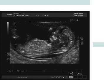
Crown–rump length
Crown–rump length (CRL; Fig. 1.8) is used to calculate gestation between 8 and 13wks. It is measured from one fetal pole to the other along its longitudinal axis in a straight line.
DATING OF PREGNANCY 39
Fig. 1.8 Ultrasound image of a 12wk fetus measuring the CRL.

40 CHAPTER 1 Normal pregnancy
Ultrasound assessment of fetal growth
Any clinical suspicion that the fetus may be small or large for gestational age should be followed by a formal ultrasound assessment of fetal growth and amount of amniotic fluid (liquor volume).
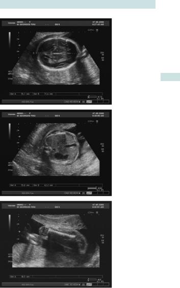
The measurements used are (Fig. 1.9):
Biparietal diameter and head circumference
• The anatomical landmarks used to ensure the accuracy and reproducibility of the measurement are a midline falx, the thalami symmetrically positioned on either side of the falx, the visualization of the cavum septum pellucidum at one-third the fronto-occipital distance, and the lateral ventricles with their anterior and posterior horns identifiable.
•The calipers are placed between the leading edge of the proximal and distal skull bones (BPD) and circumferentially around the head (HC).
Abdominal circumference
•The abdominal circumference (AC) is the single most important measurement in assessing fetal size and growth.
•It is measured where the image of the stomach and the portal vein is visualized in a tangential section.
Femur length
By convention, measurement of the FL is considered accurate only when the image shows two blunted ends.
FL can be underestimated if the correct plane is not obtained.
See bIntrauterine growth restriction: overview, p. 142.
Uterus measurement anomalies
The uterus may measure small for dates because of:
•Wrong dates.
•Oligohydramnios.
•Intrauterine growth restriction.
•Presenting part deep in the pelvis.
•Abnormal lie of the fetus.
The uterus may measure large for dates because of:
•Wrong dates.
•Macrosomia.
•Polyhydramnios.
•Multiple pregnancy.
•Presence of fibroids.
ULTRASOUND ASSESSMENT OF FETAL GROWTH 41
Fig. 1.9 Ultrasound measurement of biparietal diameter, abdominal circumference, and femur length.

42 CHAPTER 1 Normal pregnancy
Booking visit
The needs of each pregnant woman should be assessed at the first appointment and a plan of care made for her pregnancy. This should be reassessed at each appointment as new problems can arise at any time. Many women in the UK have ‘shared obstetric care’ whereby the woman’s GP and community midwife undertake most of the obstetric care, with a limited number of visits to the hospital.
Routine involvement of an obstetrician in the care of women with uncomplicated pregnancy does not appear to improve perinatal outcomes compared with involving obstetricians when complications arise.
There should be continuity of care throughout the AN period and this should be provided by a small group of carers with whom the woman feels comfortable. The environment in which AN appointments take place should enable women to discuss sensitive issues, such as domestic violence, sexual abuse, psychiatric illness, and illicit drug use. Women should be given the information needed to choose between giving birth at home, in a midwifery-led unit, or in hospital.
Booking should ideally be early in pregnancy (before 12wks) in order to take full advantage of AN care. However, many women are seen for the first time in the second trimester.
Children born to very late bookers or unbooked women have a higher risk of perinatal mortality (4–5-fold) and morbidity, with an attendant increase in maternal morbidity and mortality.
Booking visit: history
A comprehensive history should be elicited (see b Obstetric history: current pregnancy, p. 2) and a full physical examination undertaken (see bObstetric physical examination, p. 6).
•Risk factors from past history should be highlighted.
•It is essential to obtain past obstetric notes if it is thought that this information may change the management.
•History of inheritable diseases in close relatives should be sought, and history of migration and travel may identify risk for diseases such as haemoglobinopathies, some forms of hepatitis, and HIV infection.
•Histories of alcohol abuse, smoking, and addictive drug use are useful behavioural markers of potential risks (e.g. fetal abnormalities, impaired fetal growth, preterm labour, and neonatal drug withdrawal problems).
•It is important to identify women at risk of postnatal depressive illness:
•women should be asked about previous and family history of psychiatric disorders, and social problems including domestic violence and previous self-harm
•women at risk should have full psychiatric care and social support.
•Advice and support should be given on healthy lifestyles (including diet and exercise), pregnancy care services available, maternity benefits, and sufficient information to enable informed decision-making following screening tests.

BOOKING VISIT 43
First contact with healthcare professionals
Ensure the woman is given information on:
•Folic acid supplementation.
•Lifestyle advice.
•Antenatal screening.
•Booking appointment.
Booking appointment
•Identify high risk women who need additional care.
•Calculate BMI.
•Measure BP.
•Dipstick test urine (protein, glucose, blood, etc.).
•Ultrasound for gestational age and gross structural anomalies.
•Take blood tests for:
•haemoglobinopathies
•rubella
•Venereal Disease Research Laboratory test (VDRL)
•HIV
•hepatitis B virus
•red cell allo-antibodies
•Hb for anaemia.
•Give information on:
•AN classes
•pregnancy care pathway
•nutrition, diet, and vitamin supplementation
•maternity benefits
•how baby develops.

44 CHAPTER 1 Normal pregnancy
Antenatal care: planning
The basic aims of AN care are:
•To provide evidence-based information and support to women and their partners, to enable them to make informed decisions regarding their care.
•To advise on minor problems and symptoms of pregnancy.
•To assess maternal and fetal risk factors at the onset of pregnancy.
•To facilitate provision of prenatal screening and subsequent management of any abnormalities detected.
•To monitor fetal and maternal well-being throughout pregnancy and screen for commonly occurring complications (most notably BP and urine check at every visit to detect signs of developing pre-eclampsia and diabetes).
•To determine timing and mode of delivery when complications arise or if pregnancy continues after the EDD.
2The needs of each pregnant woman should be assessed at each appointment as new problems can arise at any stage in pregnancy.
Urine should be dipstick tested and BP measured at every AN visit.
Screening for chromosomal and structural abnormalities
Ideally, screening should be offered to all women at the time of booking. Detailed, unbiased, written information should be provided about the conditions being screened for, types of test available, and the implications of the results.
It is important for a woman to understand that a negative result in any screening test does not guarantee that her baby does not have that or another abnormality.
For full details on current UK screening see b Prenatal diagnosis: overview, p. 108.

ANTENATAL CARE: PLANNING 45
Antenatal appointment schedule
A schedule of AN appointments and what needs to be done at each visit has been described in the recent NICE Antenatal Care Guidelines and is summarized below.
Second trimester
•16wks:
•discuss screening results
•investigate if Hb level <11g
•offer information and arrange anomaly scan (18–20wks).
•25wks—nulliparaous women only: BP, urine dip, plot symphysis SFH.
•28wks:
•screening for anaemia and atypical red cell allo-antibodies
•anti D prophylaxis to rhesus (Rh) –ve women
•BP, urine dip, plot SFH.
Third trimester
•31wks—nulliparaous women only: BP, proteinuria, and plot SFH.
•34wks:
•discuss labour and birth (including pain relief and birth plan)
•give anti-D if rhesus –ve
•BP, proteinuria, plot SFH.
•36wks—discuss:
•breast-feeding
•vitamin K prophylaxis
•postnatal self-care
•awareness of baby blues and postnatal depression
•BP, proteinuria, plot SFH.
•38wks: BP, proteinuria, plot SFH.
•40wks:
•BP, proteinuria, plot SFH
•give information about prolonged pregnancy.
Membrane sweep at 41wks and induction of labour at 42wks.
Further reading
NICE. (2008). Antenatal care: routine care for the healthy pregnant woman. Antenatal care CG62.
Mwww.nice.org.uk/CG62
46 CHAPTER 1 Normal pregnancy
Antenatal care: routine blood tests
Antenatal care starts once the pregnancy is confirmed, and a referral is made by the GP to the community midwife for booking.
Routine blood tests
Full blood count (FBC)
•Physiological anaemia means lower limit for a ‘normal’ Hb is 10.5g/dL in pregnancy.
•Commonest cause of anaemia is iron deficiency. Investigate by assessment of haematinic indices, such as ferritin and total iron-binding capacity (TIBC) in microcytic hypochromic anaemia, serum and red cell folate, and serum B12 levels in macrocytic anaemia.
Blood grouping and antibody screen
Determining the blood group makes it possible to identify rhesus –ve women who are at risk of rhesus isoimmunization and to detect abnormal antibodies such as Kell and Duff (see bRhesus isoimmunization (immune hydrops), p. 135).
Rubella screen
Around 2% of nulliparous and 1% of multiparous women are not immune to rubella. It is recommended that these women receive post-partum rubella vaccination.
Syphilis screen
•Although incidence of syphilis in the UK is low, outbreaks do occur.
•Early treatment can prevent congenital syphilis in the neonate and screening and treatment is cost effective.
Hepatitis B screen
•Screening for hepatitis B is performed on all women in pregnancy at booking so that effective postnatal intervention can be offered.
•In adults, the virus is cleared within 6mths in 90% of those infected.
•In neonates, 90% become chronic carriers with risk of post-infective hepatic cirrhosis and hepatocellular carcinoma—hence the need to screen and prevent.
•Active immunization with hepatitis vaccine may be adequate for those neonates where mother was surface (s) antigen +ve, but passive and active immunization is recommended for those who are core (e) antigen +ve.
HIV screen
•The recommendation for all maternity units in the UK is for universal screening for HIV at the booking visit (‘opt out’ policy).
•vertical transmission from mother to fetus can be significantly reduced (by two-thirds) by treatment of mother with antiretrovirals in pregnancy and labour, and infant for 6wks postnatally
•risk of transmission is reduced by Caesarean section (CS) and avoidance of breast-feeding (see bHIV and pregnancy, p. 176).

ANTENATAL CARE: SPECIFIC BLOOD TESTS 47
Antenatal care: specific blood tests
Haemoglobin electrophoresis
This should be routinely performed in women of minority ethnic or racial origins with high incidence of haemoglobinopathies.
Some ethnic origins at high risk of thalassaemia
•Cyprus.
•Eastern Mediterranean.
•Middle Eastern.
•Indian subcontinent.
•South-east Asia.
2 Women of African or Afro-Caribbean origin are at risk of sickle cell disease or trait.
2If a woman is affected, testing her partner’s status would enable appropriate counselling and further prenatal testing.
Persistent anaemia, of undiagnosed cause, may be an indication for Hb electrophoresis in any woman, regardless of racial origin.
Miscellaneous tests
A variety of other blood tests may be indicated on an individual basis. For example, thyroid function tests (TFTs, history of thyroid disease), HbA1c (to assess long-term control of diabetes), or baseline urea and creatinine (in chronic hypertensives with renal complications).
Screening for gestational diabetes
X There is little consensus as to who, when, how, or even whether to screen for gestational diabetes mellitus (GDM). At present there is insufficient evidence to support routine screening for GDM. The timing of any screening is equally controversial as the later the test is performed, the higher the detection rate since glucose tolerance progressively deteriorates. On the other hand, the earlier in pregnancy GDM is diagnosed and hyperglycaemia treated, the greater the likelihood of positively influencing the outcome. Most units currently use targeted screening based on known risk factors.
Risk factors for gestational diabetes
•Previous GDM.
•Family history of diabetes (first-degree relative with diabetes).
•Previous macrosomic baby.
•Previous unexplained stillbirth.
•Obesity (BMI>30).
•Glycosuria on more than one occasion.
•Polyhydramnios.
•Large for gestational age fetus in current pregnancy.

48 CHAPTER 1 Normal pregnancy
Antenatal care: preparing for delivery
•In the early 3rd trimester, women are seen monthly and, at each visit, BP, urinalysis, and fundal height measurement, as well as enquiry about maternal well-being and fetal activity, are recorded.
•FBC and antibody screen is repeated at 28 and 34wks gestation, and rhesus –ve women are given anti-D prophylaxis at these times.
•From 36wks onwards, fetal presentation, as well as growth are assessed, and an ultrasound assessment performed if indicated.
•Preparation for labour and delivery should be discussed.
•The final routine visit between 40 and 41wks includes discussions on induction of labour after 41wks gestation.
•In women who wish to avoid induction, the risks of prolonging pregnancy should be discussed and a plan for increased fetal surveillance with cardiotocography (CTG) and ultrasound assessment of fetal growth and liquor volume can be made.