


Chapter 2 |
49 |
|
|
Pregnancy complications
Minor symptoms of pregnancy: gastrointestinal 50
Minor symptoms of pregnancy: musculoskeletal and vascular 52 Minor symptoms of pregnancy: genitourinary and others 54 Antepartum haemorrhage: overview 55
Antepartum haemorrhage: assessment 56 Antepartum haemorrhage: management 58 Blood pressure in pregnancy: physiology 60 Blood pressure in pregnancy: hypertension 62 Pre-eclampsia: overview 64
Pre-eclampsia: clinical features and investigations 66 Pre-eclampsia: management 67
Severe pre-eclampsia: management 68
Eclampsia and haemolysis, elevated liver enzymes, and low platelets (HELLP) syndrome 70
Multiple pregnancy: overview 72 Multiple pregnancy: types 74 Multiple pregnancy: antenatal care 76 Monochorionic, diamniotic twins 78 Multiple pregnancy: labour 80 Breech presentation: overview 82 External cephalic version 84
Breech presentation: delivery 86 Transverse, oblique, and unstable lie 88
Abdominal pain in pregnancy: pregnancy related (<24wks) 90
Abdominal pain in pregnancy: pregnancy related (>24wks) 92
Abdominal pain in pregnancy: bowel related 94 Abdominal pain in pregnancy: other causes 96 Preterm labour: overview 98
Preterm labour: prevention and prediction 100
Preterm prelabour rupture of membranes: overview 102 Preterm prelabour rupture of membranes: management 103 Prolonged pregnancy: overview 104
Prolonged pregnancy: management 106

50 CHAPTER 2 Pregnancy complications
Minor symptoms of pregnancy: gastrointestinal
Minor symptoms of pregnancy are mostly related to hormonal, physiological, and increased weight-bearing aspects of pregnancy. Although usually mild and self-limiting, some women may experience severe symptoms, which can affect their ability to cope with activities of daily living (see Further reading).
Nausea and vomiting (morning sickness)
• Most common complaint, especially in the 1st trimester: nausea—80–85%; vomiting—52%.
•Believed to be caused by hormones of pregnancy especially hCG.
•Increased in multiple and molar pregnancies.
•May be severe enough to warrant hospital admission—hyperemesis gravidarum (see bHyperemesis gravidarum, p. 546).
•Not usually associated with poor pregnancy outcome.
•Tends to resolve spontaneously by 16–20wks.
•Management:
•lifestyle modification (e.g. eat small meals, increase fluid intake)
•take ginger
•acupressure (P6)
•antiemetics (prochlorperazine, promethazine, metoclopramide).
Gastro-oesophageal reflux (heartburn)
•Very common complaint at all stages of pregnancy: 1st trimester—22%; 2nd trimester—39%; 3rd trimester—72%.
•Progesterone relaxes oesophageal sphincter allowing gastric reflux, which gradually worsens with increasing intra-abdominal pressure from the growing fetus.
•Management:
•lifestyle modification (e.g. sleep propped up, avoid spicy food)
•alginate preparations and simple antacids
•if severe, H2 receptor antagonists (ranitidine).
Constipation
•Common complaint that appears to decrease with gestation:
•1st trimester 39%
•2nd trimester 30%
•3rd trimester 20%.
•Progesterone reduces smooth muscle tone, affecting bowel activity.
•Often made worse by iron supplementation.
•Management:
•lifestyle modification (e.g. increasing fruit, fibre, and water intake)
•fibre supplements
•osmotic laxatives (lactulose).
Further reading
NICE. (2008). Antenatal care: routine care for the healthy pregnant woman. Antenatal care CG62.
Mwww.nice.org.uk/CG62
This page intentionally left blank

52 CHAPTER 2 Pregnancy complications
Minor symptoms of pregnancy: musculoskeletal and vascular
Symphysis pubis dysfunction (SPD) or pelvic girdle pain (PGP)
•Describes a collection of signs and symptoms producing pelvic pain.
•Usually mild, but can present with severe and debilitating pain.
•Incidence up to 10%.
•Management:
•physiotherapy advice and support
•simple analgesia
•limit abduction of legs at delivery
•CS not indicated.
See Mwww.pelvicpartnership.org.uk
Backache and sciatica
•Common complaint, attributed to hormonal softening of ligaments exacerbated by altered posture due to the weight of the uterus.
•Prevalence estimated between 35% and 61%.
•Pressure on the sciatic nerves may also produce neurological symptoms (sciatica).
•Management:
•lifestyle modification (e.g. sleeping positions)
•alternative therapies including relaxation and massage
•physiotherapy input (e.g. back care classes)
•simple analgesia.
Carpal tunnel syndrome
•Occurs due to oedema compressing the median nerve in the wrist.
•Usually resolves spontaneously after delivery.
•Management:
•sleeping with hands over the side of the bed may help
•wrist splints may be of benefit
•if evidence of neurological deficit, surgical referral may be indicated.
Haemorrhoids
•Tend to occur in the 3rd trimester.
•Incidence 8–30% of pregnant women.
•Management:
•avoid constipation from early pregnancy
•ice packs and digital reduction of prolapsed haemorrhoids
•suppositories and topical agents for symptomatic relief
•if thrombosed, may require surgical referral.

MSP: MUSCULOSKELETAL AND VASCULAR 53
Varicose veins
•Common complaint, which increases with gestation.
•Thought to be due to progesterone relaxing the vasculature and the fetal mass effect decreasing pelvic venous return.
•Management:
•regular exercise
•compression hosiery
•consider thromboprophylaxis if other risk factors are present.

54 CHAPTER 2 Pregnancy complications
Minor symptoms of pregnancy: genitourinary and others
Urinary symptoms
•Frequency in the 1st trimester results from increased glomerular filtration rate and the uterus pressing against the bladder.
•Stress incontinence may occur in the 3rd trimester as a result of pressure on the pelvic floor.
Urinary tract infections (UTI) are common in pregnancy.
•Management:
•screen for UTI (urine dipstick testing: nitrite analysis is best)
•avoid caffeine and fluid late at night.
Vaginal discharge
•Increases due to increased blood flow to the vagina and cervix.
•Should be white/clear and mucoid;
•offensive, coloured, or itchy may indicate an infection
•profuse and watery may indicate ruptured membranes.
•Management:
•exclude ruptured membranes
•exclude sexually transmitted infection (STI) and candidiasis (common in pregnancy)
•reassurance.
Itching and rashes
•Skin changes and itching are common in pregnancy.
•Rashes are usually self-limiting and not serious.
•Management:
•full history and examination to exclude infectious causes (e.g. varicella (bVaricella (chickenpox), p. 164) and obstetric cholestasis (see bObstetric cholestasis, p. 216)
•emollients and simple over-the-counter ‘anti-itch creams’
•reassurance—most will resolve after delivery
•referral to dermatologist if severe.
Other common minor symptoms of pregnancy
•Breast enlargement and pain: may be helped with supportive underwear.
•Mild breathlessness on exertion: important to exclude pulmonary embolus and anaemia.
•Headaches: important to exclude pre-eclampsia or (rare) neurological cause.
•Tiredness.
•Insomnia.
•Stretch marks.
•Labile mood.
•Calf cramps.
•Braxton Hicks contractions.

ANTEPARTUM HAEMORRHAGE: OVERVIEW 55
Antepartum haemorrhage: overview
Women with placenta praevia or placental abruption may present with typical symptoms and signs and with recognized risk factors. However, there may be minimal or no per vaginum (PV) loss in a large abruption and an abruption is usually, but not always, painful.
Antepartum haemorrhage (APH) is bleeding from the genital tract in pregnancy at 24wks gestation before onset of labour.
Causes of antepartum haemorrhage
•Unexplained (797%): usually marginal placental bleeds (i.e. minor placental abruptions).
•Placenta praevia (71%).
•Placental abruption (71%).
•Others (71%), including:
•Maternal:
•incidental (cervical erosion/ectropion)
•local infection of cervix/vagina
•a ‘show’
•genital tract tumours
•varicosities
•trauma.
•Fetal: vasa praevia.
There may be rapid and severe haemorrhage from a placenta praevia.
Most bleeding from an abruption is concealed.
Vasa praevia
•This occurs when the fetal vessels run in membranes below the presenting fetal part, unsupported by placental tissue or umbilical cord.
•Incidence is 1:2500 to 1:2700.
•May present with PV bleeding after rupture of fetal membranes followed by rapid fetal distress (from exsanguination).
•Reported fetal mortality ranges between 33% and 100%.
•Risk factors include:
•low-lying placenta
•multiple pregnancy
•IVF pregnancy
•bilobed and especially succenturiate lobed placentas.
56 CHAPTER 2 Pregnancy complications
Antepartum haemorrhage: assessment
Initial assessment
Rapid assessment of maternal and fetal condition is a vital first step as it may prove to be an obstetric emergency.
History
A basic clinical history should establish:
•Gestational age.
•Amount of bleeding (but don’t forget concealed abruption).
•Associated or initiating factors (coitus/trauma).
•Abdominal pain.
•Fetal movements.
•Date of last smear.
•Previous episodes of PV bleeding in this pregnancy.
•Leakage of fluid PV.
•Previous uterine surgery (including CS).
•Smoking and use of illegal drugs (especially cocaine).
•Blood group and rhesus status (will she need anti-D?).
•Previous obstetric history (placental abruption/intrauterine growth restriction (IUGR), placenta praevia).
•Position of placenta, if known from previous scan.
Maternal assessment
This should include:
•BP.
•Pulse.
•Other signs of haemodynamic compromise (e.g. peripheral vasoconstriction or central cyanosis).
•Uterine palpation for size, tenderness, fetal lie, presenting part (if it is engaged, it is not a placenta praevia).
Remember, never perform a vaginal examination (VE) in presence of PV bleeding without first excluding a placenta praevia (‘No PV until no PP’).
Once a placenta praevia is excluded, a speculum examination should be undertaken to assess degree of bleeding and possible local causes of bleeding (trauma, polyps, ectropion), and to determine if membranes are ruptured. A digital examination ascertains cervical changes indicative of labour.
Fetal assessment
•Establish whether a fetal heart can be heard.
•Ensure that it is fetal and not maternal (remember, the mother may be very tachycardic).
•If fetal heart is heard and gestation is estimated to be 26wks or more, FHR monitoring should be commenced.

ANTEPARTUM HAEMORRHAGE: ASSESSMENT 57
Placenta praevia (PP) (see Fig. 2.1)
Definition
When the placenta is inserted, wholly or in part, into the lower segment of the uterus.
Major (grade III or IV)
The placenta lies over the cervical os.
Cervical effacement and dilatation would result in catastrophic bleeding and potential maternal and therefore fetal death.
Minor (grade I or II)
The placenta lies in the lower segment, close to or encroaching on the cervical os.
Incidence
About 0.5% of pregnancies at term.
Diagnosis
Transvaginal USS is safe and is more accurate than transabdominal USS in locating the placenta.
Management
•Women with major PP who have previously bled should be admitted from 34wks gestation.
•Women with asymptomatic major PP may remain at home if they:
•are close to the hospital
•are fully aware of the risks to themselves and their baby
•have a constant companion
•have telecommunication and transport.
Delivery is likely to be by CS if the placental edge is <2cm from the internal os, especially if it is posterior or thick.
Normal placenta |
Minor placenta |
Major placenta |
|
praevia |
praevia |
Fig. 2.1 Placenta praevia. |
|
|

58 CHAPTER 2 Pregnancy complications
Antepartum haemorrhage: management
Limited antepartum haemorrhage (APH)
If bleeding was minor, is settling, and there are no signs of compromise, investigations in Box 2.1 should be undertaken.
Box 2.1 Assessment
Following assessment, women will fall into one of two categories:
• Bleeding heavy and continuing, mother or fetus is/soon will be compromised (see bMassive obstetric haemorrhage: causes, p. 380).
•Bleeding minor, or settling, and neither mother nor fetus compromised: see section below.
Maternal management
•FBC.
•Kleihauer testing, if woman known to be RhD –ve, to determine extent of feto-maternal haemorrhage and if more anti-D is required.
•Group and save serum.
•Coagulation screen may be useful in cases of suspected abruption.
2 In the event of APH, all RhD –ve women require 500IU of anti-D immunoglobulin, unless they are already sensitized. More anti-D may be required based on the result of the Kleihauer test.
Fetal management
•Ultrasound to establish fetal well-being (growth/volume of amniotic fluid) and to confirm placental location.
•Umbilical artery Doppler measurement (the function of the placenta may be compromised by small abruptions).
Ongoing antenatal management
•Most units admit women who have had an APH for 24h, as the risk of further bleeding is estimated to be greatest during that time.
•If the bleeding settles and mother is discharged, a clear plan for the remaining pregnancy should be made including extra fetal surveillance of growth and well-being.
•Surveillance after due date may need to be increased.
2 Management must be individualized according to suspected cause of bleeding, gestation, fetal assessment, and continuing maternal risk factors.
X Management of women with a minor placenta praevia and minimal or no ongoing PV bleeding at an early gestation is controversial.

ANTEPARTUM HAEMORRHAGE: MANAGEMENT 59
All women who have had an APH are high-risk. i Surveillance of both mother and fetus.
History of APH i risk of bleeding at delivery ‘APH=post-partum haemorrhage (PPH)’.
Placental abruption
Definition
Placenta separates partly or completely from uterus before delivery of fetus. Blood accumulates behind placenta in uterine cavity or is lost through cervix.
Types
•Concealed: no external bleeding evident (<20%).
•Revealed: vaginal bleeding.
Presentation
•Usually present with abdominal pain.
•Typically sudden onset, constant, and severe.
•Posterior placentas may give rise to severe backache.
•The uterus is tender on palpation.
•Uterine activity is common.
•Uterus may later become hard (often described as ‘woody’).
•Many will be in labour (up to 50% on presentation).
•Bleeding is very variable, often dark.
•Maternal signs of shock.
•Fetal distress is common and precedes fetal death.
Remember, extent of the maternal haemorrhage may be much greater than apparent vaginal loss.
Incidence 0.5–1.0% of pregnancies.
Diagnosis Made clinically. Ultrasound is of use to confirm fetal wellbeing and exclude placenta praevia.
Management
•Admit all women with vaginal bleeding or unexplained abdominal pain.
•Establish immediate fetal well-being with CTG. Arrange USS as soon as possible.
•Access and bloods (see bMassive obstetric haemorrhage: causes, p. 380).
•If fetal distress or maternal compromise, resuscitate and deliver.
•If no fetal distress, and bleeding and pain cease, consider delivery by term.
Further reading
RCOG. (2011). Green-top guideline 27. Placenta praevia, placenta praevia accreta and vasa praevia: diagnosis and management. Mwww.rcog.org.uk

60 CHAPTER 2 Pregnancy complications
Blood pressure in pregnancy: physiology
Basic physiology
BP is directly related to systemic vascular resistance and cardiac output, and follows a distinct course during pregnancy:
•dIn early pregnancy until 24wks due to din vascular resistance.
•iAfter 24wks until delivery via iin stroke volume.
•dAfter delivery, but may peak again 3–4 days post-partum.
Most women book in 1st or 2nd trimester. Be aware of the pregnant woman with a high booking BP, who may have previously undetected chronic hypertension—especially important in older pregnant women.
Blood pressure measurement
•BP must be measured correctly to avoid falsely high or low readings that may influence clinical management.
•BP should be measured sitting or in the supine position with a left sided tilt (to avoid compression of the inferior vena cava by the pregnant uterus, which reduces blood flow to the heart and consequently stroke volume and leads to falsely low BP) with the upper arm at the level of the heart.
•Use the correct cuff size (a normal adult cuff is usually for an upper arm of 34cm or less). A cuff too small may lead to a falsely high reading.
•The diastolic BP should be taken as Korotkoff V (the absence of sound), rather than Korotkoff IV (muffling of sound), which was previously used, unless the sound is heard all the way down to 0.
Be aware of automated BP monitors. They may under-record BP especially in pre-eclampsia. If unsure, check with sphygmomanometer.
This page intentionally left blank

62 CHAPTER 2 Pregnancy complications
Blood pressure in pregnancy: hypertension
Pre-eclampsia
See bPre-eclampsia: overview, p. 64.
Pregnancy-induced hypertension (PIH)
Defined as hypertension (>/=140/90) in the second half of pregnancy in the absence of proteinuria or other markers of pre-eclampsia.
•Affects 6–7% of pregnancies.
•At irisk of going on to develop pre-eclampsia (15–26%).
•The risk iwith earlier onset of hypertension.
•Delivery should be aimed at the time of the EDD.
•BP usually returns to pre-pregnancy limits within 6wks of delivery.
Chronic hypertension
•It complicates 3–5% of pregnancies.
•Pregnant women who have a high booking BP (130–140/80–90 or more) are likely to have chronic hypertension.
•Increased risk of developing pre-eclampsia.
•Delivery should be planned at around the time of the EDD.
2Now more common because of an older pregnant population.
If BP very high, important to exclude a scause, rather than attributing it to essential hypertension.
Post-partum hypertension
•New hypertension may arise in the post-partum period.
•It is important to determine whether this is physiological, pre-existing chronic hypertension, or new-onset pre-eclampsia.
2Remember, BP peaks on the 3rd to 5th day post-partum.
Symptoms such as epigastric pain or visual disturbance and new-onset proteinuria are suggestive of post-partum pre-eclampsia.
Postnatal management of hypertension
•Postnatally methyldopa should be changed to a β-blocker because of the risk of postnatal depression (see Table 2.1).
•Captopril can be used (up to 50mg PO tds).
•Nifedipine (10mg PO bd up to 30mg PO qds) may also be used.
2Women should be told that breast-feeding is safe with these drugs.
•The GP can follow up the BP in the community and titrate the medication to the BP.
•Women on medication should be offered a postnatal follow-up appointment 6wks postnatally.
•The BP usually resolves by 6wks.
•If still raised after this it is important to look for secondary causes of hypertension.

BLOOD PRESSURE IN PREGNANCY: HYPERTENSION 63
|
Table 2.1 |
Antihypertensive medications |
|
|
|
|
|
|
Medication |
Dose |
Side effects |
Breast-feeding |
|
|
|
|
Labetalol |
100mg bd up to |
Avoid in asthma |
Yes |
|
|
|
|
|
600mg qds |
|
|
|
|
|
|
|
IV infusion for |
|
Yes |
|
|
|
|
|
severe refractory |
|
|
|
|
|
|
|
hypertension |
|
|
|
|
|
|
Methyldopa |
250mg bd up to |
Depression change |
Yes |
|
|
|
|
|
1g tds |
postnatally |
|
|
|
|
|
Nifedipine |
10mg bd up to |
Tachycardia, flushing, |
Yes |
|
|
|
|
|
30mg tds |
headache |
|
|
|
|
|
Hydralazine |
25mg tds up to |
Tachycardia, pounding |
Yes |
|
|
|
|
|
75mg qds |
heartbeat, headache, |
|
|
|
|
|
|
|
diarrhoea |
|
|
|
|
|
Atenolol |
50–100mg od |
Avoid in asthma |
Yes |
|
|
|
|
ACE |
Postpartum only, |
|
Captopril safe |
|
|
|
|
inhibitors |
as fetotoxic |
|
|
|
|
|
|
|
|
|
|
|
|
|
Hypertension in pregnancy treatment principles
Treatment of BP is urgently required for maternal safety at levels of160/110.
•Escalation of treatment is required until levels are below this (see Table 2.1).
•Treatment should aim for BP levels not <120/80.
2 Treatment of BP protects women from the adverse effects of i BP but does not alter the course of pre-eclampsia.
Definition of pre-eclampsia
Due to its heterogeneous nature it can be difficult to define clinically.
•It is usually taken to be a BP 140/90 and 300mg proteinuria in a 24h collection.
•In those women who are already hypertensive, a rise in the systolic BP 30mmHg or diastolic BP 15mmHg is used.
•There may be other clues such as abnormal biochemistry or fetal growth restriction.
2Several individual features may indicate diagnosis of pre-eclampsia.

64 CHAPTER 2 Pregnancy complications
Pre-eclampsia: overview
Pre-eclampsia is a multisystem disorder characterized by hypertension and proteinuria and is thought to arise from the placenta. However, it can present in a wide variety of ways and not always in the classical fashion. It has a wide spectrum of severity ranging from mild to severe (eclampsia). It is a leading cause of maternal morbidity and mortality in the UK. It is a common cause of prematurity and hospital admission and has huge economic implications.
Incidence
•Pre-eclampsia affects about 5% of pregnancies (usually in mild form).
•Severe pre-eclampsia affects up to 1% of pregnancies.
Prediction of pre-eclampsia
History:
•There is an increased (x7) chance of pre-eclampsia in subsequent pregnancies in women who have had pre-eclampsia before.
•The risk increases with earlier onset (see Box 2.2), increasing severity, and after haemolysis, elevated liver enzymes, and low platelets (HELLP) syndrome (see bEclampsia and haemolysis, elevated liver enzymes, and low platelets (HELLP) syndrome, p. 70).
•Presence of other risk factors, e.g. medical disease, family history.
•Blood tests:
•low pregnancy-associated plasma protein-A (PAPP-A) (b Common screening tests, p. 112) associated with irisk.
•raised uric acid, low platelets, and high Hb may help differentiate pre-eclampsia from PIH before proteinuria occurs.
•interest is growing in vascular endothelial growth factor (VEGF) and placental growth factor (PIGF) (dbefore pre-eclampsia develops), and soluble FM-like tyrosine kinase 1 (sFlt-1) (ibefore pre-eclampsia is manifest).
•Ultrasound: uterine artery Dopplers at 11–13 or 22–24wks are predictive of early-onset or severe pre-eclampsia.
Integrated testing: the combination of independent risk factors such as history, PAPP-A, and uterine arteries at 12wks is the most effective early predictive test.
Prevention of pre-eclampsia
Women who have had severe early-onset pre-eclampsia should be offered low-dose aspirin (75mg PO od) before 16wks in the next pregnancy as it may reduce (by 720%) the incidence of repeat severe pre-eclampsia.

PRE-ECLAMPSIA: OVERVIEW 65
Secondary causes of hypertension
•Renal disease.
•Cardiac disease, e.g. coarctation of the aorta.
•Endocrine causes, e.g. Cushing’s syndrome, Conn’s syndrome, or rarely a phaeochromocytoma.
Women with chronic hypertension are at risk of:
•Superimposed pre-eclampsia.
•Fetal growth restriction.
•Placental abruption.
Box 2.2 Risk factors for pre-eclampsia
•Previous severe/early-onset pre-eclampsia x7.
•Age >40 or teenager.
•Family history (mother or sister) x4.
•Obesity (BMI >30) x2.
•Primiparity x2–3.
•Multiple pregnancy x5.
•Long birth interval (>10yrs) x2–3.
•Fetal hydrops.
•Hydatidiform mole.
•Pre-existing medical conditions:
•hypertension
•renal disease
•diabetes
•antiphospholipid antibodies
•thrombophilias
•connective tissue disease.
Further reading
Action on Pre-Eclampsia (APEC). Mwww.apec.org.uk
66 CHAPTER 2 Pregnancy complications
Pre-eclampsia: clinical features and investigations
Pre-eclampsia can present with a wide variety of signs and symptoms. It presents to the clinician a diagnostic dilemma and never ceases to surprise. However, most women with pre-eclampsia are asymptomatic.
Symptoms
•Headache (esp. frontal) (but very common without pre-eclampsia toxaemia (PET)).
•Visual disturbance (esp. flashing lights) (but very common without
PET).
•Epigastric or right upper quadrant (of abdomen) (RUQ) pain.
•Nausea and vomiting.
•Rapid oedema (esp. face).
Symptoms usually occur only with severe disease.
Signs
•Hypertension (>140/90; severe if >/=160/110).
•Proteinuria (>300mg in 24h).
•Facial oedema.
•Epigastric/RUQ tenderness is a sign of liver involvement and capsule distension.
•Confusion.
•Hyperreflexia and/or clonus (>3 beats) is a sign of cerebral irritability.
•Uterine tenderness or vaginal bleeding from a placental abruption.
•Fetal growth restriction on ultrasound, particularly if <36wks.
Laboratory investigations
FBC
•Relative high Hb due to haemoconcentration.
•Thrombocytopaenia.
•Anaemia if haemolysis (bEclampsia and haemolysis, elevated liver enzymes, and low platelets (HELLP) syndrome, p. 70).
Coagulation profile
Mildly prolonged prothrombin time (PT) and activated partial thromboplastin time (APTT).
Biochemistry
•iUrate.
•iUrea and creatinine.
•Abnormal LFTs (itransaminases).
•iLactate dehydrogenase (LDH; a marker for haemolysis).
•iProteinuria (>300mg protein/24h).

PRE-ECLAMPSIA: MANAGEMENT 67
Pre-eclampsia: management
Pre-eclampsia has a number of severe complications (see Box 2.3). Cure is delivery of placenta. Management depends on several issues, including maternal and fetal well-being and gestational age.
Box 2.3 Severe complications of pre-eclampsia
•Eclampsia.
•HELLP.
•Cerebral haemorrhage.
•IUGR and fetal compromise.
•Renal failure.
•Placental abruption.
Outpatient management of pre-eclampsia
•Appropriate if:
•BP <160 systolic and <110 diastolic and can be controlled
•no or low ( 1+/<300mg/24h) proteinuria
•asymptomatic.
•Difficult to distinguish from gestational hypertension.
•Warn about development of symptoms.
•1–2/wk review of BP and urine.
•Weekly review of blood biochemistry.
Mild–moderate pre-eclampsia:
•BP <160 systolic and <110 diastolic with significant proteinuria and no maternal complications.
•Once significant proteinuria occurs, admission is advised:
•2+ protein
•>300mg proteinuria/24h
•a split protein:creatinine ratio can be a useful screening test for proteinuria—check with your lab for their normal values, but in general >30 equates to >300mg proteinuria/24h.
•4-hourly BP.
•24h urine collection for protein.
•Daily urinalysis.
•Daily fetal assessment with CTG.
•Regular blood tests (every 2–3 days unless symptoms or signs worsen).
•Regular ultrasound assessment (fortnightly growth and twice weekly Doppler/liquor volume depending on severity of pre-eclampsia).
2 If BP increases (>160 systolic or >110 diastolic) antihypertensive therapy should be started (see Table 2.1). Medication does not cure the condition, but aims to prevent hypertensive complications of pre-eclampsia.

68 CHAPTER 2 Pregnancy complications
Severe pre-eclampsia: management
Defined as the occurrence of BP 160 systolic or 110 diastolic in the presence of significant proteinuria ( 1g/24h or 2+ on dipstick), or if maternal complications occur.
2 Senior obstetric, anaesthetic, and midwifery staff should be informed and involved in the management of a woman with severe pre-eclampsia.
Treatment
• The only treatment is delivery, but this can sometimes be delayed with intensive monitoring if <34wks.
• Pre-eclampsia often worsens for 24h after delivery.
Indications for immediate delivery
•Worsening thrombocytopaenia or coagulopathy.
•Worsening liver or renal function.
•Severe maternal symptoms, especially epigastric pain with abnormal LFTs.
•HELLP syndrome or eclampsia.
•Fetal reasons such as abnormal CTG or reversed umbilical artery end diastolic flow.
Management
Blood pressure
•BP needs to be stabilized with antihypertensive medication (must aim for <160 systolic and <110 diastolic).
•Initially use PO nifedipine 10mg: can be given twice 30min apart.
•If BP remains high after 2–3 nifedipine doses:
•start IV labetalol infusion
•iinfusion rate until BP is adequately controlled.
•Start maintenance therapy, usually labetalol; methyldopa if asthmatic.
Other management
•Take bloods for FBC, urea and electrolytes (U&E), LFTs, and clotting profile.
•Strict fluid balance chart: consider a catheter.
•CTG monitoring of fetus until condition stable.
•Ultrasound of fetus:
•evidence of IUGR, estimate weight if severely preterm
•assess condition using fetal and umbilical artery Doppler.
If <34wks, steroids should be given and the pregnancy may be managed expectantly unless the maternal or fetal condition worsens.
This page intentionally left blank

70 CHAPTER 2 Pregnancy complications
Eclampsia and haemolysis, elevated liver enzymes, and low platelets (HELLP) syndrome
Eclampsia
Eclampsia is defined as the occurrence of a tonic-clonic seizure in association with a diagnosis of pre-eclampsia.
•Complicates approximately 1–2% of pre-eclamptic pregnancies.
•May be the initial presentation of pre-eclampsia, and may occur before hypertension or proteinuria.
•Fits may occur antenatally (38%), intrapartum (18%), or postnatally usually within the first 48h (44%).
Eclampsia is an obstetric emergency. Every hospital in the UK should have an eclampsia protocol and eclampsia box with all the drugs for treatment.
When new to a hospital, familiarize yourself with protocol and whereabouts of the drug box.
Eclampsia is a sign of severe disease: most women who die with preeclampsia or eclampsia do so from other complications, such as blood loss, intracranial haemorrhage, or HELLP.
HELLP syndrome
This is a serious complication regarded by most as a variant of severe pre-eclampsia which manifests with haemolysis (H), elevated liver enzymes (EL), and low platelets (LP).
•Incidence is estimated at 5–20% of pre-eclamptic pregnancies.
•Maternal mortality is estimated at 1%, with perinatal mortality estimates of 10–60%.
•Liver enzymes iand platelets dbefore haemolysis occurs.
•Syndrome usually self-limiting, but permanent liver or renal damage may occur.
•Symptoms include:
•epigastric or RUQ pain (65%)
•nausea and vomiting (35%)
•urine is ‘tea-coloured’ due to haemolysis.
•Signs include:
•tenderness in RUQ
•iBP and other features of pre-eclampsia.
•Eclampsia may co-exist.
•Delivery is indicated.
•Treatment is supportive and as for eclampsia (magnesium sulfate (MgSO4) is indicated).
•Although platelet levels may be very low, platelet infusions are only required if bleeding, or for surgery and <40.

ECLAMPSIA AND HELLP SYNDROME 71
Beware of epigastric pain in any pregnant and immediately postnatal women: always check the BP, urine, and liver enzymes.
Management of eclampsia
3 Call for help—obstetric specialist registrar (SpR), senior house officer (SHO), and consultant, anaesthetic SpR and consultant, delivery suite coordinator.
•Basic principles of airway, breathing, and circulation plus IV access.
•Most eclamptic fits are short-lasting and terminate spontaneously.
•MgSO4 is the drug of choice for both control of fits and preventing
(further) seizures.
• A loading dose of 4g should be given over 5–10min followed by an infusion of 1g/h for 24h.
•If further fits occur a further 2g can be given as a bolus (the therapeutic range for Mg is 2–4mmol/L).
•In repeated seizures use diazepam (if still fitting the patient may need intubation and ventilation and imaging of the head to rule out a cerebral haemorrhage).
•Strict monitoring of the patient is mandatory.
•Pulse, BP, respiration rate, and oxygen saturations every 15min.
•A urometer and hourly urine.
•Assessment of reflexes every hour for Mg toxicity (usually knee reflexes, but use biceps if epidural in situ).
•Mg toxicity is characterized by confusion, loss of reflexes, respiratory depression, and hypotension.
•Half/stop infusion if oliguric (<20mL/h) or raised creatinine and seek senior/renal advice.
•If toxic give 1g calcium gluconate over 10min.
•If hypertensive (BP >160/110) give BP-lowering drugs:
•oral nifedipine
•IV labetalol (avoid in asthmatics).
•Fluid restrict the patient to 80mL/h or 1mL/kg/h due to the risk of pulmonary oedema (even if oliguric the risk of renal failure is small); monitor the renal function with the creatinine.
•A CVP line may be needed if there has been associated maternal haemorrhage and fluid balance is difficult or if the creatinine rises.
•The fetus should be continuously monitored with CTG.
•Deliver fetus once the mother is stable.
•Vaginal delivery is not contraindicated if cervix is favourable.
•If HELLP syndrome coexists, consider high-dose steroids and involvement of renal and liver physicians.
Third stage should be managed with 5–10U oxytocin, rather than syntometrine® or ergometrine because of increase in BP.
Further reading
NICE. (2010). The management of hypertensive disorders during pregnancy. Hypertension in pregnancy, CG107. Mwww.nice.org.uk/cg107

72 CHAPTER 2 Pregnancy complications
Multiple pregnancy: overview
Incidence
•About 1 in 34 babies born in the UK is a twin or triplet. Incidence of multiple pregnancy was rising, but now appears to be stable at:
•twins—15:1000
•triplets—1:5000
•quadruplets—1:360 000.
•Higher multiples than this are extremely rare, but do occur: a surviving set of quintuplets was born in the UK in 2007.
Aetiology
Multiple predisposing factors including:
•Previous multiple pregnancy.
•Family history.
•Increasing parity.
•Increasing maternal age:
•<20yrs: 6:1000
•>35yrs: 22:1000
•>45yrs: 57:1000.
•Ethnicity:
•Nigeria: 40:1000
•Japan: 7:1000.
•Assisted reproduction—incidence of multiple pregnancy:
•clomiphene—10%
•intrauterine insemination (IUI)—10–20%
•IVF with 2-embryo transfer—20–30%.
2 In an attempt to decrease this complication, the Human Fertilization and Embryology Authority (HFEA) recommend that no more than two embryos should be transferred per IVF cycle.
Further reading
Mwww.hfea.gov.uk
Mwww.multiplebirths.org.uk
This page intentionally left blank

74 CHAPTER 2 Pregnancy complications
Multiple pregnancy: types
Dizygotic twins
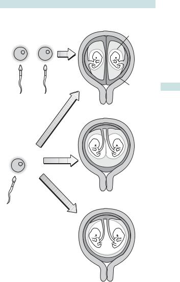
Dizygotic twins result from two separate ova being fertilized by different sperm, simultaneously implanting and developing. Consequently, these fetuses will have separate amniotic membranes and placentas (dichorionic and diamniotic—DCDA). Twins may be different sexes. This mechanism of twinning accounts for two-thirds of multiple pregnancies; this type is most affected by predisposing factors, such as age and ethnicity.
Monozygotic twins
Monozygotic twins result from division into two of a single, already developing, embryo and will be genetically identical and, therefore, always the same sex. Whether they share the same amniotic membrane and/or chorion depends on the stage of development when the embryo divides. About two-thirds are monochorionic diamniotic.
See Fig. 2.2 for an explanation of the mechanism of twinning.
Timing of division in monozygotic twins
•<3 days lDCDA 30%.
•4–7 days lmonochorionic, diamniotic (MCDA) 70%.
•8–12 days lmonochorionic, monoamniotic (MCMA) <1%.
•>12 days lconjoined twins (very rare).
The worldwide monozygotic twining rate appears to be constant at about 3.5 per 1000. However, the rate is slightly greater than expected with IVF treatment.
Diagnosis
There are several signs and symptoms associated with multiple pregnancy including:
•Hyperemesis gravidarum.
•Uterus is larger than expected for dates.
•Three or more fetal poles may be palpable at >24wks.
•Two fetal hearts may be heard on auscultation.
However, the vast majority are diagnosed on ultrasound in the 1st trimester (at a dating or nuchal translucency scan). As most women in the UK now have USS at some stage in their pregnancy, diagnosis is rarely missed.
Chorionicity
Determining chorionicity allows risk stratification for multiple pregnancy and is best done by ultrasound in the 1st trimester or early in the 2nd. The key indicators are:
•Obviously widely separated sacs or placentae—DC.
•Membrane insertion showing the lambda (λ) sign—DC.
•Absence of λ sign <14wks diagnostic of MC.
•Fetuses of different sex—DC (dizygotic).
(a) |
Dichorionic |
|
diamniotic twins |
|
Dizygotic twins |
|
MULTIPLE PREGNANCY: TYPES 75 |
|
Amnion |
Chorion
<3 days
Monochorionic diamniotic
(b)
Monozygotic twins |
4–7 days |
Monochorionic monoamniotic
8–12 days
(c)
Fig. 2.2 Mechanism of twinning. Dizygotic twins (a) are always DCDA, but with monozygotic twins (a, b, and c), the type will depend on the time of the division of the conceptus.
76 CHAPTER 2 Pregnancy complications
Multiple pregnancy: antenatal care
•All multiple pregnancies are by definition ‘high risk’ and the care should be consultant led.
•Establish chorionicity—most accurately diagnosed in 1st trimester (absence of sign diagnostic), so an early USS should be considered with any indications of multiple pregnancy (e.g. fundus palpable before 12wks or exaggerated symptoms of early pregnancy).
•Routine use of iron and folate supplements should be considered.
•A detailed anomaly scan should be undertaken.
•Advise aspirin 75microgram od if additional risk factors for pre eclampsia.
•Serial growth scans at 28, 32, and 36wks for DC twins.
•More frequent antenatal checks because of irisk of pre-eclampsia.
•Discuss mode, timing and place of delivery.
•Establish presentation of leading twin by 34wks.
•Offer delivery at 37–38wks: induction or lower segment Caesarean section (LSCS).
Surveillance needs to be more intensive for MC twins particularly <24wks, or higher multiples, so referral to a specialist fetal medicine team is advisable.
Preterm delivery and multiple pregnancy
See bPreterm labour: prevention and prediction, p. 100).
•Incidence increased: principal cause of morbidity and mortality.
•Predictable with transvaginal cervical scanning.
•Not thought to be preventable by cervical cerclage.
•Beneficial effect of progesterone limited at best.
Maternal risks associated with multiple pregnancy
The risks of pregnancy appear to be heightened with twins compared with singletons, leaving mothers at increased risk of:
•Hyperemesis gravidarum.
•Anaemia.
•Pre-eclampsia (5× greater risk with twins than singletons).
•Gestational diabetes.
•Polyhydramnios.
•Placenta praevia.
•Antepartum and post-partum haemorrhage.
•Operative delivery.

MULTIPLE PREGNANCY: ANTENATAL CARE 77
Fetal risks associated with multiple pregnancy
•All fetal risks increased with MC twins.
•iRisk of miscarriage: especially with MC twins.
•Congenital abnormalities more common only in MC twins including:
•neural tube defects
•cardiac abnormalities
•gastrointestinal atresia.
•IUGR: up to 25% of twins.
•Preterm labour: main cause of perinatal morbidity and mortality:
•40% twins deliver before 37wks
•10% twins deliver before 32wks.
•iPerinatal mortality:
•singletons 5:1000
•twins 18:1000
•triplets 53:1000.
•iRisk of intrauterine death (stillbirth):
•singletons 8:1000
•twins 31:1000
•triplets 84:1000.
•iRisk of disability (mainly, but not entirely, due to prematurity and low birth weight).
•iIncidence of cerebral palsy (CP):
•singletons 2:1000
•twins 7:1000
•triplets 27:1000.
•Vanishing twin syndrome: one twin apparently being reabsorbed at an early gestation (1st trimester).

78 CHAPTER 2 Pregnancy complications
Monochorionic, diamniotic twins
The shared circulation of MC twins can lead to several problems.
Twin-to-twin transfusion syndrome (TTTS)
This affects about 5–25% of MC twin pregnancies and left untreated has an 80% mortality rate. It may occur acutely at any stage or more commonly take a chronic course, which, at its worst, leads to severe fetal compromise at a gestation too early to consider delivery. It is caused by aberrant vascular anastamoses within the placenta, which redistribute the fetal blood. Effectively, blood from the ‘donor’ twin is transfused to the ‘recipient’ twin.
2 MC twins require intensive monitoring, usually in the form of serial USS every 2wks from 16–24wks and every 3wks until delivery. This is best performed in a specialist fetal medicine unit. The treatment options potentially available include:
•Laser ablation of the placental anastamoses. This method is associated with lowest risk of neonatal handicap.
•Selective feticide by cord occlusion is reserved for refractory disease.
TTTS managed by laser treatment leads to survival of at least one in 80% and both in 50%.
Selective intrauterine growth restriction
•Growth discordance, even without TTTS, is more common.
•A very variable pattern of umbilical artery Doppler signals (intermittent absent/reversed end diastolic flow: iAREDF) indicates a high risk of sudden demise.
•Treatment: if >28wks—delivery is safest; if <28wks, selective termination or laser ablation should be considered.
Termination of pregnancy issues
•Although MC twins may be discordant for structural abnormalities, genetically they are identical.
•Selective termination of pregnancy requires closure of the shared circulation so is normally performed using diathermy cord occlusion.
Twin reversed arterial perfusion (TRAP)
In this rare condition, one of an MC twin pair is structurally very abnormal with no or a rudimentary heart, and receives blood from the other (umbilical artery flow direction is reversed), which is called the ‘pump twin’.
This normal twin may die of cardiac failure, and unless the abnormal twin is very small or flow to it ceases, selective termination using radiofrequency ablation or cord occlusion is indicated.

MONOCHORIONIC, DIAMNIOTIC TWINS 79
Effects of twin-to-twin transfusion on the fetus
Donor twin
•Hypovolaemic and anaemic.
•Oligohydramnios: appear ‘stuck’ to the placenta or uterine wall.
•Growth restriction.
Recipient twin
•Hypervolaemic and polycythaemic.
•Large bladder and polyhydramnios.
•Cardiac overload and failure.
•Evidence of fetal hydrops (ascites, pleural, and pericardial effusions).
• This twin is often more at risk than the donor.
Intrauterine death of a twin
•Dichorionic: the death of one twin in the 1st trimester or early part of the 2nd does not appear to adversely affect the remaining fetus. Loss in the late part of the 2nd or 3rd trimester usually precipitates labour, with 90% having delivered within 3wks.
•Monochorionic: because of the shared circulation, subsequent death or neurological damage from hypovolaemia follows in up to 25%, where one of the pair dies. Delivery does not decrease the risk of brain injury.
Further reading
Mwww.eurofoetus.org

80 CHAPTER 2 Pregnancy complications
Multiple pregnancy: labour
•For all multiple pregnancies mode of delivery is debated.
•The second twin is at increased risk of perinatal mortality, but it is not currently the case that all twins are delivered by CS.
•For labour, the leading twin should be cephalic (780%), and there should be no absolute contraindication (e.g. placenta praevia).
•Triplets and higher-order multiples are usually delivered by CS.
•Some authorities advise CS for MC twins.
For management of labour and delivery see Box 2.4.
Intrapartum risks associated with multiple pregnancy
•Malpresentation.
•Fetal hypoxia in second twin after delivery of the first.
•Cord prolapse.
•Operative delivery.
•Post-partum haemorrhage.
•Rare:
•cord entanglement (MCMA twins only)
•head entrapment with each other: ‘locked twins’
•fetal exsanguination due to vasa praevia.

MULTIPLE PREGNANCY: LABOUR 81
Box 2.4 Management of labour and delivery for twins
•Twins are usually induced at 738wks gestation, but many will have delivered spontaneously before then.
•The woman should have IV access and a current Group and Save.
•Fetal distress is more common in twins; continuous fetal monitoring with CTG is important throughout labour.
•This becomes imperative after the first twin has delivered to avoid hypoxia in the second.
•It may be helpful to monitor the leading twin with a fetal scalp electrode and the other abdominally.
•An epidural may be helpful, especially if there are difficulties delivering the second twin, but is not essential.
•Many units choose to deliver twins in theatre as there is more space available and it provides immediate recourse to surgical intervention if required.
•Importance of support for mother cannot be overestimated.
•Leading twin should be delivered as for a singleton, but with care to ensure adequate monitoring of the second throughout.
•After delivery of first baby, the lie of the second twin should be checked and gently ‘stabilized’ by abdominal palpation while a VE is performed to assess the station of the presenting part.
•It may be helpful to have an ultrasound scanner available in case of concerns about malpresentation of the second twin.
•Once the presenting part enters the pelvis the membranes can be broken and the second twin is usually delivered within 20min of the first.
•Judicious use of oxytocin may help if the contractions diminish after delivery of the first twin.
•If fetal distress occurs in the second twin, delivery may be expedited with either forceps or ventouse.
•If this is inappropriate, the choice is between CS and breech extraction (often after internal podalic version).
•Breech extraction involves gentle and continuous traction on one or both feet, and must only be performed by an experienced obstetrician.
It is never used to deliver singleton breeches.
•As there is an increased risk of uterine atony, syntometrine and prophylactic oxytocin infusion is recommended.

82 CHAPTER 2 Pregnancy complications
Breech presentation: overview
Breech presentation occurs when the baby’s buttocks lie over the maternal pelvis. The lie is longitudinal, and the head is found in the fundus. This becomes decreasingly common with gestation, such that breech presentation at term occurs with only 3–4% of fetuses, but is much more common preterm.
Types of breech
•Extended breeches (70%):
•both legs extended with feet by head; presenting part is the buttocks.
•Flexed breeches (15%):
•legs flexed at the knees so that both buttocks and feet are presenting.
•Footling breeches (15%):
•one leg flexed and one extended.
Causes and associations of breech presentation
•Idiopathic (most common).
•Preterm delivery.
•Previous breech presentation.
•Uterine abnormalities, e.g. fibroids and Müllerian duct abnormalities.
•Placenta praevia and obstructions to the pelvis.
•Fetal abnormalities.
•Multiple pregnancy.
Consequences of breech presentation
Fetal
•There is an increased risk of hypoxia and trauma in labour.
•Irrespective of the mode of delivery, neonatal and longer-term risks are increased. The reasons for this are incompletely understood but may be due in part to:
•association with congenital abnormalities
•many preterm babies are breech at the time of delivery.
Maternal
Most breeches are delivered by CS.
Diagnosis of breech presentation
•Before 36wks breech presentation is not important unless the woman is in labour.
•Breech presentation is commonly undiagnosed before labour (30%).
•On examination:
•lie is longitudinal
•the head can be palpated at the fundus
•the presenting part is not hard
•the fetal heart is best heard high up on the uterus.
•Ultrasound confirms the diagnosis and should also assess growth and anatomy because of the association with fetal abnormalities.
This page intentionally left blank

84 CHAPTER 2 Pregnancy complications
External cephalic version
External cephalic version (ECV) is a method for manually turning a breech or transverse presentation into a cephalic one. It is performed from 36wks in nulliparous women and 37wks in multiparous ones. The intention is to reduce the need for delivery by CS.
•Method: after USS, a forward roll technique is used. The breech is elevated from the pelvis, and pushed to the side where the back is; the head is then pushed forward and the roll completed. Excessive force must not be used. After the attempt, CTG is performed and anti-D given if the mother is Rh –ve. See Fig. 2.3.
•Efficacy: the success rate is about 50%. Spontaneous reversion to breech presentation occurs in 3%. Attempting ECV halves the chance of non-cephalic presentation at delivery and greatly reduces the risk of CS. Nulliparity, difficulty palpating the head, high uterine tone, an engaged breech, less amniotic fluid, and white ethnicity are associated with more difficulty.
•Facilitation: success rates are increased by the use of tocolysis, such as salbutamol, given either electively or if a first attempt fails. Epidural or spinal analgesia are not usually used.
•Safety: approximately 0.5% will require immediate delivery by CS due to fetal heart rate abnormalities or vaginal bleeding. Theoretical or minor risks include pain, precipitation of labour, placental abruption, fetomaternal haemorrhage, and cord accidents. The chances of CS during labour are slightly higher than with a fetus that has always been cephalic.
•Other methods: so-called natural methods of version (postural methods, acupuncture, moxabustion) remain unproven.
Contraindications to ECV
Absolute
•Caesarean delivery already indicated.
•Antepartum haemorrhage.
•Fetal compromise.
•Oligohydramnios.
•Rhesus isoimmunization.
•Pre-eclampsia.
Relative
•One previous CS.
•Fetal abnormality.
•Maternal hypertension.

EXTERNAL CEPHALIC VERSION 85
1. Gently disimpact the breech from the pelvis, guiding it towards the iliac fossa.
2. Continue to guide the breech upwards until the baby is transverse, then gentle pressure on the occiput helps to complete the forward roll.
Fig. 2.3 External cephalic version.
86 CHAPTER 2 Pregnancy complications
Breech presentation: delivery
Mode of delivery of breech presentation
•If ECV is declined or fails, or the breech is undiagnosed, the parents should be appraised of the evidence about breech birth.
•Most breech deliveries in the UK, USA, and Europe are by CS, because meta-analysis of RCTs has shown this to reduce neonatal mortality and short-term morbidity, although not longer-term morbidity.
•Elective CS appears protective even where the ideal conditions for vaginal delivery are present and the attendant is highly experienced.
•This policy does not increase maternal morbidity because attempting a
vaginal delivery still carries a considerable risk of emergency CS, which is a more risky procedure.
•These findings have been criticized because of the trials’ methodology and the way that breech labours were managed; nevertheless this is the best evidence currently available.
•The breech in advanced labour, or who is a second twin, or preterm is not necessarily best delivered by CS.
Vaginal delivery of the breech fetus
Knowledge and experience of this remains important because breech delivery requires skill and will occasionally be inevitable because of diagnosis in advanced labour or because of the mother’s wishes.
Ideal selection for vaginal breech delivery
•Fetus is not compromised.
•Estimated fetal weight is <4kg.
•Spontaneous onset of labour.
•Extended breech presentation.
•Non-extended neck.
There is a risk of cord prolapse which is greatest in footling breeches (15%).
Oxytocin augmentation is not advised and failure of the buttocks to descend after full dilatation is a sign that delivery may be difficult.

BREECH PRESENTATION: DELIVERY 87
Vaginal breech delivery technique
•Maternal effort should be delayed until the buttocks are visible.
•After delivery of the buttocks the baby is encouraged to remain back upwards but should not otherwise be touched until the scapula is visible.
•The arms are then hooked down by the index finger at the fetal elbow, bringing them down the baby’s chest.
•The body is then allowed to hang.
•If the arms are stretched above the chest and cannot be reached, Lovset’s manoeuvre is required.
•This involves placing the hands around the body with the thumbs on the sacrum and rotating the baby 180° clockwise and then counterclockwise with gentle downward traction.
•This allows the anterior shoulder and then the posterior shoulder to enter the pelvis and for the arm to be delivered from below the pubic arch.
•When the nape of the neck is visible, delivery is achieved by placing two fingers of the right hand over the maxilla and two fingers of the left at the back of the head to flex it (Mauriceau–Smellie–Veit manouevre) and maternal pushing is encouraged.
•If this fails to deliver the head, forceps should be applied before the next contraction.
•Delivery of the head should be gentle and controlled to avoid rapid decompression which could cause intracranial bleeding.
•The upright position for delivery is advocated by some experienced attendants but there is no proof that this makes delivery safer.
Further reading
Hannah ME, Hannah WJ, Hewson SA, et al. (2000). Planned Caesarean section versus planned vaginal birth for breech presentation at term: a randomized multi-centre trial. Lancet 356: 1375–83.

88 CHAPTER 2 Pregnancy complications
Transverse, oblique, and unstable lie
Definition
•A transverse or oblique lie occurs when the axis of the fetus is across the axis of the uterus. This is common before term, but occurs in only 1% of fetuses after 37wks.
•Unstable lie occurs when the lie is still changing, usually several times a day, and may be transverse or longitudinal lie, and cephalic or breech presentation. See Fig. 2.4.
Assessment
•Ascertain stability from the history: has the presentation been changing?
•Ascertain fetal lie by palpation.
•Neither the head nor buttocks will be presenting.
•Also assess the laxity of the uterine wall.
•Does the presenting part move easily?
•Ultrasound should be performed to help ascertain the cause.
Management of abnormal lie
•Admission to hospital from 37wks is usually recommended with unstable lie, so that CS can be carried out if labour starts or the membranes rupture and the lie is not longitudinal.
•Whilst the lie remains unstable, the woman should remain in hospital.
•With increasing gestation the lie will usually revert to longitudinal and in these circumstances she can be discharged.
•If the lie does not stabilize, a CS is usually performed at 41wks.
•Some advocate a stabilizing induction whereby the fetus is turned to cephalic and an amniotomy immediately performed. This requires expertise.
•If the lie is stable but not longitudinal, a CS should be considered at 39wks.
Risks of abnormal lie
•Labour with a non-longitudinal lie will result in obstructed labour and potential uterine rupture.
•Membrane rupture risks cord prolapse because with longitudinal lie, the presenting part usually prevents descent of the cord through the cervix.

TRANSVERSE, OBLIQUE, AND UNSTABLE LIE 89
Longitudinal |
Oblique lie |
Transverse lie |
|
|
|||
(normal) lie |
|
|
|
Fig. 2.4 Longitudinal, oblique, and transverse fetal lie.
Causes and associations of abnormal fetal lie
•Multiparity (particularly >para 2) with lax uterus (common).
•Polohydramnios.
•Uterine abnormalities, e.g. fibroids and Müllerian duct abnormalities.
•Placenta praevia and obstructions to the pelvis.
•Fetal abnormalities.
•Multiple pregnancy.

90 CHAPTER 2 Pregnancy complications
Abdominal pain in pregnancy: pregnancy related (<24wks)
The diagnosis of acute abdominal pain in pregnancy can be challenging. It is often difficult to differentiate between gynaecological, non-gynaecological, and pregnancy-related causes of abdominal pain. Some of the routine surgical investigations and procedures carry a risk to the fetus but this needs to be balanced against the risk of delayed diagnosis and treatment which would be harmful to both mother and child.
Miscarriage (see bChapter 15, p. 530)
• Can be associated with lower abdominal dull ache to severe continuous or colicky pain.
•Vaginal bleeding is present in most cases.
•Positive urine pregnancy test, pelvic examination, and USS are helpful in diagnosis.
Ectopic pregnancy (see bChapter 15, p. 534)
•Usually unilateral lower abdominal pain at <12wks gestation.
•Associated with brownish vaginal bleeding.
•Shoulder tip pain is suggestive of haemoperitoneum (bleeding ectopic).
•Serum hCG, USS, and laparoscopy are diagnostic.
Constipation
Physiological changes in pregnancy result in the slowing of gut peristalsis.
Signs and symptoms Varied but colicky lower abdominal pain (L>R) is the most common.
Management
•High-fibre diet.
•Osmotic laxatives.
•Glycerin suppositories.
Round ligament pain
This pain is attributed to stretching of the round ligaments.
Incidence 20–30% of pregnancies.
Signs and symptoms
•Commonly presents in 1st and 2nd trimester.
•Pain is often bilateral and located on the outer aspect of the uterus.
•Radiating to the groin.
•Aggravated by movement (especially getting up from a chair or turning over in bed).
Treatment
•Reassurance.
•Simple analgesia.
•Support belts may help.

ABDOMINAL PAIN: PREGNANCY RELATED (<24WKS) 91
Urinary tract infection
UTIs are more common in pregnancy and are an important association of preterm labour.
Signs and symptoms
•Suprapubic/lower abdominal pain.
•Dysuria, nocturia, and frequency.
Investigations
•Urine dipstick:
•nitrites strongly suggest a UTI
•blood, leucocytes, and protein raises index of suspicion.
•Midstream sample urine (MSU).
Management
•Antibiotics.
•Analgesia.
•iFluid intake.
Fibroids—red degeneration
Uterine fibroids occur in 20% of women of reproductive age. They may increase in size during pregnancy, compromising blood supply to central areas and causing pain. This is known as red degeneration.
Incidence
15% of pregnant women who have fibroids.
Signs and Symptoms
•Usually occurs between the 12th and 22nd week of pregnancy.
•Constant pain localized to one area of the uterus coinciding with the site of the fibroid (may be severe pain).
•May have a low-grade pyrexia.
Investigations
•USS (identifies fibroids but cannot confirm red degeneration).
•FBC (may show leucocytosis).
Treatment
•Analgesia (pain should resolve in 4–7 days; however, it may be severe and prolonged, so advice from pain specialists should be sought).
2 Placental abruption differs in that the fibroid uterus is soft except at the site of the fibroid and the FH is normal.
Myomectomy must not be performed in pregnancy as it will bleed ++ (the only exception being for a torted pedunculated fibroid).

92 CHAPTER 2 Pregnancy complications
Abdominal pain in pregnancy: pregnancy related (>24wks)
Labour
Signs and symptoms
•Usually presents with regular painful contractions.
•Preterm labour may present with a history of vague abdominal pain which the woman may not associate with uterine activity.
Consider a VE in pregnant women with abdominal pain.
Braxton Hicks contractions
These are spontaneous benign contractions of the uterus, commonly occurring in the 3rd trimester.
Signs and symptoms
•Painless and infrequent tightenings of the uterus.
•VE reveals uneffaced and closed cervix.
Investigations
•Exclusion of precipitants of preterm labour (dipstick/MSU for UTI).
•Fibronectin assay if uncertain whether preterm labour (see bPreterm labour: overview, p. 98).
Treatment
• Reassurance.
Symphysis pubis dysfunction
Signs and symptoms
Pubic pain relating to upper thighs and perineum.
•Aggravated by movement.
•Difficulty walking resulting in a waddling gait.
Treatment
• Analgesia and physiotherapy.
Reflux oesophagitis
Relaxation of the oesophageal sphincter occurs in pregnancy and the pressure of the gravid uterus on the distal end of the oesophagus results in an increased incidence of reflux oesophagitis. Gastric ulceration is less common due to decreased gastric acid secretion.
Incidence
60–70% of pregnant women.
Risk factors
•Polyhydramnios.
•Multiple pregnancy.
Signs and symptoms
• Epigastric/retrosternal burning pain exacerbated by lying flat.

ABDOMINAL PAIN: PREGNANCY RELATED (>24WKS) 93
Management
•Exclude pre-eclampsia.
•Antacids, H2 receptor antagonists.
•Dietary and lifestyle advice (avoidance of supine position).
Uterine rupture
This usually occurs during labour but has been reported antenatally.
Risk factors
•Previous CS or other uterine surgery.
•Congenital abnormalities of the uterus.
•Induction or use of oxytocin in labour.
•Failure to recognize obstructed labour.
Signs and symptoms
•Tenderness over sites of previous uterine scars.
•Fetal parts may be easily palpable.
•Fetus not palpable on VE.
•Vaginal bleeding may be evident.
•Signs of maternal shock may be present.
CTG may show fetal distress and change in apparent uterine activity (contractions may seem to disappear on the tocograph).
Investigations
•FBC.
•Cross-match blood.
Management
•Maternal resuscitation.
•Urgent laparotomy to deliver fetus and repair uterus.
Other causes of abdominal pain in pregnancy
•Placental abruption.
•Pre-eclampsia/HELLP.

94 CHAPTER 2 Pregnancy complications
Abdominal pain in pregnancy: bowel related
Appendicitis
This is the most common surgical emergency in pregnant patients. Its incidence is 1:1500–2000 pregnancies with equal frequency in each trimester. Pregnant women have the same risk of appendicitis as non-pregnant women.
Signs and symptoms
• Classically periumbilical pain shifting to right lower quadrant.
Pain moves towards the right upper quadrant during the 2nd and 3rd trimesters due to displacement of the appendix by a gravid uterus.
• Nausea and vomiting.
• Anorexia.
• Guarding and rebound tenderness present in 70% of patients.
Rovsing’s sign and fever are often absent in the pregnant patient.
Investigations
•White cell count (WCC) and C-reactive protein (CRP) are often i.
•USS: to exclude other causes of pain; CT/MRI may be considered.
Management
Diagnostic laparoscopy/laparotomy and appendicectomy.
Fetal loss is 3–5% with an unruptured appendix, ito 20% if ruptured.
Intestinal obstruction
It is the third most common non-obstetric reason for laparotomy during pregnancy. It complicates 1:1500–3000 pregnancies. Incidence increases as the pregnancy progresses. Adhesions are the commonest cause.
Signs and symptoms
•Acute abdominal pain.
•Vomiting.
•Constipation.
•Pyrexia.
Diagnosis
•Erect abdominal X-ray (AXR) showing gas-filled bowel with little gas in large intestine.
•USS (abdominal and pelvic).
Treatment
•Conservative treatment (‘drip and suck’).
•Surgery for any acute obstructive cause or when not responding to conservative management.

ABDOMINAL PAIN IN PREGNANCY: BOWEL RELATED 95
Causes of intestinal obstruction
•Adhesions.
•Volvulus.
•Intussusception.
•Hernia.
•Neoplasm.
96 CHAPTER 2 Pregnancy complications
Abdominal pain in pregnancy: other causes
Acute cholecystitis
This is the second most common surgical condition in pregnancy (progesterone diminishes smooth muscle tone and predisposes to cholestasis leading to gallstone formation). The incidence of gallstones is 7% in nulliparous and 19% in multiparous women. The incidence of acute cholecystitis is 1–8:10 000 pregnancies.
Signs and symptoms
•Colicky epigastric/right upper quadrant pain.
•Nausea and vomiting.
•Murphy’s sign may be positive in acute cholecystits.
•Jaundice (indicating obstruction of the common bile duct).
•Signs of systemic infection (fever and tachycardia).
Investigations
•FBC, LFTs, CRP (WCC and alkaline phosphatase are iin pregnancy).
•iBilirubin (identify patients with concomitant biliary tree obstruction).
•USS biliary tract (may demonstate calculi or a dilated biliary tree).
Management
•Conservative approach is the most common management.
•Analgesics and antiemetics.
•Hydration.
•Antibiotics.
•Cholecystectomy preferably by laparoscopic approach may be indicated in patients with recurrent biliary colic, acute cholecystitis, and obstructive cholelithiasis (usually after delivery).
Adnexal torsion
This occurs when an enlarged ovary twists on its pedicle.
Torsion of the ovary and other adnexal structures is more common in pregnant than non-pregnant women.
Signs and symptoms
•Sudden-onset unilateral colicky lower abdominal pain.
•Nausea and vomiting.
•There may be systemic symptoms such as fever.
Investigations
•WCC and CRP: may be elevated.
•USS of pelvis may show an adnexal mass and Doppler studies may show impaired blood flow.
Management
If suspected, urgent laparotomy should be performed to either remove or untwist the adnexa. This may either preserve the ovary or present a nonviable ovary from becoming gangrenous.

ABDOMINAL PAIN IN PREGNANCY: OTHER CAUSES 97
Pancreatitis
This occurs more frequently in the 3rd trimester and immediate post-partum period. It can occur in early pregnancy associated with gallstones.
Although rare, it is more common in pregnancy than in non-pregnant women of a similar age.
Incidence 1:5000 pregnancies.
Risk factors
•Gallstone disease.
•High alcohol intake.
•Hyperlipidaemia.
Signs and symptoms
•Epigastric pain commonly radiating to the back.
•Pain exacerbated by lying flat and relieved by leaning forwards.
•Nausea and vomiting.
Investigations
•Serum amylase and lipase levels.
•USS to establish presence of gallstones.
Management
Conservative treatment is the mainstay:
•IV fluids.
•Electrolyte replacement.
•Parenteral analgesics, e.g. morphine (pethidine is contraindicated).
•Bowel rest with or without nasogastric suction.
Early surgical intervention is recommended for gallstone pancreatitis in all trimesters as >70% of patients will relapse before delivery.
•Laparoscopic/open cholecystectomy.
•Endoscopic reterograde cholangio-pancreatography (ERCP) has a limited role in pregnancy because of radiation exposure to the fetus.
If pancreatitis is severe, liaise with high dependancy unit/intensive care unit (HDU/ITU).
Non-abdominal causes of abdominal pain
Other conditions unrelated to abdominal structures may also present with abdominal pain:
•Lower lobe pneumonia.
•Diabetic ketoacidosis.
•Sickle cell crisis.
2 Women with social problems and domestic abuse may repeatedly attend with undiagnosable pain and it is important to ask them about this directly but sympathetically.

98 CHAPTER 2 Pregnancy complications
Preterm labour: overview
Preterm birth is defined as delivery between 24 and 37wks.
•Delivery <34wks is more useful as adverse outcomes are rare after then.
•1/3 is medically indicated (e.g. PET), and 2/3 spontaneous.
•Accounts for 5–10% of births but 750% of perinatal deaths.
•It also causes long-term handicap—blindness, deafness, and cerebral palsy. The risk is higher the earlier the gestation.
•The incidence is iover the years.
•>50% of women with painful preterm contractions will not deliver preterm: fetal fibronectin/transvaginal USS may help in diagnosis.
Risk factors for preterm delivery
•Previous preterm birth or late miscarriage.
•Multiple pregnancy.
•Cervical surgery.
•Uterine anomalies.
•Medical conditions, e.g. renal disease.
•Pre-eclampsia and IUGR (spontaneous and iatrogenic).
Acute preterm labour
•Preterm labour associated with cervical weakness (avoid the term ‘incompetence’) classically presents with increased vaginal discharge, mild lower abdominal pain, and bulging membranes on examination.
•Preterm labour associated with factors such as infection, inflammation, or abruption presents with lower abdominal pain, painful uterine contractions, and vaginal loss.
•Spontaneous rupture of membranes (SROM) is a common presentation of/antecedent for preterm labour.
2 In practice it is often less clear-cut than this, and infection and cervical weakness are related and often coexist.
History
•Ask about pain/contractions—onset, frequency, duration, severity.
•Vaginal loss: SROM or PV bleeding.
•Obstetric history (check hand-held notes).
Examination
•Maternal pulse, temperature, respiratory rate.
•Uterine tenderness (suggests infection/abruption).
•Fetal presentation.
•Speculum: look for blood, discharge, liquor. Takes swabs.
•Gentle VE.
Investigations
•FBC, CRP (raised WCC and CRP suggest infection).
•Swabs, MSU.
•USS for fetal presentation (malpresentation common) and estimated fetal weight (EFW).
•Consider fetal fibronectin/transvaginal USS if available
(see bMethods for prediction of preterm labour, p. 101).

PRETERM LABOUR: OVERVIEW 99
Management of preterm labour
•Establish whether threatened or ‘real’ preterm labour:
•transvaginal cervical length scan (>15mm unlikely to labour)
•fibronectin assay: if –ve, unlikely to labour.
•Admit if risk high.
•Inform neonatal unit.
•Arrange in utero transfer if no suitable beds available.
•Check fetal presentation with USS.
•Steroids (12mg betametasone IM—two doses 24h apart).
2Antenatal steroids reduce rates of respiratory distress, intraventricular haemorrhage, and neonatal death (Royal College of Obstetricians and Gynaecologists (RCOG)) grade A recommendation).
•Consider tocolysis (drug treatment to prevent labour and delivery) not >24hrs.
•Allow time for steroid administration and/or inutero transfer.
•Currently used tocolytics include nifedipine, and atosiban IV.
X Aim should be not just prolongation of gestation (a surrogate measure) but improvement in perinatal morbidity and mortality. Trials of tocolysis have not shown improvement in these substantive outcome measures, so some prefer to avoid them.
•Liaison with senior obstetricians and neonatologists is essential, especially at the margins of viability (23–26wks). A clear plan needs to be made about:
•mode of delivery
•monitoring in labour
•presence of pediatrician/appropriate intervention at delivery.
•Give IV antibiotics but only if labour confirmed.
Further reading
RCOG. (2004). Green-top guideline no.7. Antenatal corticosteroids to prevent respiratory distress syndrome. Mwww.rcog.org

100 CHAPTER 2 Pregnancy complications
Preterm labour: prevention and prediction
Prevention
Treatment of bacterial vaginosis (BV)
Some evidence suggests this may reduce the incidence of preterm prelabour rupture of membranes (PPROM) and low birth weight in women with previous preterm birth. Clindamycin rather than metronidazole is used.
Progesterone
•In high risk women (e.g. previous history of late miscarriage/preterm birth), reduces recurrence.
•In low risk women with a short cervix, reduces preterm birth by about 50%. As a result, screening for preterm birth with cervical scanning may become universal.
•Effect absent/very limited in twin pregnancies.
•Cream or pessaries used.
Cervical sutures (cerclage)
X May be of benefit in selected cases. Can be inserted vaginally or, in extreme cases, abdominally. Not thought to be useful in multiple pregnancies.
•Elective (women with previous loss from cervical weakness).
•Ultrasound-indicated (in response to short cervix on transvaginal scan (TVS)).
•Rescue (in response to cervical dilatation).
Cervical pessary
These are used more often in Europe but evidence suggests they are effective.
Reduction of pregnancy number
Selective reduction of triplet or higher-order multiple pregnancies (to 2) reduces the risk of preterm labour while slightly increasing the risk of early miscarriage.

PRETERM LABOUR: PREVENTION AND PREDICTION 101
Methods for prediction of preterm labour
Transvaginal USS of cervix
•In asymptomatic women with a singleton pregnancy:
•risk of delivering before 32wks is 4% if cervix is >15mm long at 23wks
•increasing exponentially to 78% if cervix is 5mm.
•In symptomatic women with a singleton pregnancy:
•cervix <15mm, risk of delivery within 7 days is 49%
•cervix >15mm, risk of delivery within 7 days <1%.
Fetal fibronectin (FFN)
•FFN is a protein not usually present in cervicovaginal secretions at 22–36wks.
•Those with a +ve FFN test are more likely to deliver (test for FFN with swab and commercially available kit).
•Predicts preterm birth within 7–10 days of testing (+ve likelihood ratio (LR) of 5.42 and –ve LR of 0.25).

102 CHAPTER 2 Pregnancy complications
Preterm prelabour rupture of membranes: overview
•This complicates 1/3 of preterm deliveries.
•About 1/3 is associated with overt infection (more common at earlier gestations).
History
•Ask about vaginal loss.
•Gush.
•Constant trickle or dampness.
Chorioamnionitis may cause few symptoms but is associated with significant neonatal morbidity and mortality.
1 Chorioamnionitis is also associated with significant risks to the mother.
Features suggestive of chorioamnionitis
History:
•Fever/malaise.
•Abdominal pain, including contractions.
•Purulent/offensive vaginal discharge. Examination:
•Maternal pyrexia and tachycardia.
•Uterine tenderness.
•Fetal tachycardia.
•Speculum: offensive vaginal discharge—yellow/brown.
2Avoid VE as this increases the risk of introducing infection.
Investigations
•FBC, CRP (raised WCC and CRP indicate infection).
•Swabs (high vaginal swab (HVS), low vaginal swab (LVS)).
•MSU.
•USS for fetal presentation, EFW, and liquor volume.

PRETERM PRELABOUR RUPTURE OF MEMBRANES: MANAGEMENT 103
Preterm prelabour rupture of membranes: management
•If evidence of chorioamnionitis:
•steroids (betametasone 12mg IM)
•deliver whatever the gestation
•broad spectrum antibiotic cover.
•If no evidence of chorioamnionitis, manage conservatively:
•admit
•inform special care baby unit (SCBU) and liaise with neonatologists
•steroids (12mg betametasone IM—two doses 24h apart)
•antibiotics (erythromycin).
2Use of antibiotics reduces major markers of neonatal morbidity but without long-term benefits. The ORACLE trial showed erythromycin to be beneficial.
Co-amoxiclav is associated with an increased risk of necrotizing enterocolitis (NEC) and should be avoided.
Prognosis
Depends on:
•Gestation at delivery.
•Gestation at PPROM:
•PPROM at <20wks—few survivors
•PPROM at >22wks—survival up to 50%.
•Reason for PPROM:
•prognosis better if PPROM secondary to invasive procedure (e.g. amniocentesis), rather than spontaneous.
Risks to fetus from PPROM
•Prematurity.
•Infection.
•Pulmonary hypoplasia.
•Limb contractures.
Table 2.2 Data from EPICure study (cohort of babies born in the UK and Ireland in 1995)
Gestation |
Survival to discharge |
Survival (severe disability) |
(weeks) |
(as % of live births) |
(as % of live births) |
|
|
|
<23 |
1 |
50 |
23–23+6 |
11 |
31 |
24–24+6 |
26 |
24 |
25–25+6 |
44 |
22 |
|
|
|
Further reading
Costeloe K, Hennessy E, Gibson AT, et al. (2000). The EPICure study: outcomes to discharge from hospital for babies born at the threshold of viability. Pediatrics 106(4): 659–71.
Kenyon SL, Taylor DJ, Tarnow-Mordi W; ORACLE Collaborative Group. (2001). Broad-spectrum antibiotics for preterm, prelabour rupture of fetal membranes: the ORACLE 1 randomised trial.
The Lancet 357(9261): 979–88.

104 CHAPTER 2 Pregnancy complications
Prolonged pregnancy: overview
Prolonged pregnancy is a cause of anxiety for both women and obstetricians. It is a common occurrence and is a recognized cause of increased fetal morbidity and mortality.
Definition of prolonged pregnancy
According to the International Federation of Gynaecology and Obstetrics (FIGO), prolonged pregnancy is defined as any pregnancy that exceeds 42wks (294 days) from the first day of the LMP in a woman with regular 28-day cycles. Different terminologies are used generally in day-to-day practice, such as postdates, post-term, and postmaturity.
Incidence
The incidence of pregnancy lasting 42wks or more is 3–10%. With one previous prolonged pregnancy there is a 30% chance of another one. With a history of two this rises to 40%. Incidence also varies depending on whether EDD is based on LMP or dating USS. Women who book in the 1st trimester and have an early dating scan have an incidence of prolonged pregnancy of <5%.
Dates cannot be relied upon in the following circumstances:
•Uncertainty of LMP (10–30% of women).
•Irregular periods.
•Recent use of COCP.
•Conception during lactational amenorrhea.
Maternal risks
•Maternal anxiety and psychological morbidity.
•Increased intervention:
•induction of labour
•operative delivery with irisk of genital tract trauma.

PROLONGED PREGNANCY: OVERVIEW 105
Fetal risks of prolonged pregnancy
Perinatal mortality i after 42wks of gestation
•Intrapartum deaths are 4 times more common.
•Early neonatal deaths are 3 times more common.
Other risks
•Meconium aspiration and assisted ventilation.
•Oligohydramnios.
•Macrosomia, shoulder dystocia, and fetal injury.
•Cephalhaematoma.
•Fetal distress in labour.
•Neonatal—hypothermia, hypoglycaemia, polycythaemia, and growth restriction.
Fetal postmaturity syndrome
•This is used to describe post-term infants who show signs of intrauterine malnutrition.
•The neonate has a scaphoid abdomen, little subcutaneous fat on the body or limbs, peeling skin over the palm and feet, overgrown nails, and an anxious, alert look.
•The baby’s skin is also stained with meconium.
•This condition constitutes only a small proportion of babies born after 42wks.
These features can be seen at an earlier gestation in babies with IUGR. Hence, the term prolonged pregnancy is preferred to postmaturity for pregnancy beyond 42wks.
106 CHAPTER 2 Pregnancy complications
Prolonged pregnancy: management
•Attempt to confirm the EDD as accurately as possible: EDD based on 1st trimester USS is accurate to within a week.
•Assess any other risk factors which may be an indication to induce close to the EDD:
•pre-eclampsia
•diabetes
•antepartum haemorrhage
•IUGR associated with placental insufficiency.
•Offer ‘stretch and sweep’ at 41wks.
•Offer induction of labour between 41 and 42wks:
•this slightly reduces perinatal mortality
•it also reduces the risk of CS
•but it ‘medicalizes’ many labours
•if declined, ensure adequate fetal surveillance.
Fetal monitoring
This should include an initial USS assessment of growth and amniotic fluid volume. Women should be offered daily CTGs after 42wks. They should also be advised to report any decrease in fetal movements.
(See bMonitoring the high-risk fetus: cardiotocography, p. 153.)
Counselling
Most units in the UK advise induction of labour by 42wks because of the increased perinatal mortality and morbidity beyond this time. However, many women regard elective induction of labour as interference with the natural phenomenon of childbirth. It is therefore important to discuss the issue sensitively and respect the woman’s decision. Written information should be provided clearly outlining the arguments for and against induction to ensure the woman is able make an informed decision. Any specific risk factors complicating her pregnancy, such as pre-eclampsia, should be clearly explained and the conversation carefully documented in the notes.
Those mothers who prefer to await spontaneous onset of labour should have appropriate counselling regarding increased fetal mortality and morbidity. They should also have adequate fetal surveillance, which should include a USS assessment of fetal growth and liquor volume.
Further reading
Department of Health (1993). Changing childbirth, report of the Expert Maternity Group. Cumberledge Report. HMSO, London.