


Chapter 14 |
457 |
|
|
Gynaecological anatomy and development
Gynaecological history: overview 458 Gynaecological history: other relevant details 460 Gynaecological examination 462
Anatomy: female reproductive organs 464 Anatomy: blood supply and relationship to other
structures 466
Anatomy: external genitalia 468 Female genital mutilation: overview 470
Female genital mutilation: management 471 Malformations of the genital tract: overview 472 Malformations of the genital tract: management 474 Disorders of sex development 476
Congenital adrenal hyperplasia 480 Androgen insensitivity syndrome 482 Disorders of growth and puberty 484
Delayed puberty and primary amenorrhoea 486 Vaginal discharge: in childhood 488
Vaginal discharge: in adolescence 490
Dermatological conditions in children and adolescents 492 Gynaecological disorders: in adolescence 494 Gynaecological cancers: in childhood 496
Fertility implications of childhood cancer 498

458 CHAPTER 14 Gynaecological anatomy and development
Gynaecological history: overview
2 Always introduce yourself fully and explain what you are going to do; patients are often very apprehensive and nervous.
Personal information
•Name, date of birth, age.
•Relationship status.
•Occupation.
•Partner’s details and occupation (relevant in subfertility patients).
Current problem
•Description of the problem.
•Severity, duration, relationship to menstrual cycle.
•Aggravating and relieving factors.
•Any previous investigations or treatment.
Menstrual history
• Date of first day of LMP.
1Always think: is this patient pregnant or at risk of pregnancy?
Every woman you see (10–60yrs old) should be considered potentially pregnant until proved otherwise—then you will not miss it!
•Age at menarche/menopause.
•Menstrual pattern (number of days bleeding/length of cycle).
•Amount/character of bleeding (flooding, clots, double protection).
1Always ask about intermenstrual (IMB) + postcoital bleeding (PCB).
1Always ask about any postmenopausal bleeding (PMB).
• Any associated pain + pattern (dysmenorrhoea). Has this changed?
•Ask about pain at other times including dyspareunia.
•Current and recent contraception (or not!) and details.
•Current/future pregnancy plans—this may alter/limit therapeutic options, as many treatments are contraceptive.
Past obstetric history
All pregnancies must be recorded, including successful ones, miscarriages, ectopic pregnancies, TOPs, and molar pregnancies. Outcomes, gestation and mode of delivery, complications, birth weight, and current health of child(ren) should all be documented (see b Obstetric history: current pregnancy, p. 2).
Past gynaecological history
•History of any other gynaecological problems especially endometriosis, fibroids, polycystic ovaries, and subfertility.
•All previous gynaecological surgery.
•Date of last cervical smear and result. Were they always normal?

GYNAECOLOGICAL HISTORY: OVERVIEW 459
Sexual history
•Dyspareunia: superficial on penetration or deep pain.
•Sexually transmitted infections or pelvic inflammatory disease (PID).
•Any abnormal vaginal discharge.
Key things to achieve in a gynaecological history
•A clear understanding of the presenting problem(s) including effect on quality of life. Most diagnoses are clear from a good history alone.
•A good history will inform your examination and investigative rationale.
•Exclude or confirm current pregnancy or risk of it. Offer contraceptive advice to the latter if they do not desire pregnancy!
•Discover what current and near future pregnancy plans are.
•Identify women at higher risk of malignancy or other serious pathology.
1IMB, PCB, and PMB are all red flag symptoms warranting examination and investigation.
Allow disclosure of a hidden agenda. Many women will disclose other issues regarding sex or abuse or fertility concerns if you establish a good rapport; if you sense there is another concern don’t be afraid to ask (‘You seem concerned about something. Is there anything else you would like to discuss?’).

460 CHAPTER 14 Gynaecological anatomy and development
Gynaecological history: other relevant details
Micturition
General enquiry
If urinary symptoms disclosed then explore:
•Frequency (day and night).
•Pain or burning sensation (dysuria).
•Urgency.
•Urinary incontinence (stress or urge).
•Haematuria.
•Presence of ‘something coming down’ (prolapse related symptoms).
Bowel habit
General enquiry
If bowel symptoms are disclosed then explore:
•Regularity.
•Associated bloating, pain, or difficulty defecating.
•Use of laxatives.
•Any rectal bleeding.
Medical and surgical history
•All medical conditions, especially diabetes, hypertension, asthma, thrombo-embolism. Major effect if surgery is being considered.
•All previous abdominal surgery is important also.
Drugs and allergies
•Details of all medication (doses and duration of use).
•Allergies to medications and severity (anaphylaxis or rash?).
•Use of folic acid in early pregnancy.
2Consider the risks for all drugs in relation to pregnancy (see bDrugs in pregnancy, p. 261).
•Possible teratogenesis.
•Altered pharmacodynamics and pharmacokinetics.
•Toxicity in breastmilk where appropriate.
Family history
•Especially diabetes, iBP, and thrombo-embolism.
•Familial cancers should always be considered, as well as others with a genetic association including:
•breast
•ovarian
•endometrial
•bowel.

GYNAECOLOGICAL HISTORY: OTHER RELEVANT DETAILS 461
Social history
•Home conditions and relationships.
•Occupation.
•Smoking and alcohol intake.
•Lifestyle issues such as use of recreational drugs.
1Subtle symptoms of gynaecological malignancy
•Change of urinary and/or bowel habit.
•Persistent bloating.
•Non-specific discomfort.
•Even upper GI dyspeptia-type.
These should always prompt further investigation, particularly in women >50yrs, when persistent.

462 CHAPTER 14 Gynaecological anatomy and development
Gynaecological examination
General examination
•Height and weight.
•BMI (=weight (kg)/[height (m)]2). 1 iRisks with iBMI.
•General, e.g. signs of anaemia, thyroid disease.
Abdominal examination
•Inspection: skin quality, abdominal distension, surgical scars (umbilical or Pfannensteil), any visible masses or distension.
•Palpation:
•superficial palpation for guarding, tenderness, rigidity
•deep palpation for any masses; if present determine if arising from the pelvis (‘can I get below the mass?’)
•pelvic masses are compared to the equivalent sized pregnant uterus (e.g. 20/40 sized, firm, mobile fibroid uterus).
•Percussion: dull if the mass is solid, tympanic if distended bowel, shifting dullness and fluid thrill in cases of ascites.
•Auscultation: usually used postoperatively to detect bowel sounds.
Good practice for intimate examinations
•Full explanation of procedure and reasons for it should precede examination.
•Verbal consent should be obtained.
•A trained chaperone is mandatory.
•1Do NOT use partners, friends, or children as chaperones.
•The patient must be able to undress and dress in privacy and cover herself at all other times.
•Any students or extra personnel present should be introduced and consent obtained for their presence BEFORE procedure.
Further GMC guidance on intimate examinations can be found at: Mhttp://www.gmc-uk.org
Pelvic examination
•All equipment must be ready (speculum, KY jelly, swabs, cytobrush, pipelle, etc.) before the patient is exposed.
•Position the woman:
•dorsal (most common in gynaecological outpatient setting)
•lithotomy (used for vaginal surgery, the feet suspended from poles)
•Sim’s (examination of pelvic prolapse, type of the left lateral).
•Inspection: describe any swelling, inflammation, skin changes, lesions, or ulceration seen anywhere on the vulva. Do the same for the vagina and cervix once the speculum is passed.
•Speculum examination: see Box 14.1 for description of technique. Describe findings in vagina and on cervix.
2Don’t forget to take any swabs required such as HVS for vaginal pathogens and flora or endocervical for Chlamydia +/or Gonorrhoea.
• Bimanual (VE): see Box 14.2 for description of technique.

GYNAECOLOGICAL EXAMINATION 463
Box 14.1 How to do a speculum examination
•Cusco’s bivalve speculum is more frequently used, but Sim’s speculum normally used in examination of pelvic organ prolapse.
•Use a warm and well-lubricated speculum.
•Part labia minora adequately with the left hand.
•Insert speculum upwards and backwards (direction of vagina).
•Advance into vagina fully (until it cannot advance any further).
•Directly visualize as you open blades exposing cervix: only open enough to see cervix fully.
•If cervix not seen: close blades, withdraw slightly, change direction (usually more anterior), and open again.
•Speculum removal: ensure the blades are open while sliding over cervix, avoiding trapping it—watch what you are doing!
•Blades should be closed at introitus, not trapping any vagina.
Common problems to avoid
•Obvious non-familiarity with the speculum: patients spot this a mile off and will automatically tense up.
•Inadequate labial parting leads to inversion and pain (start badly and all patient confidence quickly disappears).
•The speculum is only partially inserted ‘so as not to cause pain’: the cervix will usually not be seen, leading to repeated insertion.
•Failure to find cervix first time: likely to be more anterior and closer to the introitus—pull back and move anterior as above.
•Not watching for adequate opening of blades and continuing unnecessary wide opening.
•Not having control of closure and pulling out a still-open speculum.
Box 14.2 How to do a bimanual vaginal examination
•The lubricated index and middle fingers of the right hand are introduced into the vagina. The fingers of the left hand are on the abdomen above the symphysis pubis, and the uterus and adnexae are palpated between the two hands (‘bimanual palpation’).
•Cervix:
•consistency (soft and smooth or irregular and hard)
•tenderness
•external os (?open during miscarriage).
•Uterus:
•axis (anteverted, axial, or retroverted)
•size (equivalent to gestational weeks of a gravid uterus)
•consistency (soft in a gravid uterus, firm, or hard with fibroids)
•mobility (may be fixed in endometriosis/adhesions).
•Adnexae:
•normal ovaries are usually not palpable
•any masses (cystic/solid) and describe approximate size.
•Direct digital pressure into the fornices assesses tenderness, and cervical excitation is elicited by moving the cervix laterally right and left.
2Uterine masses usually move with cervix, ovarian masses do not.
2Obese patients are usually difficult to palpate—consider ultrasound.

464 CHAPTER 14 Gynaecological anatomy and development
Anatomy: female reproductive organs
See bThe female pelvis, p. 10 for anatomy of the bony pelvis.
Vagina
•Fibromuscular tube, 7–10cm long.
•The cervix enters through the anterior wall.
•In the resting state the anterior and posterior walls are opposed.
Uterus
•Approximately 8 × 5 × 3cm in size (non-pregnant).
•Composed mainly of smooth muscle.
•Divided into the corpus and cervix uteri.
•Cylindrical and joins the uterine cavity at the internal os and the vagina at the external os.
•Anteverted in 80% of women (the remainder are retroverted or rarely axial).
Uterine (fallopian) tubes
•10cm long; lie in the upper part of the broad ligament.
•Divided anatomically into:
•isthmus (medial)—opens into the uterus at the ostia
•infundibulum (lateral) with fimbrial end closely applied to the ovary
•ampulla—in between (where fertilization takes place).
Ovaries
•Approximately 3 × 2cm during reproductive years.
•Attached to the posterior surface of the broad ligament by the mesovarium.
•Situated in the ovarian fossa at the division of the common iliac artery (the ureter runs immediately underneath).
See Fig. 14.1.
Supports of the uterus, vagina, and pelvic floor
•Middle:
•transverse cervical ligaments (cardinal ligaments)
•pubocervical ligament
•uterosacral ligaments.
•Lower:
•levator ani muscles and coccygeus
•urogenital diaphragm
•the superficial and deep perineal muscles with the perineal body.
2Defects and weaknesses of these supporting structures due to fascial tearing and denervation during parturition and surgery can cause organ prolapse and problems with urinary incontinence.

|
ANATOMY: FEMALE REPRODUCTIVE ORGANS 465 |
||
|
Mesovarium |
|
|
|
(ovarian |
|
Fallopian |
|
ligament) |
|
|
|
|
tube |
|
|
|
|
|
Uterus |
|
|
Fimbriae |
|
|
|
|
|
Corpus |
Ovary |
Mature follicle |
|
albicans |
|
|
|
|
|
|
Cervix |
Corpus luteum |
|
|
Vagina
Fig. 14.1 Basic coronal view of the female pelvis. Adapted from Pocock G, Richards C. (2004). Human physiology: the basics of medicine, 2nd edn. Oxford: OUP. By permission of Oxford University Press.
466 CHAPTER 14 Gynaecological anatomy and development
Anatomy: blood supply and relationship to other structures
Blood supply
Uterus
•The uterine artery:
•branches from the internal iliac
•runs behind the peritoneum to enter the lateral border of the uterus, through two layers of the broad ligament
•anastomoses with the ovarian and vaginal arteries.
•The venous drainage is to the internal iliac vein.
Ovaries
•The ovarian arteries: branches of the abdominal aorta from below the renal arteries.
•The right ovary drains directly into the inferior vena cava.
•The left ovary drains into the left renal vein.
Vagina
Supplied by
•Vaginal artery.
•Inferior vesical artery.
•Clitoral branch of the pudendal artery.
Urinary tract
Ureters
•Retroperitoneal throughout.
•Enter the pelvis in the base of the ovarian fossa.
•Run above the levator ani in the base of the broad ligament.
•Insert into the bladder posterolaterally.
1The ureters are very close to the uterine artery near the lateral fornix and can be injured at hysterectomy.
Bladder
•Lies anterior to the uterus.
•Three layers: serous (peritoneal), muscular (detrusor smooth muscle), and mucosa (transitional epithelium).
•Supplied by superior and inferior vesical arteries (internal iliac artery).
Rectum
•Lies posterior to the uterus (separated from it by loops of small bowel lying in the pouch of Douglas).
•A thin rectovaginal septum separates the vagina and rectum.
•Supplied by superior, middle, and inferior rectal arteries (from the inferior mesenteric, internal iliac, and pudendal arteries respectively).
See Fig. 14.2.

ANATOMY: BLOOD SUPPLY 467
Lymphatic drainage of the pelvic organs
•Vulva and lower vagina linguinofemoral lexternal iliac nodes.
•Cervix lcardinal ligaments lhypogastric, obturator, internal iliac l common iliac, and para-aortic nodes.
•Endometrium lbroad ligament liliac and para-aortic nodes.
•Ovaries linfundibulopelvic ligament lpara-aortic nodes.
1 Knowledge of lymphatic drainage is important when considering metastatic spread from genital tract cancer.
Sacrum
Fallopian
tube Ovary
Uterus |
|
|
Bladder |
Cervical |
|
Symphysis |
||
canal |
||
pubis |
||
Rectum |
||
Urethra |
|
|
Clitoris |
|
|
Labium |
Vagina |
|
majus |
|
|
|
Labium |
|
|
minus |
Fig. 14.2 Basic sagittal view of female pelvis demonstrating relationship to other pelvic organs. Adapted from Pocock G, Richards C. (2004). Human physiology: the basics of medicine, 2nd edn. Oxford: OUP. By permission of Oxford University Press.

468 CHAPTER 14 Gynaecological anatomy and development
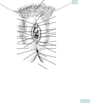
Anatomy: external genitalia
Perineum
•The area inferior to the pelvic diaphragm can be divided into:
•anterior urogenital triangle (pierced by the vagina and the urethra)
•posterior anal triangle.
•The superficial and deep perineal fascias are continuous with the labia majora and are attached:
•anteriorly to the pubic symphysis
•laterally to the body of the pubis.
•The superficial perineal muscles are:
•superficial transverse perineus
•ischiocavernosus
•bulbocavernosus.
Vulva
The external genital organs are known collectively as the vulva and are composed of the mons pubis, labia majora and minora, and clitoris.
•Labia majora: lateral boundary of the vulva from the mons pubis to the perineum.
•Labia minora:
•anteriorly join to cover the clitoris
•posteriorly form the fourchette.
•Clitoris:
•composed of erectile tissue covered by a prepuce
•supplied by a branch of the internal pudendal artery.
•The vestibule:
•lies between the labia minora and the hymen
•the urethra lies anterior in the vestibule
•posteriorly and laterally lie the vestibular or Bartholin’s glands.
See Fig. 14.3.
ANATOMY: EXTERNAL GENITALIA 469
Mons pubis
|
Clitoris |
|
Prepuce |
body |
|
|
Glans |
|
Frenum |
Urethral |
|
|
||
Labium minus |
orifice |
|
Vestibule |
||
|
||
Labium majus |
Position of |
|
|
Bartholin’s |
|
Vaginal orifice |
gland |
|
|
Posterior |
|
Hymen |
fourchette |
|
|
Perineum |
|
|
Anus |
Fig. 14.3 External female genitalia. Reproduced from Collier J, Longmore M, Brinsden M. (2006). Oxford handbook of clinical specialties, 7th edn. Oxford: OUP. By permission of Oxford University Press.

470 CHAPTER 14 Gynaecological anatomy and development
Female genital mutilation: overview
Female genital mutilation (FGM) is defined by the World Health Organization (WHO) and the United Nations (UN) agencies as “the partial or total removal of the female external genitalia or other injury to the female genital organs for non-medical reasons” (See Table 14.1 for classification and Box 14.3 for complications).
Table 14.1 Classification of FGM (WHO)
Type I ‘Sunna’ or traditional circumcision with removal of prepuce with or without part or the entire clitoris.
Type II Clitoridectomy with removal of prepuce and clitoris together with partial or total excision of labia minora.
Type III Infibulation or ‘clasp circumcision’ with removal of part or all of external genitalia and stitching/narrowing of vaginal opening leaving a small aperture for passing urine and menstrual blood.
Type IV |
Unclassified. |
Box 14.3 Complications of FGM
Immediate complications
•Death.
•Shock and pain.*
•Haemorrhage.*
•Infection including septicaemia.*
•Adjacent organ damage.*
•Acute urinary retention.
Long-term complications affecting pelvic organs
•Failure of healing.
•Recurrent UTI and renal/bladder calculus formation.*
•Urethral obstruction and difficulty in passing urine.
•Pelvic infections and abscess formation.*
•Menstrual abnormalities and associated infertility.
•Sexual dysfunction.*
•Fistulae.
Long-term impact on reproductive health
•AIDS, HIV, and other blood borne diseases.
•Problems with pregnancy and childbirth.
•Psychological or psychiatric problems.
* Common complications.

FEMALE GENITAL MUTILATION: MANAGEMENT 471
Female genital mutilation: management
The management of girls and women affected by FGM is really determined by the complication that they present with, principally:
•Problems with sexual intercourse and/or micturition: de-infibulation under GA.
•Problems during and/or following delivery: obstructed labour and/or major tears or urethral injury—de-infibulation in the second stage of labour under local anaesthetic (LA)/regional block.
•Individual problems: such as infection, adjacent organ damage, and fistulae can be managed on an individual basis.
De-infibulation
•Obstructing skin divided in the middle.
•Anterior/upward episiotomy in labour.
•Edges of incised surfaces freshened and sutured.
•The urethra needs to be protected to avoid injury.
•Extensive reconstruction may be needed in severe cases.
•De-infibulation should be carried out by people experienced in dealing with this problem.
FGM overview
•Deeply rooted cultural tradition in 28 countries particularly northern, eastern, and western Africa.
•Highly complex social, religious, and political problem.
•WHO estimates 130–140 million women affected annually.
•Can occur at birth, infancy, childhood, teenage years, or in adulthood.
•May be carried out by a wide range of ‘practitioners’ mostly
untrained with a variety of ‘instruments’.
•Complications (as above) are common.
•Management is related to the individual complications/presenting symptoms: usually de-infibulation.
•Prevention of FGM is an ongoing major human rights issue.
1 An illegal practice in the UK and most parts of the world including areas where it is commonly practiced.

472 CHAPTER 14 Gynaecological anatomy and development
Malformations of the genital tract: overview
These congenital malformations range from asymptomatic minor defects to complete absence of the vagina and uterus. The prevalence is estimated to be as high as 3%.
Aetiology
They arise from failure of the paramesonephric (müllerian) ducts to form, fuse in the midline, or fuse with the urogenital sinus:
•Complete failure to form: Rokitansky syndrome.
•Partial failure to form: unicornuate uterus.
•Failure of the ducts to fuse together properly:
•longitudinal vaginal septae
•bicornuate uterus
•uterus didelphys (complete double system).
•Failure to fuse with the urogenital sinus: transverse vaginal septae.
•Remnants of the mesonephric (Wolffian) ducts may be present as lateral vaginal wall or broad ligament cysts: usually trivial incidental findings and rarely of clinical significance.
1 Always look for renal and urinary tract anomalies (up to 40% co-existence).
Clinical features
Presentation often depends on whether it causes obstruction of menstrual flow.
•Mayer–Rokitansky–Küster–Hauser (MRKH): painless 1° amenorrhoea, normal 2°sexual characteristics, blind ending or absent vagina (dimple only).
•Imperforate hymen: cyclical pain, 1°amenorrhoea, bluish bulging membrane visible at introitus.
•Transverse vaginal septum: cyclical pain, 1°amenorrhoea, possible abdominal mass +/– urinary retention due to haematocolpos, endometriosis due to retrograde menstruation, not all obstructed, may present with dyspareunia.
•Longitudinal vaginal septae and rudimentary uterine horns: dyspareunia alone if no obstruction, but if one hemi-uterus or hemi-vagina is obstructed then increasing cyclical pain in the presence of normal menses +/– abdominal mass from haematocolpos, and endometriosis.
•Uterine anomalies (bicornuate uterus, arcuate uterus, uterine septae): often asymptomatic, incidental finding at CS, may present with 1° infertility, recurrent miscarriage, preterm labour, or abnormal lie in pregnancy (a causal relationship with these conditions is controversial).

MALFORMATIONS OF THE GENITAL TRACT: OVERVIEW 473
Embryology of the female genital tract in a nutshell
•Genetic sex is determined at fertilization.
•Gender becomes apparent in the normal fetus by the 12th week of development.
•By the 6th week of life the following structures start to develop either side of the midline:
•genital ridges (induced by primordial germ cells from the yolk sac)
•mesonephric (Wolffian) ducts (lateral to the genital ridge)
•paramesonephric (müllerian) ducts (lateral to the mesonephric ducts).
•In the female fetus the mesonephric ducts regress.
•The paramesonephric ducts go on to develop into:
•the fallopian tubes (upper and middle parts)
•the uterus, cervix, and upper 4/5 of the vagina (this results from the lower part of the ducts fusing together in the midline).
•The lower 1/5 of the vagina develops from the sinovaginal bulbs of the urogenital sinus, which fuses with the paramesonephric ducts.
•The muscles of the vagina and uterus develop from the surrounding mesoderm.
2 Development of male genitalia is instigated by a single transcription factor encoded on the Y chromosome (SRY gene). This leads to the differentiation of the gonad to a testis, and production of testosterone and anti-müllerian hormone (AMH) with subsequent masculinization. In the absence of the SRY gene, fetus will develop female phenotype.
2 The mesonephric ducts also sprout the ureteric buds (which go on to form the kidneys and ureters) and caudally develops into trigone of the bladder. Hence, close association between genital tract and urinary tract abnormalities.

474 CHAPTER 14 Gynaecological anatomy and development
Malformations of the genital tract: management
Investigations
•A thorough history and examination are required.
•Abdominal and TVS are invaluable, but not appropriate if not sexually active.
•MRI is the gold standard, especially if complex surgery is planned.
•Examination under anaesthesia +/– vaginoscopy, cystoscopy, and hysteroscopy may be required.
•Karyotyping to exclude 46XY female (androgen insensitivity syndrome) if uterus and upper vagina are absent.
1 Renal tract ultrasound +/– IV urography should always be undertaken because of high incidence of related renal tract abnormalities.
Aims for the management of genital tract malformations
•Minor anomalies usually need nothing more than reassurance, particularly if an incidental finding, as most are of no clinical significance.
•Management should be a multidisciplinary approach including psychological help for the patient and her parents, as well as arranging correction of anomaly.
•The aim of any treatment should be well defined.
Treatment
•Imperforate hymen: easily corrected by a cruciate incision in the obstructive membrane.
•Vaginal septae: should be removed surgically. Resection of longitudinal septae usually straightforward; transverse septae can be more complex, especially if high and thick, requiring surgical vaginoplasty.
•Obstructive uterine anomalies should also be surgically corrected or removed. This is usually performed laparoscopically. These procedures can be technically difficult and should only be performed in centres with expertise in this area.
•Rokitansky: vaginal dilation is first-line treatment for creating a functional vagina. If this fails, surgical vaginoplasty can be performed by several techniques. Timing should be related to when sexual activity is anticipated.
•Patients should be given information regarding their condition; support groups are often very helpful.
Aims for the treatment of genital tract malformations
•Creation of a vagina suitable for penetrative sexual intercourse.
•Relief of menstrual obstruction and associated pain.
•Prevention of long-term sequelae of endometriosis due to obstruction and retrograde menstruation.
•Restoration or optimization of fertility wherever possible.
This page intentionally left blank
476 CHAPTER 14 Gynaecological anatomy and development
Disorders of sex development
Sex determination occurs in embryo, with female phenotype being default setting. Male genitalia require testosterone to develop; sex determining region (SRY) gene on Y chromosome principally responsible for development of testis, which in turn secretes AMH, causing regression of paramesonephric ducts. If any part of this process fails, resulting offspring may be genetically male, but phenotypically female.
Causes of disorders of sex development (DSD), classified according to karyotype
46XX karyotype
•Virilizing forms of congenital adrenal hyperplasia (CAH).
•Ovo-testicular DSD (previously termed true hermaphroditism).
•Maternal virilizing condition or ingested drugs.
•Placental aromatase deficiency.
46XY karyotype
•Androgen insensitivity syndrome (AIS).
•Defects of testosterone biosynthesis (e.g. 5α-reductase deficiency, 17β-hydroxysteroid dehydrogenase deficiency).
•Swyer syndrome (pure gonadal dysgenesis).
•Partial gonadal dysgenesis 2°to single gene mutations.
•Leydig cell hypoplasia.
Abnormal karyotype
•Turner syndrome (45XO): aneuploidy or mosaicism.
•XO/XY mixed gonadal dysgenesis.
Later presentations of DSD
•DSD is not synonymous with ambiguous genitalia. Many conditions will present much later.
•Androgen insensitivity, Swyer syndrome, and Turner syndrome often present with 1°amenorrhoea.
•Although often associated with a degree of genital ambiguity, 5α-reductase deficiency and 17β-hydroxysteroid dehydrogenase deficiency may present with virilization at puberty.
•Ambiguous genitalia at birth.
Genitalia are said to be ambiguous when their appearance is neither that expected for a girl nor for a boy. Incidence approximately 1:4000 births. The extent ranges from mild clitoral enlargement to micropenis with hypospadias.
0Never guess the sex of a baby.
A full family history, drug history, and whether the mother has experienced any virilization during pregnancy should be ascertained.

DISORDERS OF SEX DEVELOPMENT 477
Investigations
•Full assessment of the infant should occur looking for:
•evidence of life-threatening salt-losing crisis (adrenal insuffiency), including hypovolaemia, hypoglycaemia, and hyperpigmentation
1U&E are essential and must be sent urgently.
•features of Turner syndrome or other congenital anomalies
•full inspection of genitalia carefully recording the position of orifices.
•Urgent serum 17-hydroxyprogesterone (raised in CAH).
•24h urine collection for steroid analysis.
•Karyotyping with urgent FISH for fragments of the Y chromosome.
•Ultrasound to locate gonads and presence of a uterus.
•Further investigations as deemed appropriate by multidisciplinary team.
Corrective surgery
XFull disclosure is advocated and parents should be fully informed of the risks of surgery and anaesthesia. These include:
•Surgery as an infant may not be definitive.
•Each episode of surgery increases the risk of damage to sensitivity of the genitalia and dissatisfaction with sexual function in adult life.
•Such children may one day want to be the opposite sex to that assigned, because of hormonal influences on the fetal brain.
The need for gonadectomy should be discussed openly with regards to the risk of malignancy, especially for patients with gonadal dysgenesis (30% lifetime risk) or the presence of a Y fragment. In other conditions it may be advocated to prevent further virilization. In AIS, it is advised to delay gonadectomy until after puberty as the malignancy risk is much lower. In all cases parents should be given time to think.
Such children should be given age and developmentally appropriate information regarding their condition at an early stage, with psychological support as required leading up to full disclosure so they can be involved in decisions regarding their care.

478 CHAPTER 14 Gynaecological anatomy and development
Coping with a child with ambiguous genitalia
•Keeping parents informed and psychologically supported at a very difficult time is of prime importance.
•Referral to a dedicated multidisciplinary team is essential.
•Pressure to decide on sex of rearing should not be allowed to interfere with giving time to allow parents to come to terms with their child’s condition or reach the correct diagnosis.
•Parents must be full partners in allocation of sex of rearing.
•Access to relevant support groups is invaluable.
Intersex Society of North America (ISNA). Mhttp://www.isna.org
Androgen Insensitivity Syndrome Support Group (AISSG). Mhttp://www.aissg.org
CAH Climb support group. Mwww.livingwithcah.org
Surgery for ambiguous genitalia
X Timing of surgery is a difficult decision. Traditionally, surgery as an infant was advocated; however, emerging evidence from research and adult patients has led to surgery being deferred until adolescence.
•Aim of surgery is to improve cosmetic appearance of genital area and to provide potentially normal sexual function during adulthood.
•Feminizing genitoplasty is a very complex procedure that requires highly experienced surgeons in a specialized unit.
•As there is a risk of damaging clitoral sensation with surgery consideration must be given to deferring clitoral surgery, especially in mild or moderate clitoromegaly.
•Vaginoplasty can be achieved by a variety of techniques, including a ‘pull-through’ technique, skin flaps, skin grafts, or the use of bowel substitution. To avoid post-operative stenosis regular dilator use may be required. This is not recommended in children, so delaying surgery may be more appropriate.
This page intentionally left blank

480 CHAPTER 14 Gynaecological anatomy and development
Congenital adrenal hyperplasia
•An autosomal recessive condition of enzyme defects in the adrenal steroidogenesis pathways leading to:
•cortisol deficiency
•iACTH secretion with build-up of cortisol precursors
•iAndrogen production.
•90% is due to deficiency of 21-hydroxylase.
•If severe, aldosterone production is also affected leading to salt wasting.
•Incidence 71:14 000 births (carrier rate of 1:80).
•The gene responsible is Cyp21, located on chromosome 6 (but up to 20% cases have no mutation detectable).
Clinical features of CAH (46XX)
CAH is the commonest cause of ambiguous genitalia at birth, responsible for up to 50% of cases (ranges from mild clitoral enlargement to a near normal male appearance). There is a wide spectrum of presentations including:
•Neonatal salt wasting crisis and hypoglycaemia.
•Childhood virilization and accelerated growth with early epiphyseal closure lrestricted final height.
•Late-onset with hirsutism and oligomenorrhoea.
2 Diagnosis is by detection of elevated plasma 17-hydroxyprogesterone levels and 24h urinary steroid analysis.
Fertility and CAH
•Menstrual irregularity occurs in:
•730% of non-salt-losers
•750% of salt-losers.
•Natural fertility:
•~60% women with non-salt-losing CAH
•710% women with salt-losing CAH.
•Almost all have polycystic ovaries on ultrasound.
•Fertility treatment should be the same as for women without CAH.
•High levels of progesterone in poorly controlled CAH may be contraceptive by blocking implantation.

CONGENITAL ADRENAL HYPERPLASIA 481
Management of CAH
•A multidisciplinary approach by paediatric urologists, endocrinologists, psychologists, and gynaecologists is required.
•Treatment requires replacement glucocorticoid to suppress ACTH and dexcess androgen production (whether dexamethasone, hydrocortisone, or prednisolone is used is a balance between risk of iatrogenic Cushing’s syndrome and compliance, especially with teenagers).
•Salt-losing CAH requires fludrocortisone to replace aldosterone.
•Antiandrogens may be used to combat the effects of raised androgens with lower doses of glucocorticoids.
•In pregnancy, requirement is ifor both mineralocorticoid and glucocorticoid (placental aromatase converts testosterone to oestradiol protecting the fetus from virilization and destroys excess therapeutic hydrocortisone).
•Prenatal diagnosis is available if a previous child has CAH:
•dexamethasone is started with a positive pregnancy test (it crosses the placenta and suppresses the fetal adrenal dthe severity of ambiguous genitalia)
•if CVS then shows the fetus is male or negative for the gene mutation, it can be stopped.

482 CHAPTER 14 Gynaecological anatomy and development
Androgen insensitivity syndrome
•Caused by a mutation in the androgen receptor gene causing resistance to androgens in the target tissues:
•in the embryo the testis develops normally, but the testosterone-dependent Wolffian structures do not
•AMH is still secreted by the fetal testis, so regression of the müllerian structures also occurs.
•It has an X-linked recessive pattern in 2/3 of cases (up to 30% de novo mutations).
•If the mutation can be identified in a family then prenatal diagnosis can be offered with CVS.
•It can be complete (complete androgen insensitivity syndrome, CAIS) or partial (partial androgen insensitivity syndrome, PAIS).
•It is the commonest form of under-masculinization in an XY individual.
•Incidence of CAIS is thought to be about 1:20 000, that of PAIS is unclear.
Clinical features of AIS (46XY but appear female)
•CAIS individuals have:
•female external genitalia
•a short blind-ending vagina
•absent uterus and fallopian tubes
•normal breast development
•sparse pubic and axillary hair.
•Presentation can be:
•Prenatally—fetal karyotype (XY) does not match ultrasound findings
•after birth—inguinal hernias or labial swellings, found to contain testes
•at puberty: pamenorrhoea.
•PAIS includes a broad spectrum of under-masculinization ranging from ambiguous genitalia to simple hypospadias.
•The mildest form (mild androgen insensitivity syndrome (MAIS)) will not present until puberty with a high-pitched voice and gynaecomastia.
1In CAIS physical appearance and core gender identity are both female.
2 Individuals with PAIS raised as female have a higher than average dissatisfaction with gender identity (some studies show that >40% request gender reassignment).
2 Diagnostic tests should include karyotype and pelvic ultrasound (to exclude müllerian structures and locate testes).

ANDROGEN INSENSITIVITY SYNDROME 483
Management of AIS
1 The lifetime risk for malignancy within the testes is thought to be about 2% and therefore there is no need for immediate gonadectomy.
•If CAIS is diagnosed before puberty the testes may be left in to allow natural puberty without the need for hormone replacement therapy (HRT) in a child.
•After puberty:
•gonadectomy should be offered because of the difficulty in monitoring intra-abdominal testes
•HRT with oestrogens should be started following gonadectomy
•some may require testosterone replacement to feel their best.
•Bone mineral density should be checked as, even with good compliance to HRT, a degree of osteopaenia is noted.
•Once sexual activity is anticipated then vaginal lengthening with the use of dilators should be offered.
•If dilators fail then consider surgical vaginoplasty.
Coping with the diagnosis of AIS
•The patient should be referred to a multidisciplinary team experienced in the management of DSD.
•Input from a psychologist should be offered with an open door policy (disclosure may need to be repeated on subsequent visits).
•The clinician should offer to explain the condition to the patient’s relatives or boyfriend.
•Information should be given regarding her diagnosis and referral to patient support groups offered.
Androgen Insensitivity Syndrome Support Group (AISSG). Mhttp://www.aissg.org

484 CHAPTER 14 Gynaecological anatomy and development
Disorders of growth and puberty
Puberty is the development of s sexual characteristics in response to an iin the pulsatile secretion of LH. In girls, breast budding with accelerated growth is usually the first sign, followed by development of pubic and axillary hair, with menarche occurring 72yrs after breast budding. The average age for menarche is 12.7yrs.
Precocious puberty
This is the onset and progression of signs of puberty before the age of 8 or menarche before the age of 10yrs. Precocious puberty leads to early accelerated linear growth with premature epiphyseal closure resulting in restricted final height.
Causes of precocious puberty
•Central precocious puberty (gonadotrophin-dependent):
•mostly idiopathic (74%)
•congenital (e.g. cerebral palsy)
•CNS space-occupying lesion.
•Peripheral precocious puberty (gonadotrophin-independent):
•1°hypothyroidism
•hormone-secreting ovarian cysts
•McCune–Albright syndrome
•late-onset CAH (premature pubic hair).
Full history and examination
Should include documenting Tanner stage and enquiring about:
•Cerebral palsy.
•Previous diagnosis of intracranial space-occupying lesion.
•Exposure to sex steroids.
Investigations
Should include:
•Bone age (X-ray wrist).
•Cranial MRI.
•Pelvic USS.
•FSH/LH/oestradiol/17-hydroxyprogesterone.
•TFTs.
•Gonadotrophin-releasing hormone (GnRH) stimulation test.
Treatment
Should be for the underlying cause. If idiopathic chronic pelvic pain (CPP), injectable GnRH analogues are used as they:
•Have minimal side effects in children.
•Enable achievement of normal final height.
•Cause breast, uterine, and ovarian regression (so the child resembles its peers).
•Have no long-term effect on bone mineral density in this age group.
•Are safe to use for 4–5yrs.

DISORDERS OF GROWTH AND PUBERTY 485
Tanner stages
I Prepubertal, basal growth rate, no breast or pubic hair development. II Accelerated growth, breast budding, sparse straight pubic hair.
IIIPeak growth velocity, elevation of breast contour, coarse, curly pubic hair spreading on to mons pubis, axillary hair.
IV Growth slowing, areolae form 2° mound, adult pubic hair type, but no spread to inner thigh.
VNo further increase in height, adult breast contour, adult pubic hair type and distribution.
2Menarche usually occurs in stage III or IV.
486 CHAPTER 14 Gynaecological anatomy and development
Delayed puberty and primary amenorrhoea
Definition
The absence of menstruation and s sexual characteristics by age 14. p amenorrhoea also includes the absence of menstruation with normal s sexual characteristics by age 16 (see b Menstrual disorders: amenorrhoea, p. 506).
Causes of delayed puberty
•Constitutional delay.
•Chronic systemic disease.
•Weight loss/excessive exercise.
•Hypothalamo-pituitary disorders (hypogonadotrophic hypogonadism, pituitary tumours).
•Ovarian failure (Turner syndrome, Swyer syndrome, iatrogenic).
History
Should include details of:
•Chronic illnesses.
•Anorexia.
•Excessive exercise.
•Family history of similar problems.
Examination
Should include assessment of:
•Height and weight.
•Pubertal (Tanner) stage.
•Visual fields (pituitary tumours).
•Hirsutism.
•Any stigmata of chronic disease.
•Signs of Turner syndrome.
Investigations
•LH/FSH, testosterone, TFTs, and prolactin.
•Karyotype.
•Pelvic ultrasound or MRI if müllerian anomaly suspected.
•Cranial MRI if prolactin >1500mU/L.
1With hCG never forget pregnancy as a cause of amenorrhoea, even 1°.
2Puberty can be induced with low-dose oestrogen (oral or patches) and growth hormone. This is a specialist area for a paediatric endocrinologist.

DELAYED PUBERTY AND PRIMARY AMENORRHOEA 487
Management
•Referral to an appropriate specialist is critical.
•Input may be required from endocrinologists, psychologists, and neurosurgeons.
•Treatment will depend on diagnosis.

488 CHAPTER 14 Gynaecological anatomy and development
Vaginal discharge: in childhood
Vaginal discharge is the commonest symptom in young girls and is often associated with itching or soreness. The history is usually from the carer, but the child should be engaged in the conversation and asked questions about her complaint, which should include:
•Duration, frequency, and quantity of the discharge.
•Colour and odour.
•Blood staining.
•Whether the child wipes ‘front to back’.
•Use of bubble baths, soaps, washing powders.
•Previously tried creams or ointments.
Examination should be done carefully with the carer present. Frog-leg position or knee–chest position can be used and often seated in the mother’s lap can be most reassuring for the child. A cotton-tipped swab may be used to collect a sample of discharge, for microbiological assessment, from the posterior vulva.
2 If the discharge is bloodstained, particularly purulent or profuse, then examination under anaesthesia and vaginoscopy (with removal of any foreign body) are appropriate.
Differential diagnosis
•Vulvovaginitis.
•Foreign body (commonly small bits of toilet paper).
•Trauma (including sexual abuse).
•Rare tumours.
•Skin disease.
Vulvovaginitis
• Most common cause of vaginal discharge and soreness.
•Often occurs when girl starts to be responsible for going to toilet.
•Normally no specific organisms are isolated.
•Treatment based on simple measures:
•wiping front to back
•avoidance of perfumed soaps, bubble bath, and biological washing powder for underwear
•loose cotton underwear (avoid tights, leggings, and pants at night)
•a simple emollient such as nappy cream may be helpful.
1 Antifungal, antibiotic, or steroid creams are unhelpful and may cause further irritation.
•If these measures are unhelpful, a short course of oestrogen cream may be beneficial.
•The symptoms always improve at puberty.

VAGINAL DISCHARGE: IN CHILDHOOD 489
Sexual abuse
1 Always needs to be considered, but it is an area fraught with difficulty. Seek senior advice if you have any concerns.
2Many chronically sexually abused girls show no signs on examination.
•Inspection of the hymen can be misleading for inexperienced doctor as irregularities, notches, and hymenal tags can all be normal findings.
•If STI is detected in a young girl it is normally an indicator of abuse, but not always.
•If abuse is suspected child should be referred to lead doctor responsible for child protection.
•Child should be examined by most experienced doctor available; if possible, refer to a local dedicated centre.
If abuse is suspected
1It is your duty to disclose confidential information if there is an issue of child protection.
1 If swabs are to be useful medico-legally, set protocols for a chain of evidence needed to be followed. Seek senior advice urgently.

490 CHAPTER 14 Gynaecological anatomy and development
Vaginal discharge: in adolescence
Vaginal discharge in adolescents may be:
•Physiological leucorrhoea requiring explanation and reassurance only.
•A foreign body, such as a retained tampon.
•Due to any of the infections that affect adult women (see bSexually transmitted infections, p. 552).
The adolescent consultation
The adolescent consultation differs from that of an adult patient as obtaining a history may be more complicated.
•Usually the girl will be accompanied by a parent and unwilling to disclose information in front of them.
•It is important to give her an opportunity to talk to you away from her parent; this may be easily achieved by asking the parent to sit outside for the examination.
•Your manner should be frank and non-judgemental.
•She may need advice regarding contraception, as well as treatment for her presenting symptom.
2 The girl may be very anxious about the examination and may be much more forthcoming with information once this is completed.
2Always explain what you are going to do, as this helps to allay anxiety. The British Association for Sexual Health and HIV has specific guidelines for the treatment of infections and has a pro forma for consultations with
the under 16s. See Mhttp://www.bashh.org.
Sexually transmitted infections in adolescents
•The rates of STIs in teenagers are increasing rapidly.
•Teenagers are likely to have unprotected intercourse and are biologically more susceptible to infections than adults.
1The risk of pelvic inflammatory disease in a sexually active 15yr-old may be up to 10 times that of a sexually active 25yr-old.
1Always remember that a teenager having consensual sex may also be the victim of abuse.
This page intentionally left blank

492 CHAPTER 14 Gynaecological anatomy and development
Dermatological conditions in children and adolescents
Many dermatological conditions affect children and may well present on the vulva. Children will generally present with itching and soreness, with skin changes being noticed by a carer. Adolescents may be slow to present due to embarrassment and uncertainty of what are normal changes associated with puberty.
Labial adhesions
•The labia minora stick together due to the hypo-oestrogenic state.
•Usually asymptomatic:
•noticed at nappy changing or bathing by the carer
•occasionally may be associated with soreness (if an element of vulvovaginitis is present) or dysuria.
•Usually resolves spontaneously at puberty with no long-term problems.
•Treatment is not usually required. A short course of daily topical oestrogen cream can be useful if there is associated dysuria or pain. It may also be reassuring for the carer to see the adhesions disappear; however, they must understand that the adhesions are likely to reappear when treatment is stopped.
•Surgery is not indicated, unless the adhesions persist after puberty.
•USS to check for müllerian structures can be offered for reassurance if the adhesions are severe.
Lichen sclerosus
•Chronic inflammatory condition.
•Occurs in about 1:900 prepubertal girls.
•Usually presents with severe itching associated with dysuria and surface bleeding, but can be asymptomatic.
•Shiny, white crinkly plaques are classically distributed in a ‘butterfly’ pattern around the anogenital area. The vagina is spared.
•Diagnosis is usually by inspection alone in children.
See bVulval dermatoses: lichen sclerosus, p. 694.
1Rubbing and scratching by the child leads to telangectasia, purpura, fissures, and bleeding, with possible s bacterial infection. This can wrongly lead to suspicions of sexual abuse.
•Can be associated with other autoimmune diseases (careful examination is required for other signs of illness).
•Treatment is symptomatic relief with use of topical corticosteroids.
•Symptoms generally improve at puberty, although the condition will still be present.
•Long-term follow-up is required (association with squamous cell carcinoma in adulthood).

DERMATOLOGICAL CONDITIONS IN CHILDREN & ADOLESCENTS 493
Other common dermatoses found in young people
Molluscum contagiosum
•Caused by Molluscum contagiosum virus, a member of the poxviruses.
•Common in nursery and primary school children.
•Lesions are typically 1–5mm, shiny pale pink, domed papules with a central depression, found on the trunk and limbs, but anogenital spread is common.
•Destruction of the papules is painful and can lead to scarring so is not recommended.
•Resolves spontaneously in 6–18mths (but may take up to 3yrs).
Irritant dermatitis
•Trigger factor is dependent on age group:
•urine and faeces in infants.
•bubble bath, soap, and sand in toddlers and young girls
•shampoo and shower gels in adolescents.
•Check no secondary infection with Candida.
•Advise avoidance of triggers and use of a simple barrier cream.
Threadworms
•Common in schoolchildren with poor hand hygiene.
•Worms migrate from the anus and cause anogenital itching.
•Skin is excoriated and sore and can have secondary infection.
•Treat with systemic antiparasitic (such as mebendazole) and a local barrier cream.
•Emphasize the need for improved hand washing to prevent reinfection.
Eczema and psoriasis
•May present on the vulva as part of a generalized condition.
•Vulval ulceration: differential diagnosis:
•aphthous ulcers
•Behçet’s disease
•Lipschütz ulcer
•herpes simplex (in a young child, consider the possibility of abuse).
Warts
•In sexually active teenagers human papilloma virus (HPV) 6 and 11 are most common.
•In children common cutaneous warts (HPV 2) are found.
•Most will resolve untreated within 5yrs (destructive treatments may be poorly tolerated in children, but may be useful).
1Sexual abuse should be considered in children with anogenital warts, but vertical transmission can present up to 3yrs of age and transmission can occur from existing warts on the child’s fingers.

494 CHAPTER 14 Gynaecological anatomy and development
Gynaecological disorders: in adolescence
Following menarche there is a continuing change in pituitary-ovarian activity. Regular ovulatory cycles usually establish within 2–3yrs. If irregular cycles or menorrhagia persist after this time, then there may be an underlying disorder.
Menstrual disorders (bsee Chapter 15, p.501)
1Do not assume that heavy, painful periods, irregular menses, or pelvic pain are a physiological part of adolescence.
Amenorrhoea
•1°with no 2°sexual characteristics should be investigated by 14yrs.
•1°with 2°sexual characteristics by 16yrs (See bMenstrual disorders: amenorrhoea, p. 506).
•2°= no periods for at least 6mths.
•Eating disorders are common in this age group and, if missed, anorexia nervosa can have life-threatening complications.
1Don’t forget pregnancy—talk to the patient privately.
Oligomenorrhoea
Normal puberty is associated with an increase in insulin resistance.
•If associated hirsutism or excessive weight gain, consider PCOS.
•Weight loss should be strongly advised if overweight.
•Long-term risks of insulin resistance and endometrial hyperplasia are harder to get across to adolescents.
•Management can be with the COCP and advice regarding weight loss.
Norethisterone can be taken (21 days with 1wk break). 1It is important to explain this will not work as a contraceptive.
Menorrhagia
•Try to get them to quantify loss in terms of pad soakage.
•The COCP is very useful in this age group.
•Tranexamic acid is also effective.

GYNAECOLOGICAL DISORDERS: IN ADOLESCENCE 495
Ovarian cysts
Consider all types occurring in adults but with varying frequency (see bBenign ovarian tumours: diagnosis, p. 690).
•Simple unilateral, unilocular cysts are the most commonly found cysts in children and adolescents (most resolve spontaneously).
•Complex/solid ovarian tumours are most likely to be germ cell in origin, most commonly benign cystic teratomas.
110% of ovarian tumours in children are malignant.
2Epithelial tumours account for less than 20% of ovarian cysts in children and adolescents.
23–5% of ovarian tumours in children are sex-cord tumours.
2 Preservation of reproductive function should always be considered in children and adolescents undergoing treatment for ovarian masses whether benign or malignant.
Pelvic pain in adolescence
Acute pelvic pain
See bAcute pelvic pain, p. 564.
•Adolescents may be more prone to torsion of the ovary or fallopian tube than older women and this should always be considered.
•Consider acute pelvic inflammatory disease.
1Don’t forget to consider pregnancy.
Chronic pelvic pain
•1° dysmenorrhoea occurs in 80% of adolescents.
•associated with an early menarche and menorrhagia
•has a significant effect on schooling, sleep, exercise, and family life
•treat with NSAIDS and the COCP
•pain unresponsive to NSAIDS/COCP should be investigated with transabdominal pelvic ultrasound +/– diagnostic laparoscopy
•consider Mirena® intrauterine system (IUS) (may need insertion under GA).
•Endometriosis often presents atypically in adolescents and symptoms may be non-cyclical.
•Rare müllerian anomalies (e.g. obstructed rudimentary horn) may present with cyclical pelvic pain of increasing severity and predispose to endometriosis: if suspected, get an MRI.
1 Chronic pelvic pain is commonly reported in individuals who have suffered sexual abuse. Be aware of any signs of ongoing abuse.
1 Extremely rare. All should be managed in a tertiary referral centre with links to the UK Children’s Cancer Study Group (UKCCSG).
496 CHAPTER 14 Gynaecological anatomy and development
Gynaecological cancers: in childhood
The most common is an ovarian germ cell tumour with the second being a vaginal embryonal rhabdomyosarcoma (sarcoma botryiodes).
Ovarian cancer in children
•Incidence 1.7/million in children under 15yrs, 21/million in girls aged 15–19yrs.
•>80% are germ cell tumours (most are dysgerminomas).
•Others include epithelial tumours (especially in the teens) and sex-cord stromal tumours (usually <10yrs).
•Present most commonly with pain, and ovarian mass on pelvic USS.
•Hormone-producing tumours may present with vaginal bleeding and/ or precocious puberty.
•Check hormonal profile and tumour markers (see bOvarian cancer: presentation and investigation, p. 722), for details of investigations and management).
•In childhood 1°treatment is surgery with chemotherapy if required (as childhood ovarian cancer is so rare the majority will be entered into trials).
•Prognosis is good for germ cell tumours: 5yr survival >85% for all stages.
Non-ovarian cancers in children
•Most common is vaginal embryonal rhabdomyosarcoma, but this is still extremely rare, with incidence of approximately 0.5/million girls.
•Most present before the age of 5yrs with vaginal bleeding, discharge, and classically a polypoid mass in the vagina.
•Examination under anaesthetic, biopsy, cystoscopy, and rectal examination are required for diagnosis.
•Multi-agent chemotherapy is the mainstay of treatment.
•5yr survival is approximately 82% overall.
•Clear cell adenocarcinomas of the cervix and vagina are now incredibly rare, as diethylstilbestrol (DES) (a synthetic oestrogen) has not been used in pregnancy since the 1970s.
This page intentionally left blank

498 CHAPTER 14 Gynaecological anatomy and development
Fertility implications of childhood cancer
•Childhood cancer has a cumulative risk of 71:650 by age 15yrs.
•Most common are leukaemias.
•Advances in the treatment means there is an overall survival rate in excess of 70%, leading to increasing numbers of young adults affected by the reproductive consequences.
Late effects of cancer therapy
Ovary
•Premature ovarian failure can be caused by radiotherapy or chemotherapy.
•A prepubertal ovary is more resistant to damage (ireserve of primordial follicles).
•Can present as delayed puberty, samenorrhoea, or premature menopause depending on:
•age at time of treatment
•dose of radiotherapy
•chemotherapeutic agents used (some have no effect on ovarian function).
Uterus
Abdominal, pelvic, or total body irradiation (TBI) can damage uterine function causing reduced uterine volume, decreased elasticity of uterine musculature, and impaired vascularization. Successful pregnancies have been reported following radiotherapy, but there is risk of miscarriage, premature delivery, and intrauterine growth restriction. Chemotherapy does not seem to affect uterine function.
Hypothalamus/pituitary
Cranial irradiation or TBI can lead to hypogonadotrophic hypogonadism. With high-dose cranial irradiation progressive compromise occurs, 60% having gonadotrophin deficiency 4yrs after treatment. Even with low-dose cranial irradiation the presence of regular periods does not equate with fertility.
2 Early referral is essential for these women if they present with subfertility.
Further malignancy
Up to 4% of childhood cancer survivors will develop a second p malignancy within 25yrs of the initial cancer. This is thought to be the carcinogenic (stochastic) effect of radiotherapy and certain alkylating agents.

FERTILITY IMPLICATIONS OF CHILDHOOD CANCER 499
Fertility preservation in cancer
1Urgent referral to a specialist-assisted reproduction centre for advice before commencing cancer therapy is essential.
Rapid advances are being made in this field. Current techniques offered are:
•Oophoropexy: laparoscopic translocation of the ovaries away from the field of radiation to minimize exposure.
•Ovarian stimulation and cryopreservation of mature oocytes or embryos: generally not suitable for paediatric patients.
•Harvesting and cryopreservation of ovarian tissue prior to treatment: achieving fertility by in vitro maturation of oocytes followed by assisted reproductive techniques.
This page intentionally left blank