


Chapter 3 |
107 |
|
|
Fetal medicine
Prenatal diagnosis: overview 108 Trisomy 21 (Down’s syndrome) 109 Other types of aneuploidy 110
Screening for chromosomal abnormalities 111 Common screening tests 112
Other screening tests 114
Diagnosis of structural abnormalities 116 Neural tube defects 118
Cardiac defects (congenital heart disease) 120 Urinary tract defects 122
Lung defects 124 Gastrointestinal defects 126
Soft markers/normal variant screening 128 Diagnostic tests 130
Fetal hydrops: overview 132 Non-immune hydrops: treatment 134
Rhesus isoimmunization (immune hydrops) 135 Rhesus disease: management 136 Oligohydramnios 138
Polyhydramnios 140
Intrauterine growth restriction: overview 142 Intrauterine growth restriction: causes 144
Intrauterine growth restriction: management and outcome 145 Antenatal fetal surveillance: overview 146
Identifying the high-risk fetus 148
Monitoring the high-risk fetus: Doppler ultrasound 150 Monitoring the high-risk fetus: cardiotocography 153

108 CHAPTER 3 Fetal medicine
Prenatal diagnosis: overview
Benefits
Congenital abnormalities affect approximately 2% of newborn babies in the UK, and account for around 21% of perinatal and infant deaths, as well as causing significant disability and morbidity later in life. Although some pregnancies are known to be at high risk, e.g. for mothers with type I diabetes or parents with a previously affected child, the vast majority of congenital defects occur unexpectedly in otherwise uncomplicated pregnancies. Prenatal identification in such situations can help in a multitude of ways:
•Enabling decision on timing, mode, and place of delivery (e.g. in a unit that provides paediatric surgery).
•Preparing parents to cope with an affected child.
•Introducing parents to specialist neonatal services.
•Ensuring fetal surveillance, such as later USSs to monitor the condition and ensure the best possible outcome.
•Potentially allowing in utero treatment (rarely available at present).
•Giving parents the option of terminating the pregnancy in severe cases.
Counselling
The news that there is a problem with their unborn child is often devastating for parents. How they respond to the situation will vary with such factors as age, social background, and religious belief. Not all parents will wish to terminate the pregnancy: many will choose to go on, even in the face of abnormalities incompatible with life. Some parents report that the opportunity to hold their child enabled them to grieve. Counselling must be supportive, informative, and non-directional. Care must also be taken to counsel adequately before any screening tests. If parents have no intention of having the riskier diagnostic tests performed then there is little benefit in screening and much anxiety may be generated. Detailed written information should always be provided beforehand.

TRISOMY 21 (DOWN’S SYNDROME) 109
Trisomy 21 (Down’s syndrome)
Commonest identifiable cause of learning disability. Usually occurs as a result of non-disjunction of chromosome 21 at meiosis (95%). May also be due to balanced translocation in parents (4%). 1% estimated due to mosaicism. Variable penetrance, which explains wide range of features and characteristics seen in people with Down’s syndrome. Around 50% will have one or more serious congenital abnormality. Around 10% will die before age of 5yrs, and current life expectancy is 50–55yrs.
Risk of trisomy 21 iwith maternal age
•<25yrs: 1:1500
•30yrs: 1:910
•35yrs: 1:380
•40yrs: 1:110
•45yrs: 1:30
•Natural prevalence 1:600 live births, but incidence is now 6:10 000 due to termination of pregnancy.
•Typical appearance:
•flat nasal bridge
•epicanthic folds
•single palmar crease.
•Intellectual impairment:
•80% profound or severe
•mean mental age at 21yrs is 5yrs
•increased risk of early-onset dementia.
•Congenital malformations:
•cardiac abnormalities (46%, e.g. VSD, atrial septal defect (ASD), and tetralogy of Fallot)
•gastrointestinal atresias are common (e.g. duodenal atresia).
•Increased risk of other medical conditions, including:
•leukaemia
•thyroid disorders
•epilepsy.
Genetic counselling after diagnosis of trisomy 21
•If the karyotype indicates straightforward trisomy 21 from non-disjunction, the risk of recurrence is 71% above the risk from maternal age alone.
•If a chromosomal translocation is demonstrated, the recurrence risk is 1:10 if the mother, and 1:50 if the father, is carrying the abnormality.
Support groups
Down Syndrome Medical Interest Group. Mwww.dsmig.org.uk

110 CHAPTER 3 Fetal medicine
Other types of aneuploidy
Trisomy 18 (Edwards’ syndrome)
Second-most common autosomal trisomy. Most due to non-disjunc- tion at meiosis. Vast majority will die soon after birth; survival to 1yr is anecdotal.
•Incidence 1:6000 live births (prevalence estimated at 1:3000).
•Risk increases with increasing maternal age.
•Features:
•craniofacial abnormalities including small facial features, small chin, and low-set ears
•rocker bottom feet.
•Congenital malformations:
•cardiac abnormalities in almost all fetuses (usually VSD)
•gastrointestinal abnormalities
•urogenital abnormalities.
Trisomy 13 (Patau’s syndrome)
Least common autosomal trisomy; 775% due to non-disjunction. Babies die soon after birth.
•Incidence 1:10 000 live births.
•Risk increases with increasing maternal age.
•Features: craniofacial, including cyclopia with proboscis located on forehead; microcephaly.
•Congenital malformations (midline):
•holoprosencephaly (failure of cleavage of the embryonic forebrain)
•gastrointestinal abnormalities, especially exomphalos
•cleft lip and palate (midline).
See Mwww.rarechromo.org
Turner’s syndrome (45 XO)
This affects only females and is due to the loss of an X chromosome. It is one of the few chromosomal abnormalities that does not result in mental impairment. Almost all women with Turner’s syndrome will have short stature and loss of ovarian function, but the extent of the other features varies enormously.
•Incidence 1:2500 live female births.
•Features:
•short stature, webbed neck, and wide carrying angle
•non-functioning ‘streak’ ovaries
•coarctation of the aorta.
See Mwww.tss.org.uk
Klinefelter’s syndrome (47 XXY)
Affects only males and caused by non-disjunction of the X chromosomes. The individual is almost always sterile and may have hypogonadism. They are phenotypically tall with occasionally reduced IQ. Incidence 1:700 live male births.

SCREENING FOR CHROMOSOMAL ABNORMALITIES 111
Screening for chromosomal abnormalities
Ideally, screening should be offered to all women at the time of booking. In the UK, almost all units use the ‘combined test’ to screen for Down’s syndrome, the most common chromosomal abnormality. This has a detection rate of 75% with a false +ve rate (FPR) of no more than 3%. This means that a risk of 1 in 150 or less is considered ‘high risk’.
Counselling
Detailed, unbiased, written information should be provided about the condition itself, types of test available, and the implications of the results. It is important for a woman to understand that a negative result does not guarantee that her baby does not have an abnormality.
Screening relies on the integration of different independent risk factors, such as maternal age, blood hormone levels, and scan findings. These findings are slightly dependent on other factors including maternal weight, ethnicity, IVF pregnancies, smoking, multiple pregnancy, and diabetes, and calculations are modified according to these.
Screening versus diagnostic tests
Care must always be taken to explain the difference between the types of test available including their advantages, disadvantages, and limitations.
Screening tests
•Should be cheap and widely available.
•Non-invasive, safe, and acceptable.
•Have good sensitivity (high detection rate) and specificity (low FPR).
•Provide a measure of the risk of being affected by a certain disorder (e.g. 1 in 100 risk of Down’s syndrome).
•Must have a suitable diagnostic test for those identified as ‘high risk’.
Diagnostic tests
•Need to definitely confirm or reject the suspected diagnosis (e.g. the fetus does, or does not, have Down’s syndrome).
•Must be as safe as possible.
•Must have high sensitivity and specificity.
•The implications of the disorder tested for must be serious enough to warrant an invasive test.

112 CHAPTER 3 Fetal medicine
Common screening tests
Combined test
When
Scan and blood test at 11–13+6wks.
How
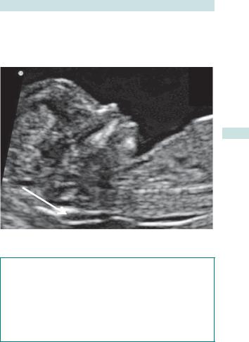
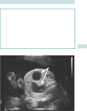
•Ultrasound (nuchal) scan measurement of the subcutaneous (SC) tissue between the skin and the soft tissue overlying the cervical spine with the fetus in the neutral position (see Fig. 3.1).
•A blood test measuring:
•PAPP-A
•β-human chorionic gonadotrophin (hCG).
Summary
•Now the recommended and most commonly used screening test.
•Performance enhanced by use of additional risk factors: nasal bone, tricuspid regurgitation.
•Careful ultrasound can identify many structural abnormalities.
Risk of trisomy 21
Calculated by multiplying the background maternal age and gestationrelated risk by a likelihood ratio derived from the nuchal translucency (NT) measurement and the two blood tests.
Combined test
Advantages
•Performance 790% detection for 5% FPR (75% for 3%).
•May detect other abnormalities such as anencephaly.
•An increased NT is also a marker for structural defects, e.g. cardiac malformations.
•Result usually available in 1st trimester, allowing surgical termination of pregnancy (TOP).
•Acceptable detection rate for all trisomies.
Disadvantages
Expensive and difficult to perform nuchal scan.
Triple and quadruple tests
When
Blood tests at 16wks.
How
•Dating scan (but not nuchal scan).
•Blood tests at 15wks measuring:
•oestriol
•hCG
•alpha-fetoprotein (AFP)
•inhibin A (not if triple).
COMMON SCREENING TESTS 113
Summary
•Recommended if NT scan not possible or gestation too advanced.
•Less operator dependent (scan) than the combined test.
Nuchal translucency
Fig. 3.1 Nuchal translucency at 11–14wks USS.
Nasal bone or tricuspid regurgitation
•Fetuses with Down’s syndrome are more likely to have an absent or hypoplastic nasal bone and have significant tricuspid regurgitation at 11–13+6wks.
•Only in conjunction with a nuchal scan can the presence or absence of these be used to modify the risk of a combined test.
•They may considerably increase the accuracy of the combined test.
•These are not commonly used because they require skill and time to detect accurately.

114 CHAPTER 3 Fetal medicine
Other screening tests
Integrated test
When
•Nuchal scan and blood tests (10–13+6wks).
•Blood tests again at 15wks.
How
•Nuchal scan.
•PAPP-A at 10wks.
•Quadruple test at 15wks measuring:
•oestriol
•hCG
• AFP
• inhibin A.
Summary
Best screening test (see Table 3.1), but expensive and seldom used.
Serum integrated test
When
Blood test at 10wks, then again at 15wks.
How
•Dating scan (but not nuchal scan).
•PAPP-A at 10wks.
•Quadruple test at 15wks measuring:
•oestriol
•hCG
•AFP
•inhibin A.
Summary
Less operator dependent (scan) than the combined test and probably as accurate; not recommended despite this (see Table 3.1).

OTHER SCREENING TESTS 115
Table 3.1 |
Comparison of screening tests available for |
|
|
|
|
|||
Down’s syndrome |
|
|
|
|
|
|
|
|
|
Combined |
Serum |
Integrated |
Triple |
Quadruple |
|
|
|
|
|
integrated |
test |
test |
test |
|
|
|
|
|
test |
|
|
|
|
|
|
|
|
|
|
|
|
|
|
|
Trimester |
1st |
1st and 2nd |
1st and 2nd |
2nd |
2nd |
|
|
|
Type |
Scan and |
Two |
Scan |
Blood |
Blood |
|
|
|
|
blood test |
blood |
and |
test |
test |
|
|
|
|
|
tests |
two |
|
|
|
|
|
|
|
|
blood |
|
|
|
|
|
|
|
|
tests |
|
|
|
|
|
Detection |
90% |
85% |
85% |
71% |
75% |
|
|
|
rate |
|
|
|
|
|
|
|
|
For a |
5% |
2.7% |
1.2% |
6% |
5% |
|
|
|
|
||||||||
given |
|
|
|
|
|
|
|
|
false |
|
|
|
|
|
|
|
|
+ve |
|
|
|
|
|
|
|
|
rate |
|
|
|
|
|
|
|
|
|
|
|
|
|
|
|
|
|
Significance of raised serum AFP
An elevated level of maternal serum AFP (greater than 2.2 multiples of median (MoM)) can also be used as a screening tool for open neural tube defects. It is also elevated in other conditions, including:
•Abdominal wall defects.
•Congenital nephrosis.
•Upper fetal bowel obstruction.
•Placental or umbilical cord tumours.
•Sacrococcygeal teratoma.
•Multiple pregnancy.
•After bleeding in early pregnancy.
An elevated AFP should trigger closer fetal and maternal surveillance as it is also a marker of adverse perinatal outcomes including:
•Fetal death.
•IUGR.
•Late pregnancy bleeding.
•Preterm delivery.
116 CHAPTER 3 Fetal medicine
Diagnosis of structural abnormalities
This should be offered to all women in the UK at the time of booking and usually takes the form of the ‘anomaly scan’, a detailed USS undertaken at around 18–21wks gestation. The aim is to identify specific structural malformations.
The detection of malformations may vary and is dependent on:
•The anatomical system affected.
•Gestational age at the time of the scan.
•Skill of the operator.
•Quality of the equipment.
•BMI of the mother.
Percentage of abnormalities detected by routine anomaly scan (these figures are better under optimum conditions)
•Central nervous system: 76%.
•Urinary tract: 67%.
•Pulmonary: 50%.
•Gastrointestinal: 42%.
•Skeletal: 24%.
•Cardiac: 17%.
NHS Screening Programmes: fetal anomaly 2010. Mhttp://fetalanomaly.screening.nhs.uk
Minimum standards required for 18–21wk anomaly scan
Fetal normality
•Skull shape and internal structures:
•cavum pellucidum
•cerebellum
•ventricular size at atrium <10mm
•nuchal fold.
•Spine—longitudinal and transverse views.
•Abdominal shape and content at the level of:
•stomach
•kidneys
•umbilicus/abdominal wall.
•bladder.
•Arms (three bones and hand).
•Legs (three bones and foot).
•Heart:
•four-chamber view
•outflow tracts—pulmonary artery and aorta
•lungs.
•Face and lips.
This page intentionally left blank

118 CHAPTER 3 Fetal medicine
Neural tube defects
Craniospinal defects occur early in development when the neural tube fails to close properly. Type and severity depend on degree and site of defect. There is growing evidence that prevalence is declining, possibly due to increased use of folate supplementation.
•Spina bifida and anencephaly make up over 95% of neural tube defects (NTDs).
•Incidence 2:1000 in England (3:1000 in Scotland).
Anencephaly
Absence of skull vault and cerebral cortex. It is incompatible with life, with babies rarely living more than a few hours if they are not stillborn.
Spina bifida
Incomplete fusion of vertebrae potentially allowing herniation of all or part of spinal cord. The malformation falls into three categories:
•Spina bifida occulta (mildest):
•split in vertebrae with no herniation of spinal cord
•varies from asymptomatic to mild neurological symptoms.
•Meningocele (least common):
•split in vertebrae with herniation of meninges and cerebrospinal fluid (CSF)
•varies from normal neurological function to moderate symptoms.
•Myelomeningocele (most severe):
•split in vertebrae allows herniation of spinal cord and meninges
•invariably have abnormal neurology at and below the lesion
•usually have an abnormal cerebellum and hydocephalus, which may result in mental impairment.
Dietary supplementation with folic acid decreases the incidence of NTDs. Recommended doses are:
• 400 micrograms/day for 3mths before conception, continued to 12wks.
• 5mg/day for women with previously affected child or those taking anticonvulsants.
Specific USS findings with some neural tube defects
•Anencephaly:
•absence of cranium and bulging eyes (‘frog-like’ appearance)
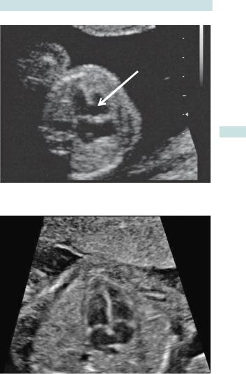
•99% will be detected by 20wks (see Fig. 3.2).
•Spina bifida: findings vary according to the severity of the lesion:
•defect seen in the vertebral bodies or tissue overlying the spine
•frontal bone scalloping (‘lemon sign’)
•abnormal shaped cerebellum (‘banana sign’)
•up to 95% detection rate for major defects
Association for Spina Bifida and Hydrocephalus. Mwww.asbah.org
NEURAL TUBE DEFECTS 119
Hand
Brain tissue
Face
Abdomen
Fig. 3.2 Ultrasound of anencephaly.

120 CHAPTER 3 Fetal medicine
Cardiac defects (congenital heart disease)
Cardiac defects are the most common major malformation in children, with an estimated incidence of 6–8:1000. They may be associated with a chromosomal abnormality, commonly trisomy 21 or 18, and with many other congenital abnormalities. Where isolated, many can be surgically corrected at birth, leading to a good quality of life. The most commonly seen abnormalities are VSDs (Fig. 3.3).
Risk factors for cardiac abnormalities
•Family history of congenital heart disease (CHD) in first-degree relative (recurrence risk 3%).
•Previous affected child (risk depends on type).
•Drug exposure, particularly anticonvulsants and lithium.
•Maternal diabetes mellitus.
•Other congenital abnormalities.
•Increased nuchal translucency (risk related to thickness).
Timing of cardiac scans
•In skilled hands many cardiac abnormalities can be seen at 12–13wks.
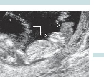
•Most cardiac abnormalities are missed at routine anomaly scans. The detection rate improves with training and specifically if ‘three-vessel’ and ‘outflow tracts’ views are found in addition to the four-chamber view (Fig. 3.4).
•Cardiac abnormalities may be better detected at 22wks than at 20wks. Evolution of defects change and later scanning, e.g. at 32wks, may be beneficial.
CARDIAC DEFECTS (CONGENITAL HEART DISEASE) 121
Ventricular septal defect
Fig. 3.3 USS of ventricular septal defect.
Fig. 3.4 USS of a normal cardiac four-chamber view.

122 CHAPTER 3 Fetal medicine
Urinary tract defects
Renal agenesis
•Bilateral renal agenesis is lethal because of anhydramnios causing lung hypoplasia.
•It may not be evident until >16wks because amniotic fluid is not all urinary before this time.
Posterior urethral valve syndrome
•This occurs in male fetuses where folds of mucosa block the bladder neck causing outflow obstruction.
•The severity is variable; back pressure may cause irreversible renal damage and oligohydramnios.
Hydronephrosis
•Accounts for 75% of fetal renal abnormalities.
•At least 40% resolve spontaneously in the neonatal period.
•It is usually due to pelviueretric obstruction, vesicoureteric reflux, utereocele, or posterior urethral valves (Fig. 3.5).
•Most cases can be treated postnatally so the prognosis is excellent.
Specific USS findings with some urinary tract defects
•Posterior urethral valves:
•thick walled dilated bladder with ‘keyhole’ sign of upper urethral dilatation
•hydronephrosis
•variable degrees of oligohydramnios according to severity.
•Ureterocele: cystic area of prolapsed ureter in bladder.
URINARY TRACT DEFECTS 123
‘Keyhole’ bladder
Fig. 3.5 Ultrasound of posterior urethral valve syndrome (keyhole bladder).

124 CHAPTER 3 Fetal medicine
Lung defects
Lung hypoplasia
•Failure to develop sufficient alveoli to permit adequate gas exchange at delivery.
•Often due to mid-trimester oligohydramnios from very preterm rupture of membranes or renal anomalies.
•May be due to compression (e.g. diaphragmatic hernia).
•Severity is increased with:
•preterm rupture of membranes at less than 25wks gestation
•severe oligohydramnios (amniotic fluid index (AFI) <4) for more than 2wks
•earlier delivery.
Estimated mortality rate 71–95%.
Diaphragmatic hernia
•A defect in the diaphragm results in the abdominal contents herniating into the chest.
•There is a 30% incidence of aneuploidy and a strong association with other malformations.
•The degree of compromise is correlated to the amount of compression of the original chest contents; prognosis is worse if the liver is in the chest and if little contralateral lung tissue is visible.
•Approximately 40% will die postnatally of lung hypoplasia; the others will require postnatal surgery.
•In utero treatment for severe cases involving tracheal obstruction (fetoscopic tracheal occlusion (FETO)) should be considered.
Congenital cystic adenomatoid malformation
•A rare form of lung disease where the normal alveolar tissue is replaced by a proliferation of cysts resembling bronchioles.
•The prognosis is good, but occasional very severe cases may cause hydrops.
XPostnatal surgery in asymptomatic cases is contentious.
Specific ultrasound findings with some lung defects
Diaphragmatic hernia
•Stomach or liver is seen within chest cavity.
•With a left-sided hernia, the heart may be deviated to the right.
•Abdominal circumference is often smaller than expected.
•Usually detectable at 20wks.
Congenital cystic adenomatoid malformation (CCAM)
•Cystic mass present within the lung parenchyma.
•Usually detectable at 20wks (Fig. 3.6).

LUNG DEFECTS 125
CCAM
Fig. 3.6 USS of (solid) congenital cystic adenomatoid malformation (CCAM)—it appears as a bright, space occupying lesion in the chest and can be solid, cystic, or a combination of both.
126 CHAPTER 3 Fetal medicine
Gastrointestinal defects
Exomphalos (omphalocele)
•Failure of the gut to return into the abdominal cavity after the normal embryological extrusion and rotation; the bowel, and sometimes other viscera, are contained within a sac and the umbilical cord arises from the apex of this sac (see Table 3.2).
•Approximately 1/3 occur with chromosomal abnormalities and up to 50% of the remainder have other malformations (e.g. cardiac).
•Prognosis depends on karyotype, presence of co-existing malformations, and presence of bowel ischaemia.
•Requires referral to a fetal medicine centre and postnatal surgery.
• Exomphalos alone is not an indication for delivery by CS.
Gastroschisis
•This involves protrusion of the gut through an anterior abdominal wall defect, usually to the right of the umbilical cord; the bowel is not covered by a sac and floats freely (see Table 3.2).
•Usually occurs in very young women. Rare after 25yrs.
•There is no increased risk of chromosomal abnormalities.
•The fetal gastrointestinal tract may become obstructed or atretic.
•Approximately 1 in 10–20 will die in utero or postnatally, but in the remainder the prognosis is good.
•Requires referral to a fetal medicine centre and postnatal surgery.
•Vaginal delivery is not necessarily contraindicated.
Gastrointestinal obstruction
•Duodenal atresia: 30% have trisomy 21.
•Oesophageal atresia: 15% have aneuploidy.
•Other atresias: may occur with syndromes or gastroschisis.
•Cystic fibrosis: common with bowel obstruction.
Table 3.2 Comparison of the features of exomphalos and gastroschisis
|
Exomphalos |
Gastroschisis |
Viscera contained within |
Yes, unless ruptured |
No |
a sac |
|
|
Insertion of umbilical cord |
At apex of sac |
Next to the defect |
Evisceration of: |
Liver ± intestinal loops, |
Intestinal loops only |
|
spleen |
|
Chromosomal |
30% |
<1% |
abnormalities |
|
|
Other malformations |
50% |
<5% |
Mortality |
40% |
5–10% |
|
|
|
GASTROINTESTINAL DEFECTS 127
Specific USS findings with some gastrointestinal defects
Anterior abdominal wall defects
•Bowel is seen outside the abdominal wall.
•Detection rates >95%.
Duodenal atresia
•Distension of stomach and proximal duodenum (‘double bubble’) (Fig. 3.7).
•May not be apparent by 20wks, but usually seen by 25wks.
•Polyhydramnios.
Oesophageal atresia
• Absence of stomach bubble and polyhydramnios.
• Often not seen due to presence of a tracheo-oesophageal fistula.
‘Double bubble’
Fig. 3.7 USS of duodenal atresia (‘double bubble’).

128 CHAPTER 3 Fetal medicine
Soft markers/normal variant screening
Some ultrasound features at 20wks may themselves be of little significance, but are nevertheless slightly more common in chromosomally abnormal fetuses. They have therefore been used to modify the risk of aneuploidy. However, these features may cause considerable parental anxiety as they are often, mistakenly, seen as abnormalities. Furthermore, they may increase the false +ve rate of screening.
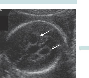
Choroid plexus cysts
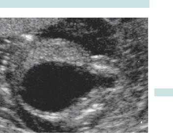
•Small benign cysts in the choroid plexus (Fig. 3.8), seen in about 1% of 20wk scans.
•Weakly associated with trisomy 18.
• NSC recommends these are not considered significant.
Echogenic intracardiac foci
•1 or more small echogenic foci (‘golf balls’) in the cardiac ventricles.
•No significance for cardiac defects.
•Slight increase (71.5-fold) in risk of chromosomal abnormalities.
•NSC recommends these are not considered significant.
Two-vessel umbilical cord
•Cord contains only one umbilical artery.
•Slight increase in chromosomal abnormalities.
•Slight increase in perinatal risk, and renal tract, and cardiac abnormalities.
•NSC recommends these are not considered significant.
Mild renal pelvic dilatation
•Dilatation of >7mm at 20wks.
•Slightly increased (71.5-fold) risk of chromosomal abnormalities.
•Repeat scan in 3rd trimester (to ensure not enlarged) and neonatal follow-up is recommended.
Echogenic bowel
•Bowel with areas of echogenicity similar in brightness to bone.
•Moderate association (75-fold increase) with chromosomal abnormalities.
•Although usually benign, occasionally associated with increased perinatal risk, cystic fibrosis, and bowel obstruction.
•Consider referral for expert opinion.
Increased nuchal fold
•20wk equivalent of nuchal translucency (>6mm in transverse section).
•Increased risk of chromosomal abnormalities (710-fold).
•Amniocentesis for chromosomal abnormalities is usually offered.
•Sign of early hydrops.
See M www.fetalanomaly.screening.nhs.uk/fetalanomalyresource/whats- in-the-hexagons1/about-the-scan/normal-variant-screening.
SOFT MARKERS/NORMAL VARIANT SCREENING 129
Bilateral choroid plexus cysts
Fig. 3.8 USS of choroid plexus cysts.

130 CHAPTER 3 Fetal medicine
Diagnostic tests
Chorionic villus sampling (CVS)
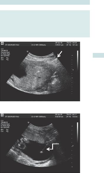
Diagnostic test usually performed between 10 and 13wks and involves aspiration of some trophoblastic cells. The amount of tissue obtained is small, but sufficient for karyotyping, and with the development of fluorescent in situ hybridization (FISH) and polymerase chain reaction (PCR), rapid analysis is possible (see Fig. 3.9). It requires ultrasound guidance and is usually performed transabdominally, occasionally transcervically.
Indications
•For karyotyping if 1st trimester screening test suggests high risk for aneuploidy.
•For DNA analysis if parents are carriers of an identifiable gene mutation such as cystic fibrosis or thalassaemia.
Benefits
Allows 1st trimester TOP if an abnormality is detected:
•Can be performed surgically.
•Can be done before the pregnancy has become physically apparent.
Risks
•Miscarriage as a result of CVS is estimated at 1%.
•Increases risk of vertical transmission of blood-borne viruses such as HIV and hepatitis B.
•False negative results (rare) from contamination with maternal cells—especially with DNA analysis requiring PCR.
•Placental mosaicism producing misleading results—estimated at <1%.
Amniocentesis
This is only undertaken from 15wks onwards. It involves aspiration of amniotic fluid which contains fetal cells shed from the skin and gut. It is performed transabdominally with ultrasound guidance (see Fig. 3.10).
Indications
•For karyotyping if screening tests suggest aneuploidy.
•For DNA analysis if parents are carriers of an identifiable gene mutation, such as cystic fibrosis or thalassaemia.
•For enzyme assays looking for inborn errors of metabolism.
•For diagnosis of fetal infections such as CMV and toxoplasmosis.
Benefits
•Lower procedure-attributed miscarriage rate than CVS (71%).
•Less risk of maternal contamination or placental mosaicism.
DIAGNOSTIC TESTS 131
Risks
•Miscarriage as a result of amniocentesis is estimated at 1%, although recent data suggest that the risk of miscarriage after amniocentesis is very little higher than would occur naturally.
•Failure to culture cells 70.5%.
•Full karyotyping may take 3wks (results for certain chromosomal abnormalities may be available more rapidly using FISH or PCR).
Needle taking
a sample from the placenta
Placenta
Fig. 3.9 Chorionic villus sampling.
Needle taking a sample of amniotic fluid
Fig. 3.10 Amniocentesis.

132 CHAPTER 3 Fetal medicine
Fetal hydrops: overview
Definition
Fetal hydrops is the abnormal accumulation of serous fluid in two or more fetal compartments. This may be pleural or pericardial effusions, ascites, skin oedema, polyhydramnios, or placental oedema. It may be divided into non-immune and immune causes.
Pathophysiology
The mechanism for the development of hydrops appears to be due to an imbalance of interstitial fluid production and inadequate lymphatic return. This can result from congestive heart failure, obstructed lymphatic flow, or decreased plasma osmotic pressure.
•Immune hydrops: results from blood group incompatibility between the mother and the fetus causing fetal anaemia.
•Non-immune hydrops: results from other causes, including fetal anaemia that is due to other causes such as fetal infection.
Incidence
Occurs in 71:2000 births.
Non-immune fetal hydrops: diagnosis and investigations
Ultrasound
•The diagnosis is made by USS.
•Associated structural abnormalities may be seen.
•Fetal echocardiography is required to diagnose cardiac lesions.
•Peak systolic velocity in middle cerebral artery shows fetal anaemia.
Fetal blood or amniotic fluid sampling
•Fetal blood sampling if anaemia is suspected (with blood ready for in utero transfusion).
•Amniotic fluid or fetal blood for chromosome analysis +/– virology.
Maternal blood testing
•Kleihauer test for feto-maternal haemorrhage.
•Antibody screen must be performed to exclude immune hydrops.
•Virology (parvovirus, CMV, toxoplasmosis).
•Consider haemoglobin electrophoresis for α-thalassaemia trait.

FETAL HYDROPS: OVERVIEW 133
Non-immune fetal hydrops: principal causes
Severe anaemia
•Congenital parvovirus B19 infection.
•α-Thalassaemia major (common in areas such as South-east Asia).
•Massive feto-maternal haemorrhage.
•Glucose-6-phosphate dehydrogenase deficiency.
Cardiac abnormalities
•Structural abnormalities.
•Fetal tachyarrhythmia (supraventricular tachycardia (SVT) or atrial flutter).
•Congenital heart block.
Chromosomal abnormalities
•Trisomies 13, 18, and 21.
•Turner’s syndrome (45 XO).
Other genetic syndromes
Multiple other syndromes, e.g. achondrogenesis, Noonan’s syndrome, Fryn’s syndrome, myotonic dystrophy.
Other infections
•Toxoplasmosis (bToxoplasmosis, p. 168).
•Rubella (bRubella (German measles), p. 156).
•CMV (bCytomegalovirus, p. 162).
•Varicella (bVaricella (chickenpox), p. 164).
Other structural abnormalities
•Congenital cystic adenomatoid malformation (CCAM).
•Diaphragmatic hernia.
•Pleural effusions.
Twin-to-twin transfusion syndrome
Recipient from volume overload and donor from anaemia (see b Twin-to-twin transfusion syndrome, p. 78).
Placental
Chorioangioma.

134 CHAPTER 3 Fetal medicine
Non-immune hydrops: treatment
The prognosis depends on the underlying cause. Where treatment is not possible, the option of TOP should be discussed. In the 3rd trimester, delivery may be a better alternative than in utero treatment.
If severe polyhydramnios is present, removal of excess amniotic fluid (amnioreduction) may reduce the risk of preterm labour. Consider giving steroids before the procedure as it carries a small risk of triggering preterm labour.
Treatable causes of non-immune fetal hydrops
Fetal anaemia
In utero blood transfusion may be performed.
Pleural effusions or large cystic CCAM
In utero percutaneous drainage and subsequent insertion of shunt into amniotic fluid may be possible.
Twin-to-twin transfusion syndrome
Laser photocoagulation of placental anastomoses.
Cardiac
•Tachyarrhythmias may be treated with maternal digoxin and flecainide.
•Valvotomy of stenotic cardiac valves is technically possible, but is usually too late if hydrops has already developed.

RHESUS ISOIMMUNIZATION (IMMUNE HYDROPS) 135
Rhesus isoimmunization (immune hydrops)
Definition Occurs when a maternal antibody response is mounted against fetal red cells. These immunoglobulin (IgG) antibodies cross the placenta and cause fetal red blood cell destruction. The ensuing anaemia, if severe, precipitates fetal hydrops, which is often referred to as immune hydrops.
Rhesus blood groups Consists of three linked gene pairs; one allele of each pair is dominant—C/c, D/d, and E/e. There are only five antigens (d is not an antigen; it merely implies absence of D). Inheritance is Mendelian. D gene is the most significant cause of isoimmunization, because 716% of white mothers are rhesus D –ve (d/d). Incidence lower in Afro-Caribbean and Asian populations. Other significant antigens include c, E, and atypical Kell antibody. Because of success of anti-D prophylaxis, these account for up to 1/2 of cases.
Pathophysiology of rhesus disease
•Fetal cells cross into the maternal circulation in normal pregnancy; the amount is increased during particular ‘sensitizing events’.
•The fetus may carry the gene for an antigen which the mother does not have—with rhesus D, the fetus may be D/d (rhesus D +ve), but the mother d/d (rhesus D –ve).
•Individuals exposed to a ‘foreign’ antigen mount an immune response (sensitization); initially, this is immunoglobulin (IgM), which cannot cross the placenta so this pregnancy is not at risk.
•Re-exposure in a subsequent pregnancy causes the primed memory B cells to produce IgG, which actively crosses into fetal circulation.
•IgG binds to fetal red cells, which are destroyed in the reticuloendothelial system.
•Causes a haemolytic anaemia (if erythropoesis is inadequate to compensate, severe anaemia causes high output cardiac failure, ‘fetal hydrops,’ and, ultimately, death).
•In milder cases, haemolysis leads to neonatal anaemia or jaundice from increased bilirubin levels.
Potential sensitizing events for rhesus disease
•TOP or evacuation of retained products of conception (ERPC) after miscarriage.
•Ectopic pregnancy.
•Vaginal bleeding 12wks, or earlier if heavy.
•ECV.
•Blunt abdominal trauma.
•Invasive uterine procedure, e.g. amniocentesis or CVS.
•Intrauterine death.
•Delivery.
136 CHAPTER 3 Fetal medicine
Rhesus disease: management
•All women should be checked for antibodies (rhesus and atypical) at booking, 28, and 34wks.
•If antibodies are detected, identifying the partner’s status will help determine the potential fetal blood group and risk to the fetus.
•PCR of fetal cells in maternal blood may also determine the fetus’s blood group, if the father is heterozygous or paternity is uncertain.
•Positive, but low levels of antibodies (<10IU/mL) should prompt repeat testing every 4wks.
•If levels are >10IU/mL, assessment for fetal anaemia is required.
2Amniocentesis or fetal blood sampling based on antibody levels or history alone is now obsolete.
•The peak systolic velocity (PSV) of the fetal middle cerebral artery (MCA) should be measured, about once a week. The MCA PSV will become abnormal before the baby is so anaemic as to be hydropic.
•If it is increased (>1.5 MoM), normograms for gestation are available) fetal blood sampling is indicated, with blood available for transfusion.
Treatment
•If the fetal haemaotocrit is <30, irradiated, Rh –ve, CMV –ve packed red cells are transfused into the umbilical vein at the cord insertion, or into the hepatic vein.
•This can be performed from 18wks onwards (beyond 35wks, delivery is preferable).
•Haemolysis will continue and the transfusion is repeated either every 2wks or when the MCA becomes abnormal again.
The risk of fetal loss, or need for urgent delivery if >26wks, is 1–3% per transfusion in skilled hands.
Postnatal management
•Anaemia can be corrected by blood transfusion.
•Severe anaemia may be accompanied by a coagulopathy from decreased platelets and clotting factors.
•Hyperbilirubinaemia and jaundice occur because in utero the mother cleared this red blood cell breakdown product, but the immature neonatal liver is unable to cope (this usually needs phototherapy, but may require exchange transfusion).
Antibodies may persist for weeks, causing continued haemolysis in the neonate; this requires careful monitoring with haematocrit measurements.

RHESUS DISEASE: MANAGEMENT 137
Prevention of rhesus (D) disease
If sufficient anti-D immunoglobulin is given to the mother it will bind to any fetal red cells in her circulation carrying the D antigen. This prevents her own immune system from recognizing them and therefore becoming sensitized.
•Anti-D (1500IU) is given to all women who are rhesus –ve (d/d):
•routinely at 28wks
•within 72h of any potentially sensitizing event
•after delivery if the neonate is found to be rhesus +ve (D/d).
•Occasionally, large feto-maternal haemorrhages occur during sensitizing events. If this is suspected a Kleihauer test should be performed as the standard dose of anti-D may not be sufficient. This should be routinely undertaken at delivery if the neonate is RhD +ve.
•Other antibodies, particularly anti-Kell and anti-c, now account for about half of all cases of fetal haemolysis.
•The use of anti-D, together with smaller family sizes, mean that rhesus disease is now rare. Only about 2% of RhD –ve women in the UK have been sensitized.

138 CHAPTER 3 Fetal medicine
Oligohydramnios
Amniotic fluid after 20wks largely consists of fetal urine. The volume depends on urine production, fetal swallowing, and absorption. Normal volume varies with gestation, and is highest between 24 and 36wks. The volume is measured by ultrasound, either by measuring the deepest vertical pool, or by adding up the deepest pools in the four quadrants of the uterus (AFI).
In oligohydramnios, the amniotic fluid volume is reduced. Normograms of both measures are available, but, as a general rule, a deepest pool of <2cm or an AFI of <8cm is considered low.
Complications
•Related to cause:
•preterm rupture of the membranes is commonly followed by delivery and/or intrauterine infection
•IUGR is an important cause of fetal and neonatal mortality and long-term morbidity.
•Related to reduced volume:
•lung hypoplasia if occurs <22wks
•limb abnormalities, e.g. talipes, if prolonged
•oligohydramnios before 22wks has a very poor prognosis.
Investigations
•USS of fetus, including Doppler.
•Speculum examination to look for ruptured membranes.
•If suspected spontaneous rupture of membranes (SROM): CRP, FBC, and vaginal swabs should be taken.
Management of oligohydramnios
If SROM at 34–36 or more weeks
Induce labour unless CS is indicated for another reason.
If SROM before 34–36wks
•Give prophylactic oral erythromycin.
•Monitor for signs of infection (4-hourly temperature and pulse).
•Daily CTG.
•Consider induction by 34–36wks.
If IUGR
Manage according to umbilical artery Doppler and CTG.
If apparently isolated oligohydramnios
•Reconsider cause.
•Intervention is not usual if umbilical artery Dopplers are normal.
If fetal renal tract abnormality
Refer to fetal medicine centre.

OLIGOHYDRAMNIOS 139
Causes of oligohydramnios
•Leakage of amniotic fluid: SROM.
•Reduced fetal urine production:
•IUGR
•fetal renal failure or abnormalities
•post-dates pregnancy.
•Obstruction to fetal urine output: fetal abnormalities such as posterior urethral valves.

140 CHAPTER 3 Fetal medicine
Polyhydramnios
The amniotic fluid is increased. In general a deepest pool of >8cm or an AFI >22 is abnormal.
Causes of polyhydramnios
Increased fetal urine production
•Maternal diabetes.
•Twin–twin transfusion syndrome (recipient twin).
•Fetal hydrops.
Fetal inability to swallow or absorb amniotic fluid
• Fetal gastrointestinal (GI) tract obstruction (e.g. duodenal atresia, tracheo-oesophageal fistula).
•Fetal neurological or muscular abnormalities (e.g. myotonic dystrophy, anencephaly).
•Other rare abnormalities or syndromes (e.g. facial obstruction).
•Idiopathic (usually mild).
Complications
•Preterm delivery, probably because of uterine stretch.
•Of the cause, e.g. duodenal atresia is associated with trisomy 21.
•Malpresentation at delivery because of increased room for fetus.
•Maternal discomfort because of abdominal distension.
Investigations
•Exclude maternal diabetes with a glucose tolerance test (GTT).
•Ultrasound examination of fetus.
Polyhydramnios: management
•Severe polyhydramnios is usually associated with fetal abnormality, if massive (e.g. AFI >40), amnioreduction (drainage of excess fluid with a needle), or non-steroidal anti-inflammatory drugs (NSAIDs).
•If fetal abnormality, refer to fetal medicine centre.
•Twin–twin transfusion syndrome is best managed in a fetal medicine centre, usually with laser ablation of placental anastomoses.
•If preterm, assess risk of delivery with cervical scan and/ or fibronectin assay, and consider steroids.
•If unstable or transverse lie at term, admit to hospital: CS if labour ensues with an abnormal lie.
NSAIDs cause fetal oliguria and can constrict the ductus arteriosus: close supervision is therefore indicated.
This page intentionally left blank

142 CHAPTER 3 Fetal medicine
Intrauterine growth restriction: overview
The fetus is believed to have an inherent growth potential which under ideal conditions should produce a healthy baby of the appropriate size. Attaining this potential relies on such factors as a healthy mother, well-functioning placenta, and the absence of pathology. If the in utero circumstances are less than ideal, the fetus will fail to achieve its full potential growth and fetal well-being may be compromised. This problem is analogous to ‘failure to thrive’ in children and is termed intrauterine growth restriction (IUGR).
Definition
2The term IUGR implies a fetus that is pathologically small.
Most epidemiological studies use small for gestational age (SGA) as a surrogate marker. The most commonly used method of identification of SGA is where the estimated weight of the fetus is below the 10th percentile for its gestational age.
This method is not perfect as it includes small, but healthy babies, and average-sized unhealthy babies that should have been born big.
Identifying inherent growth potential
To identify actual pathology and to improve detection of IUGR it is important to try to define the inherent growth potential of each individual fetus. Physiological factors known to affect growth and birth weight include:
•Maternal height and, to a lesser extent, paternal height.
•Maternal weight in early pregnancy.
•Parity.
•Ethnic origin.
•Gender of the fetus.
Some of these factors have been used to generate customized growth charts which aim to identify the optimal growth curve for an individual fetus. These are freely available at M www.gestation.net. They appear to reduce the rate of constitutionally small babies being classed as IUGR (false +ve rate) while helping to increase detection of pathologically small babies.
Defining expected growth also requires accurate dating of pregnancy, ideally by USS in the 1st trimester between 8 and 13wks gestation.
Further reading
Mwww.gestation.net
Perinatal Institute. (2011). Fetal growth assessment & implementation of customised charts. Mwww. pi.nhs.uk/growth/index_growth.htm

INTRAUTERINE GROWTH RESTRICTION: OVERVIEW 143
Importance of IUGR
For IUGR fetuses compared with normally grown population:
•Perinatal mortality is 6–10 times greater.
•Incidence of cerebral palsy is 4 times greater.
•30% of all stillborn infants are growth restricted.
IUGR fetuses are also more likely to have:
•Intrapartum fetal distress and asphyxia.
•Meconium aspiration.
•Emergency CS.
•Necrotizing enterocolitis.
•Hypoglycaemia and hypocalcaemia.

144 CHAPTER 3 Fetal medicine
Intrauterine growth restriction: causes
Causes of growth restriction may be grouped into maternal, utero-placental, and fetal.
By far the most common cause is utero-placental insufficiency.
Causes of IUGR
Maternal
•Chronic maternal disease:
•hypertension
•cardiac disease
•chronic renal failure.
•Substance abuse:
•alcohol
•recreational drug use.
•Smoking.
•Autoimmune diseases, including: antiphospholipid antibody syndrome.
•Genetic disorders, including: phenylketonuria.
•Poor nutrition.
•Low socio-economic status.
Placental (placental insufficiency)
•Abnormal trophoblast invasion:
•pre-eclampsia
•placenta accreta.
•Infarction.
•Abruption.
•Placental location: placenta praevia.
•Tumours: chorioangiomas (placental haemangiomas).
•Abnormal umbilical cord or cord insertion: two-vessel cord.
Fetal
•Genetic abnormalities, including:
•trisomy 13, 18, or 21
•Turner’s syndrome
•triploidy.
•Congenital abnormalities, including:
•cardiac, e.g. tetralogy of Fallot, transposition of the great vessels
•gastroschisis.
•Congenital infection, including:
•CMV
•rubella
•toxoplasmosis.
•Multiple pregnancy.

IUGR: MANAGEMENT AND OUTCOME 145
Intrauterine growth restriction: management and outcome
Symmetric and asymmetric IUGR
IUGR is often classified into symmetric and asymmetric, although the two groups overlap substantially
•Symmetric growth restriction: describes a fetus whose entire body is proportionately small and tends to be seen with very early onset IUGR and also with chromosomal abnormalities.
•Asymmetric growth restriction: is seen with an undernourished fetus who is compensating by directing most of its energy to maintaining the growth of vital organs such as the brain and heart at the expense of the liver, fat, and muscle. This ‘head-sparing effect’ results in a normal head size with small abdominal circumference and thin limbs. It is most often seen with IUGR secondary to placental insufficiency.
Management
Early identification and intensive fetal monitoring are the key to managing IUGR. The aim is to continue the pregnancy safely for as long as possible, thereby decreasing the problems associated with prematurity, but deliver before the fetus becomes excessively compromised. This is covered in depth in bAntenatal fetal surveillance, p. 146.
Long-term outcome
Most congenitally normal IUGR babies will go on to grow normally in infancy and childhood, but there may be more subtle long-term consequences, including up to 1/3 of children not reaching their predicted adult height, and having childhood attention and performance deficits.
The effects appear to last into adulthood, with a stimulus or insult at a critical, sensitive period of early life having permanent effects on structure, physiology, and metabolism. People who were small or disproportionate (thin or short) at birth have been found to have higher rates of coronary heart disease, high blood pressure, high cholesterol concentrations, and abnormal glucose-insulin metabolism (Barker hypothesis).
Further reading
Godfrey KM, Barker DJ. (2000). Fetal nutrition and adult disease. Am J Clin Nutr 71(5 Suppl.): 1344–52S.
146 CHAPTER 3 Fetal medicine
Antenatal fetal surveillance: overview
Facts
The fetus is vulnerable to any strain in the uteroplacental unit—its supply line for nutrition and gas exchange. Nearly 1/100 babies in developed countries are stillborn and 1/3 of all stillbirths occur in babies that are small for dates.
Any uteroplacental shortfall becomes more critical as the fetus gets bigger, and there is an extraordinary rate of growth during the last weeks of pregnancy. The stillbirth risk rises at the same stage.
2 There are no treatments to reverse uteroplacental insufficiency but, potentially, delivering the baby at the right time can prevent death and disability.
The only intervention is delivery so monitoring is not useful if the fetus could not survive if it were delivered, i.e. if it is <24wks or <500g.
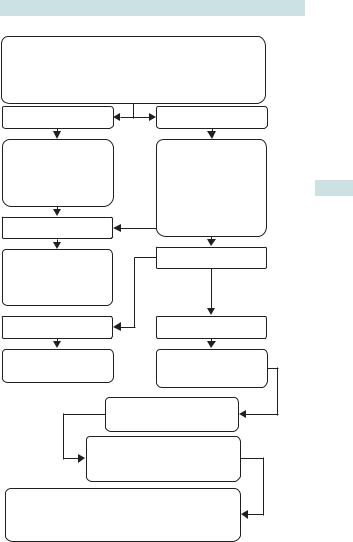
Stages in fetal surveillance
Antepartum fetal monitoring is done in two stages.
•Stage 1—assigning risk: finding normal babies developing in an abnormal situation.
•Stage 2—timing delivery:
•preterm babies should be delivered only if they show signs of distress, ensuring maximum maturity while avoiding any harm.
•after 36wks babies at high risk should be delivered.
Assigning risk
•No method of antenatal monitoring is perfect—all ‘miss’ affected fetuses (false negatives) and ‘pick up’ normal fetuses (false positives).
•Potential problems with such screening include increasing the rate of major interventions (such as CS) without benefit (see Fig. 3.11).
•Given the low prevalence of fetal compromise, the +ve predictive value of all tests when applied to low-risk populations is poor, but they perform better when applied to high-risk populations and therefore an estimation of the background risk must be made.
•Risk is assigned considering the mother’s health (e.g. pre-eclampsia) and the outcome of any previous pregnancy.
X In the future, screening for placental function using serum markers, history, and ultrasound could establish itself as a routine test, in the same way as screening for Down’s syndrome.
ANTENATAL FETAL SURVEILLANCE: OVERVIEW 147
ASSIGNMENT OF RISK
INCLUDES CONSIDERATION OF MATERNAL HISTORY,
SIGNS OF PRE–ECLAMPSIA, MEASUREMENT OF
SYMPHYSIS FUNDAL HEIGHT, AND FETAL MOVEMENTS
REMAINS LOW RISK |
HIGH RISK |
|
CONTINUE WITH |
ULTRASOUND |
|
BIOMETRY |
||
REGULAR |
||
|
||
ANTENATAL CHECK |
AMNIOTIC FLUID |
|
UPS |
||
VOLUME |
||
|
||
|
UMBILICAL |
|
NORMAL |
CORD DOPPLER |
|
|
||
REPEAT IN 2–4 WKS |
ABNORMAL |
|
|
||
(DELIVER |
|
|
TERM BABY) IF |
|
|
REMAINS HIGH RISK |
|
|
TERM BABY |
PRETERM BABY |
|
DELIVER HIGH RISK |
GROWTH |
|
RESTRICTED |
||
TERM BABY |
||
PRE TERM BABY |
||
|
||
|
BETAMETHASONE 12mg IM |
MONITOR USING UMBILICAL
CORD DOPPLER
FETAL RESCUE
DELIVER IF REVERSED DIASTOLIC
FLOW IN UMBILICAL CORD OR IF CTG ABNORMAL
Fig. 3.11 Assigning risk in antenatal monitoring.

148 CHAPTER 3 Fetal medicine
Identifying the high-risk fetus
Symphysis fundal height
•Measurement of the symphysis fundal height (centimetres).
•Sequential measurements can reveal changes in fetal growth.
•The detection rate of small for gestational age babies is improved by using ‘customized’ fundal height charts displaying curves that are specific to the mother’s height, weight, parity, and ethnic group (see bIntrauterine growth restriction: overview, p. 142).
•If growth is suspected to be abnormal, a USS should be organized.
Ultrasound assessment of fetal growth
Accurate knowledge of the age of the fetus is required. The fetal head circumference (HC) and abdominal circumference (AC) are measured together with the deepest pool of amniotic fluid using ultrasound and the results are charted against gestational age. Serial measurements are useful to assess the pattern of the fetal growth.
Late ultrasound aims to detect:
•Growth problems in the baby.
•Abnormalities in the amount of fluid around the baby.
•Problems with the placenta.
•Problems with the baby’s position.
It should be used only where growth abnormality is suspected (SFH outside the normal range) or the patient is at high risk of uteroplacental insufficiency, because its usage has not been proven to improve outcome in unselected patients.
Uterine artery Doppler
•Resistance within the placenta can be measured from the maternal side, usually as a screening test at 23wks.
•High resistance/pulsatility in the uterine artery indicates that the mother is at increased risk of developing early onset pre-eclampsia or having a baby with IUGR; therefore she should be offered extra monitoring in pregnancy.
Routine monitoring of fetal movement
•The +ve predictive value of maternal perception of reduced fetal movements is extremely low.
•Mothers should be advised to contact their midwife or the hospital for further assessment if there is a reduction in fetal movement.
Auscultation of the fetal heart
Auscultation of the fetal heart merely confirms the fetus is alive. It provides no predictive information done (with hand-held Doppler or a Pinard stethoscope) as part of a standard antenatal examination.
This page intentionally left blank

150 CHAPTER 3 Fetal medicine
Monitoring the high-risk fetus:
Doppler ultrasound
Doppler ultrasound uses sound waves to study blood circulation in the baby, uterus, and placenta.
Umbilical artery Doppler
•Increasing resistance/pulsatility in the umbilical artery is an indicator of placental failure. Using this in high-risk pregnancies reduces risk of fetal death and the need for interventions around birth, such as CS.
•Diagnostic:
•a high resistance/pulsatility can help to differentiate a normal, but small baby from one that is not reaching its full growth potential
•abnormal waveforms in the umbilical artery are an early sign of fetal impairment and tend to precede changes in CTG.
•Monitoring:
•in very preterm babies Doppler can be used to time fetal rescue
•decision to deliver will depend on gestation as well as degree of abnormality.
Middle cerebral artery Doppler
•In addition to screening for anaemia, this vessel will demonstrate a reduced resistance/pulsatility in the compromised baby as a result of head sparing.
•It may be more useful than umbilical artery Doppler at term.
•It is not usually used alone in deciding timing of delivery.
Ductus venosus Doppler
•Waveform in this vein is a surrogate for cardiac function and used in monitoring for twin–twin transfusion syndrome.
•It can also be used to time delivery in severely compromised babies as an adjunct to CTGs and umbilical artery.
Understanding umbilical artery Dopplers
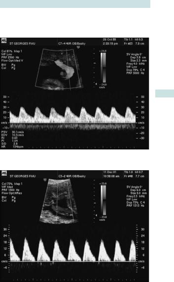
•When the placenta is functioning normally, flow through the umbilical artery is not impeded by end organ resistance; therefore blood continues to flow forwards (away from the heart) during cardiac diastole, seen on Doppler waveform as ‘end diastolic flow’ (Fig. 3.12).
•As the uteroplacental unit begins to fail, the vascular resistance rises and the forward flow in diastole begins to be reduced (this can be numerically quantified and used for monitoring).
•When the resistance is very high, blood no longer flows forwards in diastole: this is called ‘absent end diastolic flow’ (AEDF) (Fig. 3.13).
•When the situation is critical, the resistance is so great that blood may flow back towards the heart during diastole: this is called ‘reversed end diastolic flow’ (REDF) (Fig. 3.14).
These signs are used to differentiate those fetuses that are coping in an adverse situation from those that need immediate delivery.
MONITORING THE HIGH-RISK FETUS: DOPPLER ULTRASOUND 151
Uterine artery Doppler
This is most used for identification of risk pregnancy.
Fig. 3.12 Example of normal umbilical artery Doppler waveform.
Fig. 3.13 Example of umbilical artery Doppler waveform with absent end diastloic flow (AEDF).

152 CHAPTER 3 Fetal medicine
Fig. 3.14 Example of umbilical artery Doppler waveform with reversed end diastolic flow (REDF).

MONITORING THE HIGH-RISK FETUS: CARDIOTOCOGRAPHY 153
Monitoring the high-risk fetus: cardiotocography
•CTG is the output of electronic monitoring of the fetal heart rate, correlated with any uterine contractions.
•Analysis by inspecting the CTG is difficult, but it is more reproducible when done by computer systems, e.g. Oxford Sonicaid.
Normal cardiotocography
•The baseline fetal heart rate is between 110 and 160 beats/min and varies from that baseline by 5–25beats/min.
•The heart rate should speed up by at least 15 beats/min for at least 15s (accelerations). Two accelerations should be seen in 20min (reactive).
•There should be no slowing of the fetal heart rate from the baseline (decelerations).
2The most useful features in assessing the fetus’s health are the variability and presence or absence of accelerations.
Abnormal cardiotocography
Caused by a failure of autonomic regulation of the heart rate, this is an end-stage event, so the lead-time between uteroplacental insufficiency causing an abnormal CTG and fetal death or long-term damage is short, and this limits its usefulness in antenatal screening.
X Routine antenatal CTG has not been found to be useful in low-risk populations.
CTG is used to exclude current compromise:
•In acute conditions known to cause fetal compromise, e.g. abruption and in women reporting reduced fetal movements.
•Daily in the surveillance of chronic conditions that are associated with uteroplacental insufficiency such as pre-eclampsia, and in IUGR when there is AEDF.
Because the CTG only becomes abnormal at a very late stage in IUGR, less frequent than daily CTGs should not be relied upon as a method of monitoring.
Further reading
NICE. (2008). Antenatal care: routine care for the healthy pregnant woman. In: Fetal growth and wellbeing. London: NICE. Mwww.nice.org.uk/guidance/CG62
This page intentionally left blank