

Gale Encyclopedia of Genetic Disorder / Gale Encyclopedia of Genetic Disorders, Two Volume Set - Volume 2 - M-Z - I
.pdfSpinocerebellar ataxia
respiratory infection. It is therefore important to ensure that mucus does not build up in patients respiratory tracts as this could aid viral and bacterial infections.
Resources
PERIODICALS
Crawford, T. O., and C. A. Pardo. “The neurobiology of childhood spinal muscular atrophy.” Neurobiology of Disease 3 (1996): 97-110.
ORGANIZATIONS
Muscular Dystrophy Association. 3300 East Sunrise Dr., Tucson, AZ 85718. (520) 529-2000 or (800) 572-1717.http://www.mdausa.org .
WEBSITES
Families of Spinal Muscular Atrophy. http://www.fsma.org . The Andrew’s Buddies web site. FightSMA.com
http://www.andrewsbuddies.com/news.html .
Philip J. Young
Christian L. Lorson, PhD
I Spinocerebellar ataxia
Definition
The spinocerebellar ataxias (SCAs) are a group of inherited conditions that affect the brain and spinal cord causing progressive difficulty with coordination.
Description
The SCAs are named for the parts of the nervous system that are affected in this condition. Spino refers to the spinal cord and cerebellar refers to the cerebellum or back part of the brain. The cerebellum is the area of the brain that controls coordination. In people with SCA, the cerebellum often becomes atrophied or smaller. Symptoms of SCA usually begin in the 30s or 40s, but onset can be at any age. Onset from childhood through the 70s has been reported.
As of early 2001, at least 13 different types of SCA have been described. This group is numbered 1-14 and each is caused by mutations or changes in a different gene. Although the category of SCA9 has been reserved, there is no described condition for SCA9 and no gene has been found. Spinocerebellar ataxia has also been called olivopontocerebellar atrophy, Marie’s ataxia, and cerebellar degeneration. SCA3 is sometimes called MachadoJoseph disease named after two of the first families described with this condition. All affected people in a family have the same type of SCA.
Genetic profile
Although each of the SCAs is caused by mutations in different genes, the types of mutations are the same in all of the genes that have been found. Most genes come in pairs; one member of a pair comes from a person’s mother and the other one comes from their father. The genes are made up of deoxyribonucleic acid (DNA) and the DNA is made up of chemical bases that are represented by the letters C, T, G, and A. This is the DNA alphabet. The letters are put together in three letter words. The arrangement of the words are what give the gene its meaning and therefore tells the body how to grow and develop.
Trinucleotide repeats
In each of the genes that cause SCA, there is a section of the gene where a three letter word is repeated a certain number of times. In most of the types of SCA, the word that is repeated is CAG. So there is a part of the gene that reads CAGCAGCAGCAGCAG...and so on. In people who have SCA, this word is repeated too many times. Therefore, this section of the gene is too big. This is called a trinucleotide repeat expansion. In SCA8 the word that is repeated is CTG. In SCA10, the repeated word is five DNA letters long and is ATTCT. This is called a pentanucleotide expansion. The actual number of words that is normal or that causes SCA is different in each type of SCA.
In each type of SCA, there are a certain number of words that are normal (the normal range). People who have repeat numbers in the normal range will not develop SCA and cannot pass it to their children. There is also a certain number of repeats that cause SCA (the affected range). People who have repeat numbers in the affected range will go on to develop SCA sometime in their lifetime if they live long enough. People with repeat numbers in the affected range can pass SCA onto their children. Between the normal and affected ranges there is a gray range. People who have repeat numbers in the gray range may or may not develop SCA in their lifetime. Why some people with numbers in the gray zone develop SCA and others do not is not known. People with repeat numbers in the gray range can also pass SCA onto their children.
In general, the more repeats in the affected range that someone has, the earlier the age of onset of symptoms and the more severe the symptoms. However, this is a general rule. It is not possible to look at a person’s repeat number and predict at what age they will begin to have symptoms or how their condition will progress.
Anticipation
Sometimes when a person who has repeat numbers in the affected or gray range has children, the expansion
1084 |
G A L E E N C Y C L O P E D I A O F G E N E T I C D I S O R D E R S |

grows larger. This is called anticipation. This can result in an earlier age of onset in children than in their affected parent. Anticipation does not occur in SCA6. Significant anticipation can occur with SCA7. It is not unusual for a child with SCA7 to be affected before their parent or even grandparent begins to show symptoms. In most types of SCA, anticipation happens more often when a father passes SCA onto his children then when a mother passes it. However, in SCA8 the opposite is true; anticipation happens more often when a mother passes it to her children. Occasionally, repeat sizes stay the same or even get smaller when they are passed to a person’s children.
Inheritance
The SCAs are passed on by autosomal dominant inheritance. This means that males and females are equally likely to be affected. It also means that only one gene in the pair needs to have the mutation in order for a person to become affected. Since a person only passes one copy of each gene onto their children, there is a 50% or one in two chance that a person who has SCA will pass it on to each of their children. A person who has repeat numbers in the gray range also has a 50% or one in two chance of passing the gene on to each of their children. However, whether or not their children will develop SCA depends on the number of their repeats. A person who has repeat numbers in the normal range cannot pass SCA onto their children.
New mutations
Usually a person with SCA has a long family history of the condition. However, sometimes a person with SCA appears to be the only one affected in the family. This can be due to a couple of reasons. First, it is possible that one of their parents is or was affected, but died before they began to show symptoms. It is also possible that their parent had a mutation in the gray range and was not affected, but the mutation expanded into the affected range when it was passed on. Other family members may also have SCA but have been misdiagnosed with another condition or are having symptoms, but have no diagnosis. It is also possible that a person has a new mutation for SCA. New mutations are changes in the gene that happen for the first time in an affected person. Although a person with a new mutation may not have other affected family members, they still have a 50% or one in two chance of passing it on to their children.
Demographics
SCA has been found in people from all over the world. However, some of the types of SCA may be more common in certain areas and ethnic groups. SCA types 1,
K E Y T E R M S
Anticipation—Increasing severity in disease with earlier ages of onset, in successive generations; a condition that begins at a younger age and is more severe with each generation
Ataxia—A deficiency of muscular coordination, especially when voluntary movements are attempted, such as grasping or walking.
Trinucleotide repeat expansion—A sequence of three nucleotides that is repeated too many times in a section of a gene.
2, 3, 6, and 7 account for the majority of autosomal dominant SCA. SCA3 appears to be the most common type and was first described in families from Portugal. SCA3 also seems to be the most common type in Germany. SCA8 accounts for about 2-5% of all SCA. SCA types 4, 5, 10, 11, 12, 13, and 14 are rare and have each only been described in a few families. The first family described with SCA5 may have been distantly related to President Abraham Lincoln and was first called Lincoln ataxia. As of early 2001, SCA10 has only been described in Mexican families, SCA13 has only been described in one French family, and SCA14 has only been found in one family from Japan.
Signs and symptoms
Although different genes cause each of the SCAs, they all have similar symptoms. All people with SCA have ataxia or a lack of muscle coordination. Walking is affected and eventually the coordination of the arms, hands, and of the speech and swallowing is also affected. One of first symptoms of SCA is often problems with walking and difficulties with balance. The muscles that control speech and swallowing usually become affected. This results in dysarthria or slurred speech and difficulties with eating. Choking while eating can become a significant problem and can lead to a decrease in the number of calories a person can take in. The age of the onset of symptoms can vary greatly—anywhere from childhood through the seventh decade have been reported. The age of onset and severity of symptoms can also vary between people in the same family.
As the condition progresses, walking becomes more difficult and it is necessary to use a cane, walker, and eventually a wheelchair. Because of the uncoordinated walking that develops, it is not uncommon for people with SCA to be mistaken for being intoxicated. Carrying
ataxia Spinocerebellar
G A L E E N C Y C L O P E D I A O F G E N E T I C D I S O R D E R S |
1085 |
Spinocerebellar ataxia
around a note from their doctor explaining their medical condition can often be helpful.
Some of the SCA types can also have other symptoms, although not all of these are seen in every person with that particular type. SCA2: People with this type may have slower eye movements. This does not usually interfere with a person’s sight. SCA3: In this type people may develop problems with the peripheral nerves—those nerves that carry information to and from the spinal cord. This can lead to decreased sensation and weakness in the hands and feet. In SCA3 people may also have twitching in the face and tongue, and bulging eyes. SCA4: People with this type may have a loss of sensation but often have a normal lifespan. SCA5: This type often has an adult onset and is slowly progressive, not affecting a person’s lifespan. SCA6: This type often has a later onset, progresses very slowly and does not shorten a person’s life. SCA7: Progressive visual loss that eventually leads to blindness always happens with this type. SCA10: A few people with this type have had seizures. SCA11: This type is relatively mild and people have a normal lifespan. SCA12: People often have a tremor as the first noticeable symptom and may eventually develop dementia. SCA13: Some people with this type are shorter than average and have mild mental retardation.
Diagnosis
An initial workup of people who are having symptoms of ataxia will include questions about a person’s medical history and a physical examination. Blood work to rule out other causes of the ataxia such as vitamin deficiencies may also be done. Magnetic resonance imaging (MRI) of the brain in people with SCA will usually show degeneration or atrophy of the cerebellum and may be helpful in suggesting a diagnosis of SCA. A thorough family history should be taken to determine if others in the family have similar symptoms and the inheritance pattern in the family.
Since there is so much overlap between symptoms in the different types of SCA, it is not usually possible to tell the different types apart based on clinical symptoms. The only way to definitively diagnose SCA and determine a specific subtype is by genetic testing. This involves drawing a small amount of blood. The DNA in the blood cells is then examined and the number of CAG repeats in each of the SCA genes are counted. As of early 2001, clinical testing is available to detect the mutations that cause SCA1, 2, 3, 6, 7, 8, and 10.
If genetic testing is negative for the available testing, it does not mean that a person does not have SCA. It could mean that they have a type of SCA for which genetic testing is not yet available.
Predictive testing
It is possible to test someone who is at risk for developing SCA before they are showing symptoms to see whether they inherited an expanded trinucleotide repeat. This is called predictive testing. Predictive testing cannot determine the age of onset that someone will begin to have symptoms, or the course of the disease. The decision to undergo this testing is a very personal decision and one that a person can only make for his or her self. Some people choose to have testing so that they can make decisions about having children or about their future education, career, or finances. Protocols for predictive testing have been developed, and only certain centers perform this testing. Most centers require that the diagnosis of SCA has been confirmed by genetic testing in another family member. It is also strongly suggested that a person have a support person, either a spouse or close friend, be with them at all visits.
A person who is interested in testing will be seen by a team of specialists over the course of a few visits. Often they will meet a neurologist who will perform a neurological examination to see if they may be showing early signs of the condition. If a person is having symptoms, testing may be performed to confirm the diagnosis. The person will also meet with a genetic counselor to talk about SCA, how it is inherited, and what testing can and cannot tell someone. They will also explore reasons for testing and what impact the results may have on their life, their family, their job and their insurance. Most centers also require a person going through predictive testing to meet a few times with a psychologist. The purpose of this visit is to make sure that the person has thought through the decision to be tested and is prepared to deal with whatever the results may be. These visits also allow a person to make contact with someone who can help him or her deal with the results if necessary. All centers require that results are given in person and usually require that a person come in for a few follow-up visits, regardless of the testing results.
These protocols are not in place to make people go through endless steps to get testing. Rather they have been developed to make sure that people make the best decision for themselves, their life, and their family and that they are prepared to cope with the results, whatever the outcome. Once the results are given, it is not possible to give them back or forget them. People should therefore take the testing process seriously and give a great deal of consideration to making the decision to be tested.
Testing children
If a child is having symptoms, it is appropriate to perform testing to confirm the cause of their symptoms.
1086 |
G A L E E N C Y C L O P E D I A O F G E N E T I C D I S O R D E R S |
However, testing will not be performed on children who are at risk for developing SCA but are not having symptoms. The choice to know this information can only be made for oneself when they are old enough to make a mature decision. Testing a child who does not have symptoms could lead to possible problems with their future relationships, education, career, and insurance.
Prenatal testing
Testing a pregnancy to determine whether an unborn child is affected is possible if genetic testing in a family has identified a certain type of SCA. This can be done at 10-12 weeks gestation by a procedure called chorionic villus sampling (CVS) that involves removing a tiny piece of the placenta and examining the cells. It can also be done by amniocentesis after 16 weeks gestation by removing a small amount of the amniotic fluid surrounding the baby and analyzing the cells in the fluid. Each of these procedures has a small risk of miscarriage associated with it and those who are interested in learning more should check with their doctor or genetic counselor. Continuing a pregnancy that is found to be affected is like performing predictive testing on a child. Therefore couples interested in these options should have genetic counseling to carefully explore all of the benefits and limitations of these procedures.
There is also another procedure, called preimplantation diagnosis that allows a couple to have a child that is unaffected with the genetic condition in their family. This procedure is experimental and not widely available. Those interested in learning more about this procedure should check with their doctor or genetic counselor.
Treatment and management
Although there is a lot of ongoing research to try to learn more about SCA and develop treatments, no cure currently exists for the SCAs. Although vitamin supplements are not a cure or treatment for SCA, they may be recommended if a person is taking in fewer calories because of feeding difficulties. Different types of therapy might be useful to help people maintain as independent a lifestyle as possible. An occupational therapist may be able to suggest adaptive devices to make the activities of daily living easier. For example they may suggest installing bars to use in the bathroom or shower or special utensils for eating. A speech therapist might be able to make recommendations for devices that might make communication easier as the speech becomes affected. As swallowing becomes more difficult, a special swallow evaluation may lead to better strategies for eating and to lessen the risk of choking.
Genetic counseling
Genetic counseling helps people and their families to make decisions about their medical care, genetic testing, and having children by providing information and support. It can also help people to deal with the medical and emotional issues that arise when there is a genetic condition diagnosed in the family.
Prognosis
Most people with the SCAs do have progression of their symptoms that leads to full time use of a wheelchair. The duration of the disease after the onset of symptoms is about 10-30 years, but can vary depending in part to the number of trinucleotide repeats and age of onset. In general, people with a larger number of repeats have an earlier age of onset and more severe symptoms. Choking can be a major hazard because if food gets into the lungs, a life-threatening pneumonia can result. As the condition progresses, it can become difficult for people to cough and clear secretions. Most people die from respiratory failure or pulmonary complications.
Resources
PERIODICALS
Evidente, V.G.H., et al. “Hereditary Ataxias.” Mayo Clinic Proceedings (2000): 475-490.
Zohgbi, H.Y., and H.T. Orr. “Glutamine Repeats and Neurodegeneration.” Mayo Clinic Proceedings (2000): 217-247.
ORGANIZATIONS
National Ataxia Foundation. 2600 Fernbrook Lane, Suite 119, Minneapolis, MN 55447. (763) 553-0020. Fax: (763) 5530167. naf@ataxia.org. http://www.ataxia.org .
WE MOVE (Worldwide Education and Awareness for Movement Disorders) 204 E. 84th St., New York, NY 10024. (212) 875-8312 or (800) 437-MOV2. Fax: (212) 875-8389. wemove@wemove.org. http://www.wemove
.org .
WEBSITES
GeneClinics. http://www.geneclinics.org .
Online Mendelian Inheritance in Man. http://www.ncbi.nlm.nih
.gov/entrez/query.fcgi?db=OMIM .
International Network of Ataxia Friends (INTERNAF).http://www.internaf.org .
Spinocerebellar Ataxia: Making an Informed Choice about Genetic Testing. http://www.depts.washington.edu/neurogen/ AtaxiaBrochure99.pdf .
Karen M. Krajewski, MS
Spinocerebellar atrophy I see
Spinocerebellar ataxia
ataxia Spinocerebellar
G A L E E N C Y C L O P E D I A O F G E N E T I C D I S O R D E R S |
1087 |

Spondyloepiphyseal dysplasia
I Spondyloepiphyseal dysplasia
Definition
Spondyloepiphyseal dysplasia is a rare hereditary disorder characterized by growth deficiency, spinal malformations, and, in some cases, ocular abnormalities.
Description
Spondyloepiphyseal dysplasia is one of the most common causes of short stature. There are two forms of spondyloepiphyseal dysplasia. Both forms are inherited and both forms are rare.
Congenital spondyloepiphyseal dysplasia
Congenital spondyloepiphyseal dysplasia is primarily characterized by prenatal growth deficiency and spinal malformations. Growth deficiency results in short stature (dwarfism). Abnormalities of the eyes may be present, including nearsightedness (myopia) and retina (the nerve-rich membrane lining the eye) detachment in approximately half of individuals with the disorder. Congenital spondyloepiphyseal dysplasia is inherited as an autosomal dominant genetic trait.
Congenital spondyloepiphyseal dysplasia is also known as SED, congenital type; SED congenita; and SEDC.
Spondyloepiphyseal dysplasia tarda
Spondyloepiphyseal dysplasia tarda primarily affects males. It is characterized by dwarfism and hunched appearance of the spine. The disorder doesn’t become evident until five to 10 years of age. Spondyloepiphyseal dysplasia tarda is an X-linked recessive inherited disorder.
Spondyloepiphyseal dysplasia tarda is also known as SEDT; spondyloepiphyseal dysplasia, late; and SED tarda, X-linked.
Genetic profile
Both forms of the disorder are inherited, however they are inherited differently.
Congenital spondyloepiphyseal dysplasia
Congenital spondyloepiphyseal dysplasia is thought to probably always result from abnormalities in the COL2A1 gene, which codes for type II collagen. Collagen is a protein that is a component of bone, cartilage, and connective tissue. A variety of abnormalities (such as deletions and duplications) involving the COL2A1 gene may lead to the development of the disorder.
It is one of a group of skeletal dysplasias (dwarfing conditions) caused by changes in type II collagen. These include hypochondrogenesis; spondyloepimetaphyseal dysplasia, Strudwick (SEMD); and Kniest dysplasia. Type 2 collagen is the major collagen of a component of the spine called the nucleus pulposa, of cartilage, and of vitreous (a component of the eye). All of these conditions have common clinical and radiographic findings including spinal changes resulting in dwarfism, myopia, and retinal degeneration.
Congenital spondyloepiphyseal dysplasia is inherited as an autosomal dominant genetic trait. In autosomal dominant inheritance, a single abnormal gene on one of the autosomal chromosomes (one of the first 22 “nonsex” chromosomes) from either parent can cause the disease. One of the parents will have the disease (since it is dominant) and is the carrier. Only one parent needs to be a carrier in order for the child to inherit the disease. A child who has one parent with the disease has a 50% chance of also having the disease.
Autosomal recessive inheritance of congenital spondyloepiphyseal dysplasia has been considered in cases when a child with the disorder is born to parents who are not affected by the disorder. It considered more likely that in these cases the disorder resulted from germline mosaicism in the collagen Type II gene of the parent. Germline mosaicism occurs when the causal mutation, instead of involving a single germ cell, is carried only by a certain proportion of the germ cells of a given parent. Thus, the parent carries the mutation in his or her germ cells and therefore runs the risk of generating more than one affected child, but does not actually express the disease.
Spondyloepiphyseal dysplasia tarda
Spondyloepiphyseal dysplasia tarda is caused by mutations in the SEDL gene, which is located on the X chromosome at locus Xp22.2-p22.1.
Spondyloepiphyseal dysplasia tarda is inherited as an X-linked disorder. The following concepts are important to understanding the inheritance of an X-linked disorder. All humans have two chromosomes that determine their gender: females have XX, males have XY. X-linked recessive, also called sex-linked, inheritance affects the genes located on the X chromosome. It occurs when an unaffected mother carries a disease-causing gene on at least one of her X chromosomes. Because females have two X chromosomes, they are usually unaffected carriers. The X chromosome that does not have the disease-causing gene compensates for the X chromosome that does. For a woman to show symptoms of the disorder, both X chromosomes would have the disease-causing gene. That is why women are less likely to show such symptoms than males.
1088 |
G A L E E N C Y C L O P E D I A O F G E N E T I C D I S O R D E R S |

If a mother has a female child, the child has a 50% chance of inheriting the disease gene and being a carrier who can pass the disease gene on to her sons. On the other hand, if a mother has a male child, he has a 50% chance of inheriting the disease-causing gene because he has only one X chromosome. If a male inherits an X- linked recessive disorder, he is affected. All of his daughters will be carriers, but none of his sons.
Demographics
It has been estimated that spondyloepiphyseal dysplasia affects about one in 100,000 individuals.
Congenital spondyloepiphyseal dysplasia affects both males and females. Spondyloepiphyseal dysplasia tarda affects mostly males.
Signs and symptoms
Congenital spondyloepiphyseal dysplasia
Congenital spondyloepiphyseal dysplasia is characterized by these main features:
•Prenatal growth deficiency occurs prior to birth, and growth deficiencies continue after birth and throughout childhood, resulting in short stature (dwarfism). Adult height ranges from approximately 36-67 in (91170 cm).
•Spinal malformations include a disproportionately short neck and trunk and a hip deformity wherein the thigh bone is angled toward the center of the body (coxa vara). Abnormal front-to-back and side-to-side curvature of the spine (kyphoscoliosis) may occur, as may an abnormal inward curvature of the spine (lumbar lordosis). Spinal malformations are partially responsible for short stature.
•Hypotonia (diminished muscle tone), muscle weakness, and/or stiffness is exhibited in most cases.
•Progressive nearsightedness (myopia) may develop and/or retina detachment. Retinal detachment, which can result in blindness, occurs in approximately 50% of cases.
•An abnormally flat face, underdevelopment of the cheek bone (malar hypoplasia), and/or cleft palate may present in some individuals with congenital spondyloepiphyseal dysplasia.
•Additional associated abnormalities may include underdevelopment of the abdominal muscles; a rounded, bulging chest (barrel chest) with a prominent sternum (pectus carinatum); diminished joint movements in the lower extremities; the heel of the foot may be turned inward toward body while the rest of the foot is bent downward and inward (clubfoot); and rarely, hearing
K E Y T E R M S
Cleft palate—A congenital malformation in which there is an abnormal opening in the roof of the mouth that allows the nasal passages and the mouth to be improperly connected.
Coxa vara—A deformed hip joint in which the neck of the femur is bent downward.
Dysplasia—The abnormal growth or development of a tissue or organ.
Hypotonia—Reduced or diminished muscle tone.
Kyphoscoliosis—Abnormal front-to-back and side- to-side curvature of the spine.
Lumbar lordosis—Abnormal inward curvature of the spine.
Malar hypoplasia—Small or underdeveloped cheekbones.
Myopia—Nearsightedness. Difficulty seeing objects that are far away.
Ochronosis—A condition marked by pigment deposits in cartilage, ligaments, and tendons.
Ossification—The process of the formation of bone from its precursor, a cartilage matrix.
Retina—The light-sensitive layer of tissue in the back of the eye that receives and transmits visual signals to the brain through the optic nerve.
impairment due to abnormalities of the inner ear may occur.
The hypotonia, muscle weakness, and spinal malformations may result in a delay in affected children learning to walk. In some cases, affected children may exhibit an unusual “waddling” gait.
Spondyloepiphyseal dysplasia tarda
Symptoms of spondyloepiphyseal dysplasia tarda are not usually apparent until 5-10 years of age. At that point, a number of symptoms begin to appear:
•Abnormal growth causes mild dwarfism.
•Spinal growth appears to stop and the trunk is short.
•The shoulder may assume a hunched appearance.
•The neck appears to become shorter.
•The chest broadens (barrel chest).
•Additional associated abnormalities may include unusual facial features such as a flat appearance to the
dysplasia Spondyloepiphyseal
G A L E E N C Y C L O P E D I A O F G E N E T I C D I S O R D E R S |
1089 |
Spondyloepiphyseal dysplasia
face. Progressive degenerative arthritis may affect hips and other joints of the body.
Spine and hip changes become evident between 10 and 14 years of age. In adolescence, various skeletal abnormalities may cause pain in the back, hips, shoulders, knees, and ankles, a large chest cage and relatively normal limb length. In adulthood, height usually ranges from 52 to 62 inches; hands, head and feet appear to be normal size.
Diagnosis
X rays may be used to diagnose spondyloepiphyseal dysplasia when it is suspected.
Congenital spondyloepiphyseal dysplasia
Individuals with congenital spondyloepiphyseal dysplasia have characteristic x rays that show delayed ossification of the axial skeleton with ovoid vertebral bodies. With time, the vertebral bodies appear flattened. There is delayed ossification of the femoral heads, pubic bones, and heel. The coxa vara deformity of the hip joint is common.
It should be noted that x rays of individuals with spondyloepimetaphyseal dysplasia type Strudwick are virtually identical to congenital spondyloepiphyseal dysplasia. In early childhood, irregularity in the region beneath the ends of bones (metaphyseal) and thickening of the bones (sclerosis) are noted in spondyloepimetaphyseal dysplasia type Strudwick. Also, there is platyspondyly (flattened vertebral bodies) and odontoid hypoplasia.
Spondyloepiphyseal dysplasia tarda
Radiologic diagnosis cannot be established before 4- 6 years of age. Symptoms usually begin to present between five and 10 years of age. Symptomatic changes in the spine and hips usually present between 10 and 14 years of age.
In adults, vertebral changes especially in the lumbar region, may be diagnostic. Ochronosis (pigment deposits in cartilage, ligaments, and tendons) is suggested by apparent intervertebral disc calcification, and the vertebral bodies are malformed and flattened with most of the dense area part of the vertebral plate.
Genetic counseling
Genetic counseling may be of benefit for patients and their families.
In congenital spondyloepiphyseal dysplasia, only one parent needs to be a carrier in order for the child to inherit the disorder. A child has a 50% chance of having
the disorder if one parent has the disorder and a 75% chance of having the disease if both parents have congenital spondyloepiphyseal dysplasia.
In spondyloepiphyseal dysplasia tarda, if a mother has a male child, he has a 50% chance of inheriting the disease-causing gene. A male who inherits an X-linked recessive disorder is affected, and all of his daughters will be carriers, but none of his sons.
Prenatal testing
Prenatal testing may be available to couples at risk for bearing a child with spondyloepiphyseal dysplasia. Testing for the genes responsible for congenital spondyloepiphyseal dysplasia and spondyloepiphyseal dysplasia tarda is possible. Congenital spondyloepiphyseal dysplasia testing may be difficult, however, since although the gene has been located, there is variability in the mutations in the gene amongst persons with the disorder.
Either chorionic villus sampling (CVS) or amniocentesis may be performed for prenatal testing. CVS is a procedure to obtain chorionic villi tissue for testing. Examination of fetal tissue can reveal information about the defects that lead to spondyloepiphyseal dysplasia. Chorionic villus sampling can be performed at 10–12 weeks gestation.
Amniocentesis is a procedure that involves inserting a thin needle into the uterus, into the amniotic sac, and withdrawing a small amount of amniotic fluid. DNA can be extracted from the fetal cells contained in the amniotic fluid and tested. Amniocentesis is performed at 16–18 weeks gestation.
Treatment and management
Individuals with spondyloepiphyseal dysplasia should be under routine health supervision by a physician who is familiar with the disorder, its complications, and its treatment.
Congenital spondyloepiphyseal dysplasia
Treatment is mostly symptomatic, and may include:
•Orthopedic care throughout life. Early surgical interventional may be needed to correct clubfoot and/or cleft palate. Hip, spinal, and knee complications may occur, and hip replacement is sometimes warranted in adults. Additionally, arthritis may develop due to poorly developed type II collagen. Spinal fusion may be indicated if evaluation of the cervical vertebrae C1 and C2 detects odontoid hypoplasia. If the odontoid is hypoplastic or small, it may predispose to instability and spinal cord compression in congenital spondyloepiphyseal dysplasia).
1090 |
G A L E E N C Y C L O P E D I A O F G E N E T I C D I S O R D E R S |

•Ophthalmologic examinations are important for the prevention of retinal detachment and treatment of myopia and early retinal tears if they occur.
•Hearing should be checked and ear infections should be closely monitored. Tubes may need to be placed in the ear.
•Due to neck instability, persons with SEDC should exercise caution to avoid activities/sports that could result in trauma to the neck or head.
Individuals with congenital spondyloepiphyseal dysplasia should be closely monitored during anesthesia and for complications during a respiratory infection. In particular, during anesthesia, special attention is required to avoid spinal injury resulting from lax ligaments causing instability in the neck. This condition may also result in spinal injury in contact sports and car accidents. Chest constriction may also cause decreased lung capacity.
Spondyloepiphyseal dysplasia tarda
Treatment is mostly symptomatic, and may include:
•Physical therapy to relieve joint stiffness and pain.
•Orthopedic care may be needed at different times throughout life. Bone changes of the femoral head often lead to secondary osteoarthritis during adulthood and some patients require total replacement of the hip before the age of 40 years.
Some individuals with short stature resulting from spondyloepiphyseal dysplasia may consider limb-length- ening surgery. This is a controversial surgery that lengthens leg and arm bones by cutting the bones, constructing metal frames around them, and inserting pins into them to move the cut ends apart. New bone tissue fills in the gap. While the surgery can be effective in lengthening limbs, various complications may occur.
Prognosis
Prognosis is variable dependent upon severity of the disorder. Generally, congenital spondyloepiphyseal dysplasia is more symptomatic than spondyloepiphyseal dysplasia tarda. Neither form of the disorder generally leads to shortened life span. Cognitive function is generally normal.
Resources
BOOKS
Medical Genetics, edited by Lynn B. Jorde, et al. 2nd ed. St. Louis: Mosby, 1999.
PERIODICALS
Gecz, J., et al. “Gene Structure and Expression Study of the SEDL Gene for Spondyloepiphyseal Dysplasia Tarda.” Genomics 69 (2000): 242-51.
Gedeon, A.K., et al. “Identification of the Gene (SEDL) Causing X-linked Spondyloepiphyseal Dysplasia Tarda.” Nature Genetics 22 (1999): 400-404.
ORGANIZATIONS
Human Growth Foundation. 997 Glen Cove Ave., Glen Head, NY 11545. (800) 451-6434. Fax: (516) 671-4055. http:// www. hgf1@hgfound.org .
Little People of America, Inc. National Headquarters, PO Box 745, Lubbock, TX 79408. (806) 737-8186 or (888) LPA2001. lpadatabase@juno.com. http://www.lpaonline
.org .
Little People’s Research Fund, Inc. 80 Sister Pierre Dr., Towson, MD 21204-7534. (410) 494-0055 or (800) 232-5773. Fax: (410) 494-0062. http://pixelscapes.com/lprf .
MAGIC Foundation for Children’s Growth. 1327 N. Harlem Ave., Oak Park, IL 60302. (708) 383-0808 or (800) 3624423. Fax: (708) 383-0899. mary@magicfoundation.org.http://www.magicfoundation.org/ghd.html .
Short Stature Foundation. 4521 Campus Drive, #310, Irvine, CA 92715. (714) 559-7131 or (800) 243-9273.
WEBSITES
OMIM—Online Mendelian Inheritance in Man. http://www
.ncbi.nlm.nih.gov/Omim/searchomim.html .
Jennifer F. Wilson, MS
Spondyloepiphyseal dysplasia congenita see Spondyloepiphyseal dysplasia
I SRY (sex determining region Y)
Definition
The sex determining region Y (SRY) gene is located on the Y chromosome. SRY is the main genetic switch for the sexual development of the human male. If the SRY gene is present in a developing embryo, typically it will become male.
Description
The development of sex in a human depends on the presence or absence of an Y chromosome. Chromosomes are the structures in our cells that contain genes. Genes instruct the body on how to grow and develop by making proteins. For example, genes (and the proteins they make) are responsible for what color hair or eyes a person may have, how tall they will be, and what color skin they will have. Genes also direct the development of organs, such as the heart and brain. Genes are constructed
Y) region determining (sex SRY
G A L E E N C Y C L O P E D I A O F G E N E T I C D I S O R D E R S |
1091 |

SRY (sex determining region Y)
K E Y T E R M S
Cartilage—Supportive connective tissue which cushions bone at the joints or which connects muscle to bone.
Embryo—The earliest stage of development of a human infant, usually used to refer to the first eight weeks of pregnancy. The term fetus is used from roughly the third month of pregnancy until delivery.
Epididymis—Coiled tubules that are the site of sperm storage and maturation for motility and fertility. The epididymis connects the testis to the vas deferens.
Gonad—The sex gland in males (testes) and females (ovaries).
Hormone—A chemical messenger produced by the body that is involved in regulating specific bodily functions such as growth, development, and reproduction.
Nucleus—The central part of a cell that contains most of its genetic material, including chromosomes and DNA.
Ovary—The female reproductive organ that produces the reproductive cell (ovum) and female hormones.
Seminal vesicles—The pouches above the prostate that store semen.
Testes—The male reproductive organs that produce male reproductive cells (sperm) and male hormones.
Vas deferens—The long, muscular tube that connects the epididymis to the urethra through which sperm are transported during ejaculation.
out of DNA, deoxyribonucleic acid. DNA is found in the shape of a double helix, like a twisted ladder. The DNA contains the “letters” of the genetic code that make up the “words” or genes that govern the development of the body. The genes are found in the “books” or chromosomes in the cells.
Normally, there are 46 chromosomes, or 23 pairs, in each cell. The first 22 pairs are the same in men and women and are called the autosomes. The last pair, the sex chromosomes, consists of two X chromosomes in females (XX) and an X and an Y chromosome in males (XY). These 23 pairs of chromosomes contain approximately 35,000 genes.
Human males differ from human females in the fact that they have an Y chromosome and females do not. Scientists thought there must be a gene on the Y chromosome that is responsible for determining maleness. The gene for determining maleness was called TDF for testis determining factor. In 1990, the SRY gene was found and scientist believed it was the TDF gene they had been looking for. The evidence scientists had to show SRY was indeed TDF included the fact that is was located on the Y chromosome. When SRY was found in individuals with two X chromosomes (normally females) these individuals had male physical features. Furthermore, some individuals with XY sex chromosomes that had female physical features had mutations or alterations in their SRY gene. Finally, experiments were done on mice that showed a male mouse would develop when SRY was put into a chromosomally female embryo. This evidence proved that SRY is the TDF gene that triggers the pathway of a developing embryo to become male. While the SRY gene triggers the pathway to the development of a male, it is not the only gene responsible for sexual development. Most likely, the SRY gene serves to regulate the activity of other genes in this pathway.
Genetic profile
Men and women both have 23 pairs of chromo- somes—22 pairs of autosomes and one pair of sex chromosomes (either XX in females or XY in males). The SRY gene is located on the Y chromosome. When a man and woman have a child, it is the man’s chromosomes that determine if the baby will be male or female. This is because the baby inherits one of its sex chromosomes from the mother and one from the father. The mother has only X chromosomes to pass on, while the father can pass on either his X chromosome or his Y chromosome. If he passes on his X chromosome, the baby will be female. If he passes on his Y chromosome (with the SRY gene) the baby will be male. Statistically, each pregnancy has a 50% chance of being female and a 50% chance of being male. The Y chromosome is the smallest human chromosome and the SRY region contains a very small number of genes.
Signs and symptoms
Individuals with point mutations or deletions of the SRY gene have a condition known as gonadal dysgenesis, XY female type, also called Swyer syndrome. At birth the individuals with the XY female type of gonadal dysgenesis appear to be normal females (with female inner and outer genitalia), however, they do not develop secondary sexual characteristics at puberty, do not menstruate, and have “streak” (undeveloped) gonads. They
1092 |
G A L E E N C Y C L O P E D I A O F G E N E T I C D I S O R D E R S |
have normal stature and an increased incidence of certain neoplasms (gonadoblastoma and germinoma).
Normal development
In normal human sexual development, there are two stages called determination and differentiation. Determination occurs at conception when a sperm from a man fertilizes an egg from a woman. If the sperm has an Y chromosome, the conception will eventually become male. If no Y chromosome is present, the conception will become female.
Though the determination of sex occurs at conception, the differentiation of the developing gonads (future ovaries in the female and testes in the males) does not occur until about seven weeks. Until that time, the gonads look the same in both sexes and are called undifferentiated or indifferent. At this point in development, the embryo has two sets of ducts: the Mullerian ducts that form the fallopian tubes, uterus and upper vagina in females and the Wolffian ducts that form the epididymis, vas deferens, and seminal vesicles in males.
In embryos with SRY present, the undifferentiated gonads will develop into the male testes. The testes produce two hormones that cause the differentiation into maleness. Mullerian inhibiting substance (MIS), also called anti-mullerian hormone (AMH), causes the Mullerian ducts to regress and the Wolffian ducts develop into the internal male structures. Testosterone also helps with the development of the Wolffian ducts and causes the external genitals to become male.
When SRY is not present, the pathway of sexual development is shifted into female development. The undifferentiated gonads become ovaries. The Mullerian ducts develop into the internal female structures and the Wolffian ducts regress. The external genitals do not masculinize and become female.
SRY and male development
As of 2001, how the SRY gene causes an undifferentiated gonad to become a testis and eventually determine the maleness of a developing embryo is not completely understood. What scientists believe happens is that SRY is responsible for “triggering” a pathway of other genes that cause the gonad to continue to develop into a testis. The SRY protein is known to go into the nucleus of a cell and physically bend the DNA. This bending of DNA may allow other genes to be turned on that are needed in this pathway. For example, antiMullerian hormone is thought to be indirectly turned on by SRY.
It is also thought that a threshold exists that must be met at a very specific time for SRY to trigger this path-
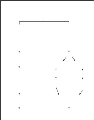
Flow chart of human sex differentiation
BIPOTENTIAL (UNDIFFERENTIATED) GONADS
XX |
|
|
XY |
|
|
|||
Absence of |
|
|
Presence of |
|||||
Y chromosome |
|
|
Y chromosome |
|||||
|
|
|
|
|
|
|
||
No TDF genes |
|
|
TDF genes |
|||||
(SRY) |
|
|
(SRY) |
|
|
|||
|
|
|
|
|
|
|
||
|
|
|
|
|
|
|
|
|
Ovary |
|
|
Testis |
|
|
|||
|
|
Leydig cells |
Sertoli cells |
|||||
|
|
|||||||
|
|
|
|
|
|
|
|
|
|
|
|
|
|
|
|
|
|
Development |
|
|
|
|
|
|
||
Testosterone |
MIS |
|||||||
of Mullerian |
||||||||
|
|
|
|
|
|
|||
ducts |
5 α |
|
|
|
|
|
||
dihydro- |
Regression |
|||||||
|
|
|||||||
|
|
testosterone (DHT) |
of Mullerian |
|||||
|
|
|
|
|
|
ducts |
||
|
|
|
|
Development of |
||||
Fallopian tubes, |
|
|
||||||
uterus, and |
|
|
Wolffian ducts |
|||||
upper vagina |
|
|
|
|
|
|
||
|
|
|
|
|
|
|||
|
|
|
|
|
|
|||
|
|
|
|
|
|
|
||
Female genitalia |
|
|
Male genitalia |
|||||
FEMALE PHENOTYPE |
|
|
MALE PHENOTYPE |
|||||
Flow chart of male and female sex differentiation from conception through development. (Gale Group)
way. This means that enough SRY protein must be made early in development (before seven weeks) to turn an undifferentiated gonad into a testis. If enough SRY is not present or if it is present too late in development, the gonad will shift into the female pathway.
Other genes in sex development
Several other genes have been found that are involved in the development of human sex, including the gene SOX9. Mutations or alterations in this gene can cause a condition called camptomelic dysplasia. People with camptomelic dysplasia have bone and cartilage changes. SOX9 alterations also cause male to female sex reversal in most affected individuals (male chromosomes and female features). As of 2001, it is not known how SRY, SOX9, and other genes in the sexual developmental pathway interact to turn an undifferentiated gonad into a testis or an ovary.
Resources
PERIODICALS
Zenteno, J.C., et al. “Clinical Expression and SRY Gene Analysis in XY Subjects Lacking Gonadal Tissue.”
American Journal of Medical Genetics 99 (March 15, 2001): 244-47.
Y) region determining (sex SRY
G A L E E N C Y C L O P E D I A O F G E N E T I C D I S O R D E R S |
1093 |