

Gale Encyclopedia of Genetic Disorder / Gale Encyclopedia of Genetic Disorders, Two Volume Set - Volume 2 - M-Z - I
.pdfSickle cell disease
The lower leg of this woman has ulcerated, necrotic tissue, resulting from sickle cell anemia. (Custom Medical Stock Photo, Inc.)
Treatment and management
There are several practices intended to prevent some of the symptoms and complications of sickle cell disease. These include preventative antibiotics, good hydration, immunizations, and access to comprehensive care. Maintaining good health through adequate nutrition, avoiding stresses and infection, and getting proper rest is also important. Following these guidelines is intended to improve the health of individuals with sickle cell disease.
Penicillin
Infants are typically started on a course of penicillin that extends from infancy to age six. Use of this antibiotic is meant to ward off potentially fatal infections. Infections at any age are treated aggressively with antibiotics. Vaccines for common infections, such as pneumococcal pneumonia, are also recommended.
Pain management
Pain is one of the primary symptoms of sickle cell anemia, and controlling it is an important concern. The methods necessary for pain control are based on individual factors. Some people can gain adequate pain control through over-the-counter oral painkillers (analgesics). Other individuals, or painful events, may require stronger methods which can include administration of narcotics. Alternative therapies may be useful in avoiding or controlling pain, including relaxation, hydration, avoiding extremes of temperature, and the application of local warmth.
Blood transfusions
Blood transfusions are not usually given on a regular basis but are used to treat individuals with frequent and severe painful events, severe anemia, and other emergencies. In some cases blood transfusions are given as a preventative measure, for example to treat spleen enlargement or prevent a second stroke (or a first stroke in an individual shown to be at high risk).
Regular blood transfusions have the potential to decrease formation of hemoglobin S, and reduce associated symptoms. However, there are limitations and risks associated with regular blood transfusions, including the risk of blood-borne infection and sensitization to proteins in the transfused blood that can make future transfusions very difficult. Most importantly, chronic blood transfusions can lead to iron overload. The body tends to store excess iron, such as that received through transfusions, in various organs. Over time, this iron storage can cause damage to various tissues and organs, such as the heart and endocrine organs.
Some of this damage can be prevented by the administration of a medication called desferoxamine that helps the body to eliminate excess iron through the urine. Alternately, some individuals receive a new, non-stan- dard treatment called erythrocytapheresis. This involves the automated removal of sickle cells and is used in conjunction with a reduced number of regular transfusions. This treatment helps to reduce iron overload.
Hydroxyurea
Emphasis is being placed on developing drugs that treat sickle cell anemia directly. The most promising of these drugs since the late 1990s has been hydroxyurea, a drug that was originally designed for anticancer treatment. Hydroxyurea has been shown to reduce the frequency of painful crises and acute chest syndrome in adults, and to lessen the need for blood transfusions. Hydroxyurea, and other related medications, seem to work by inducing a higher production of fetal hemoglobin. The major side
1054 |
G A L E E N C Y C L O P E D I A O F G E N E T I C D I S O R D E R S |
effects of the drug include decreased production of platelets, red blood cells, and certain white blood cells. The effects of long-term hydroxyurea treatment are unknown.
Bone marrow transplantation
Bone marrow transplantation has been shown to cure sickle cell anemia in some cases. This treatment is reserved primarily for severely affected children with a healthy donor whose marrow proteins match those of the recipient, namely a brother or sister who has inherited the same tissue type. Indications for a bone marrow transplant are stroke, recurrent acute chest syndrome, and chronic unrelieved pain.
Bone marrow transplantations tend to be the most successful in children; adults have a higher rate of transplant rejection and other complications. There is approximately a 10% fatality rate associated with bone marrow transplants done for sickle cell disease. Survivors face potential long-term complications, such as chronic graft- versus-host disease (an immune-mediated attack by the donor marrow against the recipient’s tissues), infertility, and development of some forms of cancer. A relatively recent advance in transplantation involves the use of donor stem cells obtained from cord blood, the blood from the placenta that is otherwise discarded following the birth of a new baby. Cord blood cells, as opposed to fully mature bone marrow cells, appear to be less likely to result in graft-versus-host disease in the recipient. This increases the safety and efficacy of the transplant procedure.
Surgery
Certain surgical interventions are utilized in the treatment of specific sickle cell-related complications. Removal of a dysfunctioning gallbladder or spleen can often lead to improvements in health. Investigations are currently underway to establish the efficacy of hip coring surgery, in which a portion of affected bone is removed to treat avascular necrosis of the hip. The hope is that this may provide an effective treatment to alleviate some pain and restore function in the affected hip.
Psychosocial support
As in any lifelong, chronic disease, comprehensive care is important. Assistance with the emotional, social, family-planning, economic, vocational, and other consequences of sickle cell disease can enable affected individuals to better access and benefit from their medical care.
Prognosis
Sickle cell disease is characteristically variable between and within affected individuals. Predicting the
course of the disorder based solely on genes is not possible. Several factors aside from genetic inheritance determine the prognosis for affected individuals, including the frequency, severity, and nature of specific complications in any given individual. The availability and access of comprehensive medical care also plays an important role in preventing and treating serious, acute complications, which cause the majority of sickle cell-related deaths. For those individuals who do not experience such acute events, life-expectancy is probably substantially greater than the average for all people with sickle cell disease. The impact of recent medical advances supports the hypothesis that current life-expectancies may be significantly greater than those estimated in the early 1990s. At that time, individuals with SS disease lived to the earlyto mid-40s, and those with SC disease lived into the upper 50s on average. With early detection and comprehensive medical care, most people with sickle cell disease are in fairly good health most of the time. Most individuals can be expected to live well into adulthood, enjoying an improved quality of life including the ability to choose a variety of education, career, and family-planning options for themselves.
Resources
BOOKS
Beutler, Ernest. “The Sickle Cell Diseases and Related Disorders.” In Williams Hematology, edited by Ernest Beutler, et al. 5th ed. New York: McGraw-Hill, 1995.
Bloom, Miriam. Understanding Sickle Cell Disease. Jackson, MS: University Press of Mississippi, 1995.
Embury, Stephen H., et al., eds. Sickle Cell Disease: Basic Principles and Clinical Practice. New York: Raven Press, 1994.
Reid, C.D., S. Charache, and B. Lubin, eds. Management and Therapy of Sickle Cell Disease. 3rd ed. National Institutes of Health Publication No. 96-2117, 1995.
PERIODICALS
Adams, R.J., et al. “Prevention of a First Stroke by Transfusions in Children with Sickle Cell Anemia and Abnormal Results on Transcranial Doppler Ultrasonography.” New
England Journal of Medicine 339 (1998): 5-11.
Davies, Sally C. “Management of Patients with Sickle Cell Disease.” British Medical Journal 315 (September 13, 1997): 656.
Golden, C., L. Styles, and E. Vichinsky. “Acute Chest syndrome and Sickle Cell Disease.” Current Opinion in Hematology
5 (1998): 89-92.
Hoppe, C., L. Styles, and E. Vichinsky. “The Natural History of Sickle Cell Disease.” Current Opinion in Pediatrics 10 (1998): 49-52.
Kinney, T.R., et al. “Safety of Hydroxyurea in Children with Sickle Cell Anemia: Results of the HUG-KIDS Study, A Phase I/II Trial.” Blood 94 (1999): 1550-1554.
Platt, O., et al. “Mortality in Sickle Cell Disease: Life Expectancy and Risk Factors for Early Death.” New England Journal of Medicine 330 (1994): 1639-1644.
disease cell Sickle
G A L E E N C Y C L O P E D I A O F G E N E T I C D I S O R D E R S |
1055 |

Simpson-Golabi-Behmel syndrome
Reed, W., and E.P. Vichinsky. “New Considerations in the Treatment of Sickle Cell Disease.” Annual Review of Medicine 49 (1998): 461.
Schnog, J.B., et al. “New Concepts in Assessing Sickle Cell Disease Severity.” American Journal of Hematology 58 (1998): 61-66.
Serjeant, Graham R. “Sickle-Cell Disease.” The Lancet 350 (September 6, 1997): 725.
Singer, S.T., et al. “Erythrocytapheresis for Chronically Transfused Children with Sickle Cell Disease: An Effective Method for Maintaining a Low Hemoglobin S Level and Reducing Iron Overload.” Journal of Clinical Apheresis 14 (1999): 122-125.
Xu, K., et. al. “First Unaffected Pregnancy Using Preimplantation Genetic Diagnosis for Sickle Cell Anemia.” Journal of the American Medical Association 281 (1999): 17011706.
ORGANIZATIONS
Sickle Cell Disease Association of America, Inc. 200 Corporate Point Suite 495, Culver City, CA 90230-8727. (800) 4218453. Scdaa@sicklecelldisease.org. http://sicklecelldisease
.org/ .
Jennifer Bojanowski, MS, CGC
Siewert syndrome see Kartagener syndrome
Silver-Russell syndrome see Russell-Silver syndrome
Simpson dysmorphia syndrome (SDYS) see
Simpson-Golabi-Behmel syndrome
I Simpson-Golabi-Behmel
syndrome
Definition
Simpson-Golabi-Behmel syndrome (SGBS) is a rare X-linked recessive inherited condition. It causes general overgrowth in height and weight. Individuals with SGBS also have characteristic facial features in childhood which tend to become less obvious in adulthood.
Description
SGBS is also known as Simpson dysmorphia syndrome (SDYS), bulldog syndrome, Golabi-Rosen syndrome, and dysplasia gigantism syndrome X-linked (DGSX). SGBS is a rare X-linked recessive inherited condition. Individuals with this condition have increased
height and weight for their age; a broad, stocky appearance; a large protruding jaw; a short, broad nose; incomplete closure of the roof of the mouth (cleft palate); and broad, short hands and fingers. Individuals with SGBS are usually taller than average. The characteristic features usually become less apparent in adulthood. There are at least two genes for SGBS. Both genes are located on the X chromosome.
Genetic profile
SGBS is caused by an alteration (mutation) in one of two genes on the X chromosome. Chromosomes are units of hereditary material passed from a parent to a child through the egg and sperm. The information on the chromosomes is organized into units called genes. Genes contain information necessary for normal human growth and development. Each cell in the body usually contains 46 chromosomes, arranged as 23 pairs. Twenty-two pairs of chromosomes are the same in males and females. The twenty-third pair is the sex chromosomes: females have two X chromosomes and males have an X and a Y chromosome. There are two genes on the X chromosome that can cause SGBS. The first gene is responsible for making a protein called glypican-3 (GPC3). The exact role of GPC3 is not known but it is thought to play a role in growth and development. When the gene for GPC3 is altered, the signs and symptoms of SGBS result. A second candidate gene, which causes a more severe form of SGBS, is also located on the X chromosome. The function of this second gene is not known. Generally, individuals who have SGBS due to a gene alteration in the GPC3 gene are said to have SGBS type 1 (SGBS1) and individuals who have SGBS due to an alteration in the second gene on the X chromosome are said to have SGBS type 2 (SGBS2).
SGBS is inherited as an X-linked recessive condition. With X-linked recessive conditions, males are usually more severely affected than females. Females have two copies of the SGBS gene (because they have two X chromosomes) while males have one copy of SGBS gene (because they have one X chromosome). Females who have an alteration in one copy of the SGBS gene are said to be carriers of SGBS. Generally, carriers show minimal or no effects of the altered gene because they have a second normal copy of the gene that is able to compensate for the altered copy. Since males have only one working copy of the SGBS gene to start, if that gene is altered, they will develop SGBS. When carrier females have children, they are at risk to have a child with SGBS. In each pregnancy, carrier females have a 25% chance of having a child (always a son) with SGBS and a 25% chance of having a child (always a daughter) whom is a carrier of SGBS. Males who are affected with SGBS cannot pass
1056 |
G A L E E N C Y C L O P E D I A O F G E N E T I C D I S O R D E R S |
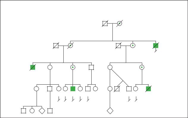
Simpson-Golabi-Behmel Syndrome
X-Linked Recessive
|
|
d.85y |
|
81y |
d.46y |
|
|
|
Pneumoria |
|
Extra vertebrae |
Unknown causes |
|
|
|
|
|
|
|
"slow" |
|
|
|
|
|
59y |
|
Stomach tumor d.32y |
|
61y |
56y |
54y |
51y |
|
of unknown type |
|
|
Prominent |
|
|
|
Mentally retarded |
|
|
chin |
|
|
|
38y |
36y |
29y 30y |
24y 21y 20y |
16y |
32y Preemie 28y 24y d.6 wks |
|
|
|
|
|
|
Unknown |
|
|
|
|
|
|
causes |
|
P |
2 |
P |
(Gale Group)
this condition to their sons (because their sons inherit the Y chromosome); however, all daughters of a male affected with SGBS will be carriers for the condition.
Demographics
SGBS is a rare inherited condition that primarily affects males from all ethnic groups. Female carriers for SGBS may show subtle features of the condition. It is not known precisely how many individuals are affected with SGBS.
Signs and symptoms
The spectrum of clinical features in SGBS is broad, ranging from very mild forms in carrier females to forms that are lethal in the newborn male. SGBS affects the face, hands, chest, abdomen, genitals, internal organs and overall growth.
Individuals with SGBS are larger than average at birth in height, weight, and head size. This overgrowth continues into adulthood with affected males being taller than average. Final height in males ranges from 74 in to 83 in (188 cm to 210 cm). There are typical facial characteristics in affected males including widely spaced
eyes, short nose, large mouth, large tongue, a groove in the lower lip, and teeth that do not align properly. Incomplete closure of the lip (cleft lip) and/or the roof of the mouth (cleft palate) can also occur. The large tongue and improperly aligned teeth can be a cause of speech difficulties.
The hands and feet of males with SGBS tend to be short and broad. Other hand abnormalities such as small nails, webbing of the skin between the fingers, and extra fingers/toes, is also common. Males with SGBS have extra nipples and some may have undescended testicles.
The internal organs are larger than average, particularly the liver, spleen, and kidneys. The kidneys may also have many cysts on them. A few individuals have been known to have lung and diaphragm abnormalities. Heart abnormalities can also occur in SGBS1 and have been a cause of death in several individuals under two years of age. These include conduction defects causing arrythmias. The stomach and intestines can also be affected, which may cause digestive problems. The bones may also be affected. Some individuals have an abnormal curving and twisting of the spine (scoliosis), extra ribs, and/or problems with the structure of the bones of the spine. The bony changes can be seen on x ray but may not cause any symptoms.
syndrome Behmel-Golabi-Simpson
G A L E E N C Y C L O P E D I A O F G E N E T I C D I S O R D E R S |
1057 |

Simpson-Golabi-Behmel syndrome
K E Y T E R M S
Amniocentesis—A procedure performed at 16-18 weeks of pregnancy in which a needle is inserted through a woman’s abdomen into her uterus to draw out a small sample of the amniotic fluid from around the baby. Either the fluid itself or cells from the fluid can be used for a variety of tests to obtain information about genetic disorders and other medical conditions in the fetus.
Chorionic villus sampling (CVS)—A procedure used for prenatal diagnosis at 10-12 weeks gestation. Under ultrasound guidance a needle is inserted either through the mother’s vagina or abdominal wall and a sample of cells is collected from around the fetus. These cells are then tested for chromosome abnormalities or other genetic diseases.
Chromosome—A microscopic thread-like structure found within each cell of the body and consists of a complex of proteins and DNA. Humans have 46 chromosomes arranged into 23 pairs. Changes in either the total number of chromosomes or their shape and size (structure) may lead to physical or mental abnormalities.
Despite their large size, newborns with SGBS tend to be floppy babies with decreased muscle tone. Due to this low muscle tone, there are several features that can result such as mouth breathing, a deformity of the chest wall (pectus excavatum), shoulders that droop, hernias, and undescended testicles.
There is an increased risk to develop tumors of the kidney (Wilms tumor) in SGBS in early childhood. This risk appears to be greatest in individuals under two years of age.
Most individuals with SGBS are of average intelligence, although some degree of mental impairment has been observed in males who are more severely affected. Individuals with SGBS may have psychological difficulties dealing with their distinctive facial appearance and speech difficulties, which often give the false impression that they are mentally impaired.
Diagnosis
The diagnosis of SGBS is based on the presence of certain clinical features and in some cases may be confirmed through genetic testing. Not all affected individuals will have all of the features associated with SGBS.
SGBS should be considered in an individual who is large in height, weight, and head circumference both before and after birth. Features of the condition that are almost always present include overgrowth; extra nipples; chest deformity; low muscle tone; and characteristic facial features including widely spaced eyes, short nose, large tongue and mouth, central groove of the lower lip, and improperly aligned teeth.
It may be possible to confirm the diagnosis of SGBS through genetic testing. Genetic testing for mutations in the GPC3 gene causing SGB1 is available. Genetic testing involves obtaining a blood sample from the affected individual in order to look for the specific disease-caus- ing mutation in the GPC3 gene. Since not all individuals with SGBS have mutations in the GPC3 gene, it may not be possible to confirm the diagnosis through genetic testing in all individuals suspected of having this condition. Genetic testing for the SGBS can be done on the developing baby before birth through amniocentesis or chorionic villus sampling if a mutation in the gene for GPC3 is first identified in an affected family member. Prenatal testing for parents of an affected individual should only be undertaken after the SGBS carrier status of the parents has been confirmed and the couple has been counseled regarding the risks of recurrence.
Treatment and management
The heart function of individuals with SGBS should be carefully monitored because it can be a cause of early death. Individuals with SGBS should be regularly followed by a heart specialist (cardiologist).
Individuals with SGBS are at increased risk to develop kidney tumors. They should be screened for possible kidney tumor development or other tumors of infancy for at least the first five years of life. Screening usually involves an ultrasound (sound wave picture) of the abdomen, including the kidneys.
The large tongue and improperly aligned teeth may lead to speech difficulties. Some individuals may require surgery to reduce the size of the tongue to aid with speech development or for cosmetic reasons. Individuals with speech difficulties may benefit from speech therapy.
Individuals with SGBS may benefit from psychological support and social support to help them reach an adequate level of self-esteem. They may also benefit from genetic counseling, which may provide them with further information on the condition itself and recurrence risks for future pregnancies.
Prognosis
The spectrum of clinical manifestations in SGBS is broad, varying from very mild forms in carrier females to
1058 |
G A L E E N C Y C L O P E D I A O F G E N E T I C D I S O R D E R S |

infantile lethal forms in affected males. As many as 50% of males affected with SGBS die in the newborn period. The cause of this high mortality is not known but may be related to heart defects. In one reported family with a severe form of SGBS causing death in the newborn period, the responsible gene was not glypican-3 but the second candidate gene on the X chromosome.
Resources
PERIODICALS
Hughes-Benzie, R.M., et al. “Simpson-Golabi-Behmel
Syndrome: Genotype/Phenotype Analysis of 18 Affected
Males From 7 Unrelated Families.” American Journal of
Medical Genetics 66 (1996): 227-234.
Neri, Giovanni, et al. “Clinical and Molecular Aspects of the
Simpson-Golabi-Behmel Syndrome.” American Journal of
Medical Genetics 79 (1998): 279-283.
ORGANIZATIONS
Beckwith-Wiedemann Support Network. 2711 Colony Rd., Ann Arbor, MI 48104. (734) 973-0263 or (800) 837-2976.http://www.beckwith-wiedemann.org .
National Organization for Rare Disorders (NORD). PO Box 8923, New Fairfield, CT 06812-8923. (203) 746-6518 or (800) 999-6673. Fax: (203) 746-6481. http://www
.rarediseases.org .
WEBSITES
“Simpson-Golabi-Behmel Syndrome, type 1.” Online Mendelian Inheritance in Man. http://www.ncbi.nlm.nih
.gov/entrez/dispomim.cgi?id=312870 . (March 9, 2001)
Nada Quercia, MS, CGC, CCGC
Fusion of the lower limbs, such as the legs of this infant, results from vital blood flow and nutrients being diverted away from the lower extremeties due to abnormal umbilical cord blood vessels. (Greenwood Genetic Center)
I Sirenomelia
Definition
Sirenomelia is a lethal birth defect of the lower body characterized by apparent fusion of the legs into a single lower limb. Other birth defects are always associated with sirenomelia, most commonly abnormalities of the kidneys, large intestines, and genitalia.
Description
This pattern of birth defects is associated with abnormal umbilical cord blood vessels. The normal fetus develops two umbilical arteries, which pump blood from the fetus to the placenta, and one umbilical vein, which returns blood from the placenta to the fetus. The umbilical arteries branch off the iliac arteries in the pelvis. The iliac arteries supply the legs and pelvic organs such as the
genitalia. Most babies with sirenomelia have only one umbilical artery and one vein. Rarely a baby with sirenomelia can have the typical two arteries and one vein with occlusion (blockage) of one artery.
In sirenomelia, the one functional artery is larger than normal and branches from the aorta high in the abdomen. Below this umbilical artery, the aorta becomes abnormally narrow. This type of single umbilical artery is known as a vitelline artery because it is thought to arise from the primitive vitelline arteries early in the life of the embryo. The vitelline arteries normally fuse a few weeks after conception to form the arteries that supply the gastrointestinal system and genitourinary system (superior mesenteric, inferior mesenteric, and celiac arteries). If the normal umbilical arteries do not form correctly as branches from the iliac arteries, then a vitelline artery might persist.
Sirenomelia
G A L E E N C Y C L O P E D I A O F G E N E T I C D I S O R D E R S |
1059 |

Sirenomelia
K E Y T E R M S
Aorta—The main artery located above the heart which pumps oxygenated blood out into the body. Many congenital heart defects affect the aorta.
Autosomal dominant—A pattern of genetic inheritance where only one abnormal gene is needed to display the trait or disease.
Iliac arteries—Arteries that supply blood to the lower body including the pelvis and legs.
Imperforate anus—Also known as anal atresia. A birth defect in which the opening of the anus is absent or obstructed.
Mermaid syndrome—Alternate name for sirenomelia, often used in older references.
Mutation—A permanent change in the genetic material that may alter a trait or characteristic of an individual, or manifest as disease, and can be transmitted to offspring.
Oligohydramnios—Reduced amount of amniotic fluid. Causes include non-functioning kidneys and premature rupture of membranes. Without amniotic fluid to breathe, a baby will have underdeveloped and immature lungs.
Stillborn—The birth of a baby who has died sometime during the pregnancy or delivery.
Teratogenic—Any agent that can cause birth defects or mental retardation in a developing fetus. Common teratogens are medications or other chemicals but they also include infections, radiation, maternal medical condition, and other agents.
Ultrasound—An imaging technique that uses sound waves to help visualize internal structures in the body.
The vitelline umbilical artery steals blood and nutrition from the lower body and diverts it to the placenta. This results in a small aorta and variable absence of the arteries that supply the kidneys, large intestine, and genitalia (renal, inferior mesenteric, and celiac arteries). Because of the loss of nutrition and blood flow, the lower limbs fail to form as separate limbs, the kidneys do not form or are malformed, the large intestine ends blindly in the abdominal cavity, the anus is imperforate, and the internal and external genitalia are absent or malformed.
The typical malformation of the lower limbs seen in babies with sirenomelia consists of apparent fusion of the legs. There is a spectrum of severity with severe cases hav-
ing one lower limb that tapers to a point with the absence of foot structures. In these severe cases there are only two bones present in the entire limb (a femur and presumably a tibia). On the mild end of the spectrum are babies with fusion of the skin of the lower limbs only. In these infants the feet may be fully formed with fusion at the ankles. All bones are fully formed and separate. Normally there are three bones in each leg—the femur in the upper leg (thigh) and the tibia and fibula in the lower leg (calf).
Other abnormalities of the upper body involving the heart, lungs, spine, brain, and arms can also be seen in this syndrome, however, not in every affected individual. It is unknown at this time why a single umbilical artery could cause these changes.
Single umbilical artery occurs in about 1% of all liveborn infants. In most of these infants the one umbilical artery is normally formed and not of vitelline origin. In these cases, the risk of other birth defects is low (about 8%). All infants born with a vitelline umbilical artery will have other malformations, the most common being sirenomelia.
Genetic profile
All cases of sirenomelia have occurred in families as isolated cases, and there is no known genetic cause. It is possible that sirenomelia is an autosomal dominant condition and because it is lethal, all cases represent a new mutation. Alternatively, it might be a multifactorial trait where multiple genes and environmental factors come together to cause this pattern of malformations. The fact that all cases have been isolated does not support this possibility. Sirenomelia is more common in twin pregnancies. This may give evidence to an environmental cause.
Demographics
Sirenomelia is rare, estimated to occur once in every 60,000 births. While the exact incidence in different populations is not known, sirenomelia has been reported in a variety of ethnic groups around the world. It is known to be more common in twin pregnancies and in babies born to mothers with diabetes mellitus.
Signs and symptoms
Abnormalities associated with sirenomelia include:
•absence of the kidneys or malformed non-functioning kidneys
•blind ending colon and imperforate anus
•small, absent, fused, or poorly formed pelvic bones
•small, absent, or poorly formed internal and external genitalia
1060 |
G A L E E N C Y C L O P E D I A O F G E N E T I C D I S O R D E R S |

•fusion of the lower limbs along the inner leg, from skin only to complete fusion with the appearance of only one leg
•death from underdeveloped and immature lungs caused by oligohydramnios
•birth defects in the upper body sometimes occur and include abnormalities in the heart, lungs, arms, spine, and brain
Diagnosis
The diagnosis is obvious at birth on examination of a baby, but prenatal diagnosis often occurs in the second trimester (weeks 13 through 26 of a pregnancy) by an ultrasound.
Treatment and management
Babies born alive with functioning kidneys may survive with appropriate surgical management. Operations to reconstruct the urinary and gastrointestinal outlet tracts are almost always needed. Other procedures and treatments depend of the extent of other birth defects. It appears that if a baby does survive, he or she will not have any mental delays.
Prognosis
Because of the birth defects involving the gastrointestinal tract and kidneys, sirenomelia is almost always fatal. About 50% of babies are stillborn (the baby has died before delivery) and 50% are liveborn with survival lasting a few minutes to a few days. There have been at least two reported cases of sirenomelia that have survived beyond the first month of life. These infants had normal functioning kidneys during their development.
Resources
PERIODICALS
De Silva, M.V., and W.D. Lakshman. “Sirenomelia Sequence (Mermaid Syndrome).” Ceylon Medical Journal 44 (March 1999): 34-5.
Stevenson, Roger E., et al. “Vascular Steal: The Pathogenic Mechanism Producing Sirenomelia and Associated Defects of the Viscera and Soft Tissues.” Pediatrics 78 (September 1986): 451-457.
Stocker, J.T., and S.A. Heifetz. “Sirenomelia. A Morphological Study of 33 Cases and Review of the Literature.”
Pespectives in Pediatric Pathology 10 (1987): 7-50. Valenzano, M., et al. “Sirenomelia: Pathological Features,
Antenatal Ultrasonographic Clues, and a Review of the Current Embryogenic Theories.” Human Reproductive Update 5 (January-February 1999): 82-6.
Randall Stuart Colby, MD
I Sjögren-Larsson syndrome
Definition
Sjögren-Larsson syndrome is an inherited disorder characterized by ichthyosis (scaly skin), speech abnormalities, mental retardation, and spasticity (a state of increased muscle tone with heightened reflexes). Severity is variable.
Description
Sjögren-Larsson syndrome is a rare genetic disorder inherited in an autosomal recessive fashion. First characterized by Swedish psychiatrist Torsten Sjögren in 1956 (and by Sjögren and Tage Larsson in 1957), they suggested that all Swedes with the syndrome are descended from one ancestor in whom a mutation (a genetic change) occurred about 600 years ago. The highest incidence of the disease occurs in northern Sweden.
In infancy, development of various degrees of scaling and reddened skin occurs, often accompanied by hyperkeratosis (thickening of the skin) on the outer skin layer. After infancy, skin on the arms, legs and abdomen often is dark, scaly, and lacking redness. Seizures and speech abnormalities may accompany skin symptoms. About half of children affected with the syndrome experience degeneration of the pigment in the retina of the eye.
Sjögren-Larsson syndrome is also sometimes known as SLS; congenital ichthyosis-mental retardation-spastic- ity syndrome; ichthyosis-spastic neurologic disorderoligophrenia syndrome; fatty aldehyde dehydrogenase deficiency (FALDH deficiency); fatty aldehyde dehydrogenase 10 deficiency (FALDH10 deficiency); or disorder of cornification 10 (Sjögren-Larsson Type). SjögrenLarsson syndrome is not to be confused with Sjögren syndrome; it is sometimes called the T. Sjögren syndrome to distinguish it from Sjögren syndrome (characterized by dry eyes and mouth), which was described by Swedish ophthalmologist Henrick Sjögren.
Genetic profile
Inheritance of Sjögren-Larsson syndrome is autosomal recessive. In autosomal recessive inheritance, a single abnormal gene on one of the autosomal chromosomes (one of the first 22 “non-sex” chromosomes) from both parents can cause the disease. Both of the parents must be carriers in order for the child to inherit the disease since recessive genes are expressed only when both copies in the pair have the same recessive instruction. Neither of the parents has the disease (since it is recessive).
syndrome Larsson-Sjögren
G A L E E N C Y C L O P E D I A O F G E N E T I C D I S O R D E R S |
1061 |

Sjögren-Larsson syndrome
K E Y T E R M S
Contracture—A tightening of muscles that prevents normal movement of the associated limb or other body part.
Diplegia—Paralysis affecting like parts on both sides of the body, such as both arms or both legs.
Hypertonia—Excessive muscle tone or tension, causing resistance of muscle to being stretched.
Ichthyosis—Rough, dry, scaly skin that forms as a result of a defect in skin formation.
Retinitis pigmentosa—Progressive deterioration of the retina, often leading to vision loss and blindness.
Spasticity—Increased muscle tone, or stiffness, which leads to uncontrolled, awkward movements.
Tetraplegia—Paralysis of all four limbs. Also called quadriplegia.
A child with both parents who carry the disease has a 25% chance having the disease; a 50% chance of being a carrier of the disease (but not affected by the disease, having both one normal gene and one gene with the mutation for the disorder); and a 25% chance of receiving both normal genes, one from each parent, and being genetically normal for that particular trait.
The gene for the Sjögren-Larsson syndrome, FALDH, is located on chromosome number 17 in band 17p11.2. The gene mutation that is responsible for the disorder is located near the center of the chromosome and is strongly associated the gene markers called D17S805 and ALDH10.
Demographics
Sjögren-Larsson syndrome is a rare disorder. The highest incidence occurs in northern Sweden. The mutation responsible for the disease is present in approximately 1% of the population in northern Sweden. All Swedes with the syndrome are believed to be descendents of one ancestor in whom a genetic change occurred about 600 years ago. (The phenomenon wherein everyone is descended from one person within what was once a tiny group of people is called founder effect.) The disease also occurs in members of families of other European, Arabic, and native American descent, but is less prevalent. Sjögren-Larsson syndrome affects both males and females.
Signs and symptoms
There are several signs and symptoms of SjögrenLarsson syndrome. The major features of the disorder are the following:
•Skin: In infancy, development of various degrees of scaling and reddened skin occurs (ichthyosis), often accompanied by hyperkeratosis (thickening of the skin) on the outer skin layer. After infancy, skin on the arms, legs, and abdomen is often dark and scaly and lacking redness. Bruises are present at birth or soon after.
•Hair: Hair may be brittle.
•Extremities: Joint contracture and hypertonia cause resistance of joints to movement and of muscles to stretching. Most individuals with the syndrome never walk.
•Eyes: About half of the individuals with this syndrome have retinitis pigmentosa (pigmentary degeneration of the retina). Glistening white or yellow-white dots on the retina (ocular fundus) are characteristic. They may be an early sign of the disease, presenting at age 1–2, and may increase with age.
•Nervous system: Spastic diplegia or tetraplegia (paralysis) affecting arms and/or legs. About half of the individuals with this disorder have seizures.
•Urogenital system: Kidney diseases may be associated with this syndrome.
•Growth and development: Individuals with the disorder tend to be unusually short in stature. Mental retardation is characteristic. Speech disorders may be present.
Speech abnormalities, mental retardation, and seizures usually occur during the first two or three years of life.
Diagnosis
The clinical features of Sjögren-Larsson syndrome are often distinctive, and a pattern of anomalies may suggest the diagnosis. In addition to ichthyosis and spasticity at birth, glistening white or yellow-white dots on the retina may be an early sign of the disease, presenting in the first or second year of life. If they occur, speech abnormalities, mental retardation, and seizures present during the first two or three years of life.
Laboratory findings are important in diagnosing Sjögren-Larsson syndrome. A laboratory test for deficiency of an enzyme (a protein that catalyzes chemical reactions in the human body) called fatty aldehyde dehydrogenase 10 (FALDH10) will determine presence of the disease. Sjögren-Larsson is due to a deficiency of FALDH10, and the gene for the Sjögren-Larsson syndrome is the same as the FALDH10 gene.
1062 |
G A L E E N C Y C L O P E D I A O F G E N E T I C D I S O R D E R S |
Positive laboratory results for Sjögren-Larsson will include the following findings:
•Hexadeconal elevated in fibroblasts.
•Fatty alcohol NAD+ deficient in Sjögren-Larsson syndrome fibroblasts.
•Fatty aldehyde dehydrogenase (FALDH) deficiency.
Genetic counseling
Individuals with a family history of SjögrenLarsson syndrome may benefit from genetic counseling to learn about the condition including treatments, inheritance, testing, and options available to them so that they can make informed decisions appropriate to their families. A child with both parents who carry the Sjögren-Larsson gene mutation has a 25% chance having the disorder. Couples who have had one affected child have a 25% risk of having another child with the disorder in each pregnancy.
Prenatal testing
Families at risk to have a child with SjögrenLarsson syndrome may have the option of prenatal diagnosis. DNA can be extracted from fetal cells obtained by either chorionic villus sampling (usually done until 12 weeks gestation) or amniocentesis (usually done at 16–18 weeks gestation) and tested to determine if the altered gene in the family is present. These techniques usually require that the alteration in the gene has been identified previously in an affected family member.
Chorionic villus sampling is a procedure to obtain chorionic villi tissue for testing. Chorionic villi are microscopic, finger-like projections that emerge from the chorionic membrane and eventually form the placenta. The cells of the chorionic villi are of fetal origin so laboratory analysis can identify a number of genetic abnormalities of the fetus. Because the villi are attached to the uterus, however, there is a chance that maternal tissue may be analyzed rather than the fetal cells. If the sample is too small, it may be necessary to repeat the procedure. In addition, the quality of the chromosome analysis is usually not as good with chorionic villus sampling as with amniocentesis. The chromosomes may not be as long, and so it may not be possible to identify some of the smaller bands on the chromosomes.
Amniocentesis is a procedure that involves inserting a thin needle into the uterus, into the amniotic sac, and withdrawing a small amount of amniotic fluid (a liquid produced by the fetal membranes and the fetus that surrounds the fetus throughout pregnancy). DNA can be extracted from the fetal cells contained in the amniotic
fluid and tested for the specific mutation known to cause Sjögren-Larsson syndrome.
Treatment and management
Individuals with Sjögren-Larsson syndrome should be under routine health supervision by a physician who is familiar with the disorder, its complications, and its treatment. Supportive resources for individuals with SjögrenLarsson syndrome and their families should be provided. Some clinical improvement has been reported to occur with fat restriction in the diet and supplementation with medium-chain triglycerides.
Other treatment of the disorder is generally symptomatic.
•For dermatologic symptoms, various skin softening ointments are useful in reducing symptoms. Plain petroleum jelly may be effective, especially when applied while the skin is still moist, such as after bathing. Salicylic acid gel may also be effective. When using the ointment, skin is covered at night with an airtight, waterproof dressing. Lactate lotion is another effective treatment for the dermatologic symptoms.
•For ocular symptoms, regular care from a qualified opthamologist is important.
•To control seizures, anti-convulsant medications may be helpful.
•Speech therapy and special education services may be helpful.
Prognosis
Prognosis is variable depending upon the severity of the disease. Sjögren-Larsson does not generally lead to shortened life span.
Resources
BOOKS
Medical Genetics, edited by Lynn B. Jorde, et al. 2nd ed. St. Louis: Mosby, 1999.
PERIODICALS
Rizzo, W.B. “Inherited Disorders of Fatty Alcohol Metabolism.”
Molecular Genetics and Metabolism (1998) 65: 63-73. Rizzo, W.B., G. Carney, and Z. Lin. “The Molecular Basis of
Sjögren-Larsson Syndrome: Mutation Analysis of the Fatty Aldehyde Dehydrogenase Gene.” American Journal of Human Genetics 65(December 1999): 1547-60.
Willemsen, M.A., et al. “Sjögren-Larsson Syndrome.” Journal of Pediatrics 136(February 2000): 261.
ORGANIZATIONS
Arc (a National Organization on Mental Retardation). 1010 Wayne Ave., Suite 650, Silver Spring, MD 20910. (800) 433-5255. http://www.thearclink.org .
syndrome Larsson-Sjögren
G A L E E N C Y C L O P E D I A O F G E N E T I C D I S O R D E R S |
1063 |