

Laser-Tissue Interactions Fundamentals and Applications - Markolf H. Niemz
.pdf188 4. Medical Applications of Lasers
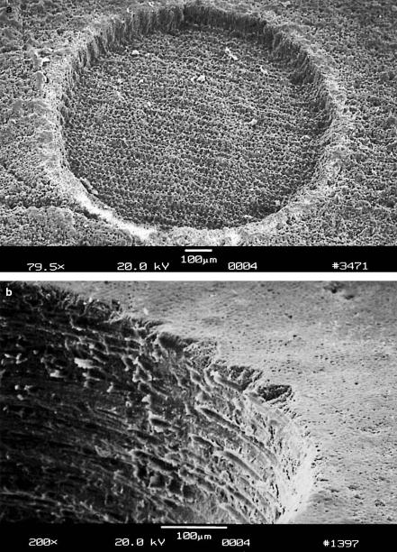
Fig. 4.31. (a) Cavity in healthy enamel achieved with 160000 pulses from a Nd:YLF laser (pulse duration: 30ps, pulse energy: 1mJ). (b) Cavity in carious enamel achieved with 16000 pulses from a Nd:YLF laser (pulse duration: 30ps, pulse energy: 1mJ)
4.2 Lasers in Dentistry |
189 |
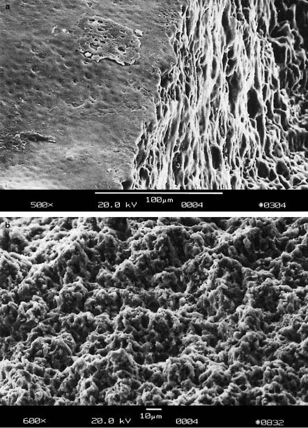
Fig. 4.32. (a) Cavity wall in healthy enamel achieved with a Nd:YLF laser (pulse duration: 30ps, pulse energy: 1mJ). (b) Cavity bottom in healthy enamel achieved with a Nd:YLF laser (pulse duration: 30ps, pulse energy: 1mJ)
190 4. Medical Applications of Lasers
Fig. 4.33. (a) Cavity in carious enamel achieved with 2500 pulses from a Nd:YLF laser (pulse duration: 30ps, pulse energy: 1mJ). (b) Cavity wall achieved with a conventional diamond drill

4.2 Lasers in Dentistry |
191 |
where K = 5.0 × 10⁴ N, and D is the impact of a diamond tip cut at an angle of 136◦ and expressed in millimeters. The results of hardness tests after exposure to picosecond Nd:YLF pulses are presented in Table 4.3. According to Niemz (1995c), no significant alteration in hardness is observed in exposed and unexposed enamel. As expected, though, dentin appears much softer due to its lower content of hydroxyapatite.
Table 4.3. Mean hardness values of teeth before and after exposure to a Nd:YLF laser (pulse duration: 30ps, pulse energy: 1mJ)
|
D (mm) |
HV (N/mm2) |
|
|
|
Exposed enamel |
5.9 |
2660 |
Unexposed enamel |
5.8 |
2760 |
Unexposed dentin |
11.5 |
700 |
|
|
|
Histology. The most important touchstone for the introduction of a new therapeutic technique is the biological response of the tissue, i.e. the survival of cells. Histologic sections enable specific statements concerning the condition of cells due to highly sophisticated staining techniques. In Fig. 4.5a (page 157), the dentin–pulp junction of a human tooth is shown. It was located underneath a 1×1mm2 area exposed to 16000 pulses from a Nd:YLF laser. Along the junction, several odontoblasts are clearly visible. They have not intruded into the dentin and have a similar appearance as in unexposed teeth. Thus, potential shock waves do not have a detectable impact on the pulp – not even on a cellular level.
Polarized Microscopy. Polarized microscopy is an e cient tool for detecting alterations in optical density which might arise from the exposure to shock waves. If these shock waves are reflected, e.g. at the enamel–dentin junction, such alterations might even be enhanced and should thus become evident. For polarized microscopy, exposed teeth are dehydrated in an upgraded series of ethanol. Afterwards, they are kept in fluid methacrylate for at least three days. Within the following period of seven days, polymerization takes place in a heat chamber set to 43◦C. Then, the embedded samples are cut into 100μm thick slices using a saw microtome. Finally, the slices are polished and examined with a polarized light microscope. In Fig. 4.5b (page 157), the enamel–dentin junction of a human tooth is shown. It was located underneath a 1×1mm2 area exposed to 16000 pulses from a Nd:YLF laser. In the top and bottom parts of the picture, dentin tubuli and enamel prisms are found, respectively. Due to the di erent optical densities of dentin and enamel, these two structures appear blue and yellow in the corresponding color photograph. However, no substantial alteration in color is observed within either the dentin or the enamel. Hence, no evidence of an altered optical density due to laser-induced shock waves is given.

192 4. Medical Applications of Lasers
From the results of the above tests, i.e. the negligibility of mechanical e ects, it can be concluded that the cavities shown in Figs. 4.31a–b were produced by means of plasma-induced ablation as discussed in Sect. 3.4. The main reason for these observations is that the applied pulse energies were just slightly above the threshold energy of optical breakdown. In Fig. 4.34, the ablation curves of healthy enamel, healthy dentin, and carious enamel are given, respectively. In healthy enamel, plasma sparking was already visible at approximately 0.2mJ. Then, when taking the corresponding focal spot size of 30μm into account, the ablation threshold is determined to be about 30J/cm2. For carious enamel, plasma generation started at roughly 0.1mJ, i.e. at a threshold density of 15J/cm2. In the range of pulse energies investigated, all three ablation curves are mainly linear. Linear regression analysis yields that the corresponding slopes in Fig. 4.34 are 1μm/0.2mJ, 3μm/0.2mJ, and 8μm/0.2mJ, respectively. Thus, the ablation e ciency increases from healthy enamel and healthy dentin to carious enamel. From the ablation volumes, we derive that – at the given laser parameters – approximately 1.5mm3 of carious enamel can be ablated per minute. To cope with conventional mechanical drills, a ten times higher ablation e ciency would be desirable. It can be achieved by increasing both the pulse energy and repetition rate. Then, the Nd:YLF picosecond laser might represent a considerable alternative in the preparation of hard tooth substances. The potential realization of such a clinical laser system is currently being evaluated.
Fig. 4.34. Ablation curves of carious enamel, healthy dentin, and healthy enamel, respectively, obtained with a Nd:YLF laser (pulse duration: 30ps, focal spot size: 30μm). Data according to Niemz (1994a) and unpublished data

4.2 Lasers in Dentistry |
193 |
The results with the Nd:YLF picosecond laser described above have proven that ultrashort laser pulses are a considerable alternative to the mechanical drill for the removal of caries. Due to the recent progress in the generation of even shorter laser pulses, femtosecond lasers have become very promising tools, as well. First experiments with these ultrashort pulse durations have been reported by Niemz (1998). The cavity shown in Fig. 4.35 was achieved with 660000 pulses from a Ti:Sapphire femtosecond laser at a pulse duration of 700fs. The geometrical accuracy of the cavity and its steep walls are fascinating.
Fig. 4.35. Cavity in healthy enamel achieved with a Ti:Sapphire laser (pulse duration: 700fs, pulse energy: 100μJ). Photograph kindly provided by Dipl.-Ing. Bauer (Hannover), Dr. Kasenbacher (Traunstein), and Dr. Nolte (Jena)
In Fig. 4.36, a similar cavity was produced with the same laser but at a larger spacing of adjacent laser pulses. The impacts of individual line scans are clearly visible at the bottom of the cavity. The cavity itself is very clean and of superior quality, if compared to results achievable with conventional diamond drills. Finally, Fig. 4.37 demonstrates the extremely high precision provided by femtosecond lasers. In between exposed areas, unexposed bars remain with a width of less than 10μm. No mechanical drill is able to achieve similar results. Furthermore, Fig. 4.37 provides the ultimate proof that mechanical shock waves are negligible when applying femtosecond laser pulses at a suitable energy.

194 4. Medical Applications of Lasers
Fig. 4.36. Cavity in healthy enamel achieved with a Ti:Sapphire laser (pulse duration: 700fs, pulse energy: 100μJ). Photograph kindly provided by Dipl.-Ing. Bauer (Hannover), Dr. Kasenbacher (Traunstein), and Dr. Nolte (Jena)
Fig. 4.37. Cavities in healthy enamel achieved with a Ti:Sapphire laser (pulse duration: 700fs, pulse energy: 100μJ). Photograph kindly provided by Dipl.-Ing. Bauer (Hannover), Dr. Kasenbacher (Traunstein), and Dr. Nolte (Jena)

4.2 Lasers in Dentistry |
195 |
One very important issue associated with dental laser systems is the temperature increase inside the pulp where odontoblasts, blood vessels, and tooth nerves are located. Only increments below 5◦C are tolerable, otherwise thermal side e ects might occur as discussed in Sect. 3.2. Moreover, the feeling of pain is induced at pulp temperatures which exceed approximately 45◦C. It is thus very important to remain below these temperatures when striving for clinical applicability. In Fig. 4.38, the temperature increments induced by a picosecond Nd:YLF laser at a repetition rate of 1kHz are summarized. For this experiment, human teeth were cut into 1mm thick slices. On one surface of these slices, the laser beam was scanned over a 1 × 1mm2 area, while the temperature was measured at the opposite surface by means of a thermocouple. The observed temperature increments depend on the number of consecutive pulses as well as on the total duration of exposure. We have already stated in Sect. 3.2 that high repetition rates can also induce an increase in temperature even when using picosecond pulses. Hence, a higher temperature is obtained when applying 30 instead of only 10 consecutive pulses before moving the focal spot to the next position. The total duration of exposure also a ects the final temperature, although the increase during the first minute is most significant. From these results, we can conclude that up to approximately 10 consecutive pulses may be applied to a tooth at a repetition rate of 1kHz if the temperature in the pulp shall not increase by more than 5◦C.
Fig. 4.38. Increase in temperature in a distance of 1mm from cavities achieved with a Nd:YLF laser (pulse duration: 30ps, pulse energy: 1mJ, repetition rate: 1kHz). Unpublished data

196 4. Medical Applications of Lasers
A novel dental application of lasers has recently been proposed by Niemz (1995d). Using a confocal laser scanning microscope, the space to be occupied by dental fillings can be very precisely determined. The confocal principle requires that only light reflected within the focal pane is detected. Thereby, very high axial resolutions can be obtained compared to conventional microscopy. With the confocal microscope, several layers of the cavity to be filled are scanned. From the reflected intensities, a three-dimensional plot of the cavity is calculated as shown in Fig. 4.39a. These data can be inverted to form a direct pattern for the milling of inlays or crowns with a CNC-machine as demonstrated in Fig. 4.39b.
Fig. 4.39. (a) Three-dimensional plot of a laser-induced cavity captured with a confocal laser scanning microscope. The cavity was achieved with a Nd:YLF laser (pulse duration: 30ps). (b) Inverted image of the same cavity as calculated with a computer. Reproduced from Niemz et al. (1995d). c 1995 Springer-Verlag
4.2 Lasers in Dentistry |
197 |
Laser Treatment of Soft Dental Tissue
Several studies have been reported on the use of a CO2 laser in the management of malignant, premalignant, and benign lesions of the oral mucosa, e.g. by Strong et al. (1979), Horch and Gerlach (1982), Frame et al. (1984), and Frame (1985). Since the oral environment is very moist, radiation from the CO2 laser is predestined for such purposes because of its high absorption. When treating a soft tissue lesion inside the mouth, the surgeon has a choice of two techniques – either excision or vaporization. It is usually preferable to excise the lesion because this provides histologic evidence of its complete removal and confirmation of the preceding diagnosis. During vaporization, a risk always remains that not all altered tissue is eliminated. Hence, if a pathologic lesion is vaporized, a biopsy should be obtained from the adjacent tissue after the treatment.
The CO2 laser is particularly useful for small mucosal lesions. Most of them can be vaporized at a power of 5–10W in pulsed or CW mode. After laser treatment, the wound is sterile and only minimal inflammatory reactions of the surrounding tissue occur. One major advantage is that there is no need to suture the wound, since small blood vessels are coagulated and bleeding is thus stopped. The wound edges can even be smoothed with a defocused beam. Wound healing usually occurs within a period of two weeks, and the process of reepithelialization is complete after about 4–6 weeks. Frame (1985) states that patients tolerate the procedure well and initially complain of little pain. However, the treated area may become uncomfortable for approximately 2–3 weeks.
Cases of leukoplakia are di cult to treat by conventional surgery, since they are frequently widespread inside the mouth. The lesion is usually outlined with a focused CO2 laser beam for easy visualization. Afterwards, it is vaporized with a defocused beam at a power of about 15–20W. According to Horch (1992), laser-treated leukoplakias heal very well, and there is only little evidence of recurrence. Even leukoplakias on the tongue and lips can be treated without losses in performance of these organs.
Malignant lesions require a higher laser power of approximately 20–30W to deal with the bulk of the tumor. Lanzafame et al. (1986) state that the recurrence of local tumors is reduced when using the CO2 laser rather than a mechanical scalpel. The thermal e ect of the radiation is made responsible for this observation. However, it is questionable whether laser treatments of malignant oral tumors are successful during a longer follow-up period, since metastases have often already spread to other parts of the body. In these cases, laser application is restricted to a palliative treatment. A specimen, for biopsy can also be excised with a CO2 laser as one would do with a conventional scalpel. More recently, Patel (1988) reported on the application of a Nd:YAG laser in the treatment of oral cancer. However, in the treatment of soft dental tissues, this laser has not gained clinical relevance so far.