

Micro-Nano Technology for Genomics and Proteomics BioMEMs - Ozkan
.pdf8
From One-Bead One-Compound Combinatorial Libraries to Chemical Microarrays
Kit S. Lam, Ruiwu Liu, Jan Marik, and Pappanaicken R. Kumaresan
University of California Davis Cancer Center, Division of Hematology/Oncology and Department of Internal Medicine, University of California Davis, 4501 X Street, Sacramento, CA 95817
8.1. INTRODUCTION
In 1984, Geysen et al. reported the synthesis of peptides on polyethylene pins in a 96-well foot-print (the multi-pin system) [45]. In 1992, Frank et al. described the synthesis of multiple different peptides on cellulose paper [40]. The number of compounds prepared by these parallel synthesis techniques is limited, but they represent the early development of synthetic combinatorial chemistry and chemical arrays. Geysen et al. applied the multi-pin system to synthesize peptide mixtures on individual pins, and applied iterative screening (enzyme-linked immunoabsorbant assay) and synthesis approaches to elucidate the chemical structure of the biologically active peptides [46]. Fodor et al. first reported the minaturization of the chemical arrays by using light-directed photolithographic chemical synthesis techniques to construct 1042 peptides on a glass chip [38]. Such spatially-addressable peptide microarrays were probed with fluorescent-labeled antibodies, and quantitated with a fluorescent scanner. About the same time, we described the use of split-mix synthesis method to generate millions of random peptide-beads such that each bead displayed only one peptide entity [84]. These “one-bead one-compound” (OBOC) peptide libraries were then screened with an enzyme-linked colorimetric assay, and positive peptide-beads were physically isolated for structural analysis. Such bead libraries can be considered as chemical microarrays that are not addressable but spatially separable. In this mini-review, the development and applications of the OBOC combinatorial library methods and chemical
284 |
KIT S. LAM ET AL. |
microarray techniques will be reviewed. For chemical microarrays, we shall focus our attention on the various peptide and small molecule microarray techniques. Protein microarrays will only be briefly addressed. Other combinatorial library methods, such as the phagedisplayed peptide libraries [24, 28, 148], positional scanning library methods [132], and affinity column library methods [186] will not be discussed here.
8.2. OBOC PEPTIDE LIBRARIES
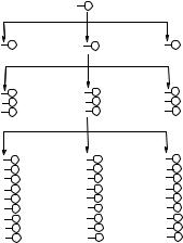
In 1991, we recognized that by using a “split-mix” synthesis method (Figure 8.1A), we could generate huge peptide libraries on beads [84]. Since each bead is exposed to only one building block at each coupling cycle and the reaction is driven to completion, each bead displays only one peptide-entity. Each 80–100 µm bead contains approximately 100 pmole or 1013copies of the same peptide. The synthesis of such OBOC combinatorial libraries is highly efficient. For example, a heptapeptide bead library consisting of 20 amino acids can be synthesized within 2 to 4 days, which has 1.28 billion permutations (Figure 8.1B). TentaGel resin (Rapp Polymere, Tubingen, Germany) is a good choice for OBOC library. This resin consists of a cross-linked polystyrene core grafted with 3000–4000 Dalton amino-polyethyleneglycol (PEG) chains. These beads can be swollen in water as well as a wide range of organic solvents such as dimethyl formamide, dichloromethane, and toluene. Therefore, TentaGel resin is compatible with both organic synthesis and biological screening. For peptide library construction, Fmoc/t-But (9-fluorenylmethoxycarbonyl/tert- butyl) chemistry is preferred because unlike Boc/Bn (tert-butoxycarbonyl/benzyl) chemistry, hydrofluoric acid (HF) is not needed. HF is highly toxic, requires a special apparatus, and partially degrades the PEG chain on the resin.
A |
|
|
|
B |
|
|
|
|
|
Coupling step |
|
Split |
|
Number of permutations |
|||||
I |
C |
D |
E |
X |
201 |
= |
20 |
||
|
C |
D |
E |
XX |
202 |
= |
400 |
||
II (9 dipeptides) |
|
Mix /Split |
|
XXX |
203 |
= |
8,000 |
||
C |
D |
E |
XXXX |
204 |
= |
160,000 |
|||
|
|||||||||
|
|
||||||||
|
C C |
D C |
E C |
XXXXX |
205 |
= |
3,200,000 |
||
|
C D |
DD |
E D |
XXXXXX |
20 |
6 |
= |
64,000,000 |
|
|
C E |
D E |
E E |
|
|||||
III (27 tripeptides) |
|
Mix /Split |
|
XXXXXXX |
207 |
= |
1,280,000,000 |
||
C |
D |
E |
|
|
|
|
|
||
|
|
|
|
|
|
||||
C C C C C D C C E
CD C
CD D
CD E
CE C
CE D
CE E
D C C D C D
DC E
DD C
DD D
DD E
DE C
DE D
DE E
E C C E C D
EC E
ED C
EDD
ED E
EE C
EE D
EE E
FIGURE 8.1. A: Synthetic scheme of the “split synthesis” method to generate a one-bead one-compound combinatorial library; B: Number of permutations for random peptide libraries constructed with 20 amino acids per coupling cycle.
FROM ONE-BEAD ONE-COMPOUND COMBINATORIAL LIBRARIES |
285 |
A B C
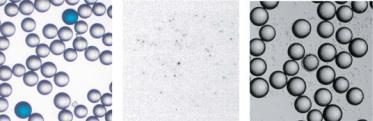
FIGURE 8.2. Various approaches to screen OBOC combinatorial libraries: (A) enzyme-linked colorimetric assay for target protein binding, (B) 33P phosphorylation functional assay for protein kinase substrates (autoradiogram, low power), and (C) whole cell binding assay for cell surface binding ligands.
The well-established on-bead screening assays used in OBOC libraries are highly efficient. Literally millions of compounds can be screened in parallel for a specific acceptor molecule (receptor, antibody, enzyme, virus, whole cell, etc) within a day or two. With various screening techniques, compound-beads with a specific biological, chemical, or physical property can be rapidly identified. We have employed an enzyme-linked colorimetric assay, similar to Western blot, to identify ligands for an anti-β-endorphin antibody [77], streptavidin [81] (Figure 8.2A), avidin [79], an anti-insulin monoclonal antibody that recognizes a discontinuous epitope [78], surface idiotype of B-cell lymphoma cell lines [83], and MHCClass I molecules [153]. We reported the use of a 32P or 33P phosphorylation assay and an autoradiographic method to identify specific and efficient peptide substrates for protein kinases (Figure 8.2B) [85, 172, 173]. We have also described the use of a whole cell binding assays in which bead-libraries are mixed with live cells (Figure 8.2C) to identify cell surface binding peptide ligands specific for prostate cancer, non-small cell lung cancer, and lymphoma cells [27, 86, 124, 129]. Meldal et al. reported using a fluorogenic quench screening method to identify protease substrates from an OBOC library [106]. Highly porous PEGbased resin (PEGA beads) was used for the peptide library construction because it allowed the enzyme to gain access to the bead interior. Those beads with the active substrates were cleaved by specific protease, resulting in the removal of the quencher and therefore appearance of fluorescent signals. These fluorescent-labeled beads were detected and isolated under a fluorescent microscope, or isolated by a fluorescent activated bead sorter (COPASTM BIOBEAD, Union Biometrica, Inc, Somerville, MA). Edman sequence analysis of positive beads reveals the substrate sequence, the cleavage point, and the degree of cleavage. This method is a powerful tool to investigate the protease activity and specificity since it gives a complete map of the substrate specificity and affinity for the cleavage site. Combining the OBOC concept and fluorogenic quench screening strategy, Meldal et al. later reported a so-called one-bead two-compounds approach to directly identify protease inhibitors. In this method, each bead contains a putative protease inhibitor along with a fluorescencequenched substrate for the protease [50, 105]. Very recently, Juskowiak et al. employed a novel on-bead screening method to identify short peptides that were capable of converting to fluorgenic compounds under an ambient photooxidative condition [63]. In this method, a random peptide bead library was illuminated with a tungsten-halogen lamp while the bead mixture was agitated with a stream of air bubbles. After illumination at 40–50 ◦C for 2 days,
286 |
KIT S. LAM ET AL. |
the resulting fluorescent beads were detected under a fluorescent microscope. Kodadek and co-workers recently reported the use of fluorescent-labeled proteins as probes to screen OBOC diverse peptoid (N-substituted oligoglycine) libraries [4]. They also reported the use of quantum dots, rather than organic fluorescent dye, as a fluorophor to label the target protein [121]. The main advantage of this latter library screening approach is that the signal to noise ratio is high.
Thus far, only a few groups have reported on the release of compounds from OBOC libraries for solution phase assays. We have described the use of the OBOC combinatorial libraries in a solution phase screening assay in which the compound-beads were immobilized in a thin layer of soft agar together with cancer cells [145]. After cleavage from the linker, compounds from each bead were released and diffused outward. A clear zone of growth inhibition around the positive bead was detected by adding 3-(4,5-dimethylthazol-2-yl)- 2,5-diphenyltetrazolium bromide (MTT) to the culture plate, in which only the live cells were stained purple. Jayawickreme et al. reported a similar ultra-high throughput assay approach termed “cell-based lawn format” utilizing an in situ photocleavage method to release the compound [59, 60]. They first grew a confluent monolayer of indicator cells (frog melanophore cells that had been transfected with a specific receptor) in a petri dish. The cells were then covered with agarose that contained OBOC combinatorial peptidebeads. In response to an agonist released from a specific bead, the frog cells surrounding that bead would turn dark. Silen et al. has also reported the use of similar lawn-based in situ photocleavage assay to screen an OBOC small molecule library (triazines) to identify novel antimicrobial agents [151]. The sensitivity of this lawn-based screen method has been modified to detect antibacterial compounds with modest potency. These in situ releasable solution phase assays have great potential but will require further development before they can be reliably used for drug screening. For instance, special solid supports need to be developed such that all compounds will diffuse freely out of all beads into the surrounding media. An alternative approach to using solution phase assays to screen OBOC library is to release compounds from an individual or small collection of compound beads in a microtiter plate. The released compounds were then subjected to standard solution phase assays [144]. However, to retrieve minute amounts of soluble compounds (100 pmol) from one single 80–100 µm bead is not easy, and often there is not enough material from one single bead for biological assays. Schreiber and his co-workers expanded on the OBOC library methods by developing releasable OBOC libraries in microtiter plates using bigger beads (500 µm macrobeads) [14, 15, 22]. However, the capacity of these macrobeads is still rather limited (<0.1 µmol/bead). Further increases in bead size leads to incomplete coupling because reagent diffusion will become a limiting factor. We have addressed this problem by developing a “one-aggregate one-compound” method [102]. The loading capacity of each bead aggregate ranges between 1–10 µmol, i.e., 10–100 fold more than that of the macrobeads used by Schreiber’s group. Bead aggregates were prepared by cross-linking the TentaGel resin beads with glutaraldehyde. Each bead aggregate contains some colored beads with an orthogonal protecting group that can be chemically encoded during the library synthesis and retrieved for decoding after biological screening. Taking full advantage of the OBOC concept, this “one-aggregate one-compound” method is highly efficient and much more economical than the IRORI Nanokan R system (Discovery Partners International Inc) [116]. Such diverse solution phase libraries can be screened with the standard solution phase assay or used to print multiple replicates of chemical microarrays (see below).
FROM ONE-BEAD ONE-COMPOUND COMBINATORIAL LIBRARIES |
287 |
As mentioned above, positive compound-beads from screening are physically isolated under a microscope. For peptide libraries that consist of natural amino acids, the straightforward approach to determine the amino acid sequence of the positive bead is to use Edman Chemistry with an automatic protein sequencer (e.g., Procise 494, Perkin-Elmer/Applied Biosystems). The inclusion of unnatural amino acids in the construction of peptide libraries not only greatly increase the diversity of the library but also renders some of the peptide library members more resistant to proteolysis. We have developed methods to sequence peptides containing many unnatural α-amino acids [93]. However, Edman sequencing is time-consuming and expensive compared with mass spectrometry analysis. Youngquist et al. introduced ‘ladder sequencing’ to determine the full peptide sequence of single beads with matrix-assisted laser desorption/ionization time-of-flight mass spectrometry (MALDITOF MS) [180]. Later, several groups have modified this method with either replacement of the capping step by the partial incorporation of a methionine residue at each coupling stage [26] or use of the dual capping groups and analysis of molecular ion redundancy to directly elucidate the structure by mass spectrometry analysis [52]. Very recently, this method has been further improved by using an isotope-labeling strategy to terminate the N terminus, and analyzing the generated ladders with ion-extraction mass spectrometry [125].
8.3. ENCODED OBOC SMALL MOLECULE COMBINATORIAL LIBRARIES
Although peptides are useful for targeting extracellular macromolecules or cell surface receptors, they often cannot penetrate cell membranes and therefore are not good drug candidates for intracellular targets. Moreover, the diversity of peptides is limited by the peptide backbone. Therefore, the major effort in combinatorial chemistry has been on small molecule libraries. In OBOC small molecule combinatorial libraries, the chemical structure of the compound on a positive bead has to be determined either directly or via an encoding strategy. Currently, there is no reliable method to directly determine the chemical structure of small molecule on one single bead (100 pmol) isolated from a huge diversity library with, e.g., 150,000 members. Several research groups have developed various physical and chemical encoding methods to encode OBOC small molecule libraries. This subject has been reviewed [2, 8, 26, 80, 149, 175].
Chemical encoding is ideal for OBOC small molecule libraries. Chemical tags are added to the bead during the synthetic steps so that the synthetic history of each compound bead in the chemical library can be recorded. These chemical codes can then be decoded by spectroscopic or chromatographic methods such as HPLC (high performance liquid chromatography), GC (gas chromatography), MS, fluorescence, IR (infrared), NMR (nuclear magnetic resonance) spectroscopy, and electron capture. Several chemical encoding methods have been reported, such as using oligonucleotide tags [19], fluorophenyl ether tags [114, 119], secondary amine tags [37, 115], peptide tags [66, 118], and trityl-based mass tags [150]. However, these methods suffer two disadvantages. First, the coding structure may interfere with the screening assay involving the testing compound. Second, these chemical encoding methods require that the chemistry of adding the tag and synthesizing the library be orthogonal, resulting in nearly doubling the number of synthetic steps. To solve these two problems, we have recently developed a novel and robust peptide-based encoding strategy for OBOC small molecule combinatorial libraries [94]. In this method,
288
A
H2N
H2N
PNH
P2 B
P3_Z _ Y _ X
B
S _A
P2
|
|
|
P2 |
P1 |
P2 P1 |
|
|
|
P3_ Z_ Y _ X |
||
|
3_ |
_ |
_ |
|
|
P |
|
Z |
Y |
X |
P1 |
PNH |
S _A |
|
P2 |
||
|
CB
H2N |
_ |
_ |
_ |
|
|
B |
|
|
|
Z |
Y X |
Edman degradation |
_A |
||||
|
S |
|||||||
B |
|
|
_ |
A |
|
|
C |
|
|
|
S |
B |
C |
|
|||
C |
|
|
Chemical structure |
|||||
|
|
|
|
PTH _ Z PTH _ Y |
PTH _ X |
|||
Identify A, B, C |
S: scaffold |
|
|
|
A, B, C: building blocks |
|
X, Y, Z: amino acids |
|
P, P1, P2, P3: protecting groups |
KIT S. LAM ET AL.
B
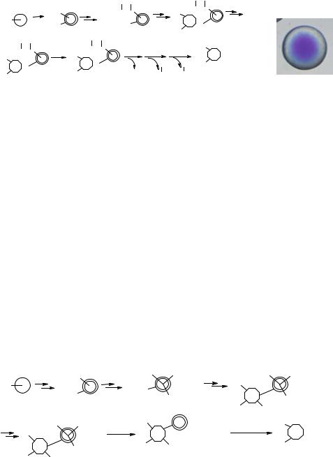
FIGURE 8.3. (A) General synthetic and decoding scheme of peptide-encoded library, (B) Photomicrograph of the topographically segregated bifunctional bead. Free amine at the core reacted with bromophenol blue.
the testing molecule is on the bead surface, and the coding tag is in the interior of the beads. Therefore, the coding tags will not interfere with screening (Figure 8.3). This encoding method is highly efficient as each of the building blocks is incorporated into the testing arm (bead surface) and coding peptide backbone (bead interior) simultaneously. Consequently, no additional synthetic steps are needed. After screening, the positive beads can be isolated, and the peptide coding tags, which consist of α-amino acids with side chains derivatized by the building blocks, can be readily decoded by Edman microsequencing.
Recently, we have further improved our encoding method by incorporating triple or quadruple cleavable coding arms in the bead interior so that mass spectrometry can be used for the decoding process (Figure 8.4) [155]. Prior to library synthesis, the inner core of each bead is derivatized with three or four different coding arms on a cleavable linker. Each of these coding arms contains a functional group that is identical or related to the functional groups on the scaffold of the testing compound to be synthesized. Similar to the abovementioned peptide-based encoding method, each building block will react with the testing and encoding arms simultaneously, thus eliminating many synthetic steps. After screening, the coding tags in the positive beads are released, followed by molecular mass determination using matrix-assisted laser desorption/ionization-Fourier transform mass spectrometry (MALDI-FTMS). This MS-decoding method may have broader applications than the
FmocNH |
P1-Linker |
|
Linker-P2 |
|
P1-Linker |
|
Linker-P2 |
|||
H2N |
BocNH |
BocNH |
|
|
P3 |
|
|
|
|
|
|
Linker-P3 |
|
S |
|
Linker-P3 |
|||||
|
|
|
|
|||||||
|
|
|
|
|
P2 |
P1 |
|
|
|
|
A-Linker |
Linker-B |
Cleave C |
S |
A-Linker |
MS analysis |
B |
S |
_ |
A |
|
C |
|
|
+ B-Linker |
|
|
|
|
|||
|
B |
Identify A, B, C |
C |
|
|
|||||
S |
Linker-C |
A |
C-Linker |
|
|
|
||||
|
|
Chemical structure |
||||||||
|
|
|
|
|
|
|
||||
BA
S: scaffold
A, B, C: building blocks
P1, P2, P3: protecting groups
FIGURE 8.4. General synthetic and encoding strategy of the MS-encoded library.
FROM ONE-BEAD ONE-COMPOUND COMBINATORIAL LIBRARIES |
289 |
peptide-based encoding method due to the following reasons: (i) MALDI mass spectrometer is readily available in many chemical laboratories, (ii) MS offers rapid analysis (e.g., over 100 compounds per day), and (iii) MS requires minute quantity of material for analysis.
8.4. PEPTIDE AND CHEMICAL MICROARRAYS
As mentioned in the introduction, peptide microarrays [38, 40, 45] preceded DNA microarrays [134]. With the tremendous success of DNA microarrays in the field of genomics in the last decade, the field of chemical microarrays has re-emerged and many researchers have developed methods to prepare microarrays composed of proteins [69], peptides [31], carbohydrates [68, 97, 107], small molecules and other biological molecules. These new techniques enable investigators to rapidly analyze, in parallel, molecular interactions between immobilized molecules and complex biological mixtures. They have been widely applied in the field of diagnostics and proteomics. The immobilized molecules on chemical microarrays are generally addressable, which means that the chemical identity of each immobilized molecule is known. The molecules can be immobilized by in situ synthesis [38, 40], chemical ligation, or non-specific adsorption. Proteins can be readily immobilized on polystyrene surfaces, and nitrocellulose or PVDF membranes through non-specific adsorption. However, immobilization of small molecules, carbohydrates, or short peptides often requires covalent ligation to the solid surface, unless these molecules are first ligated to a macromolecule prior to adsorption onto the solid support [102, 178].
An OBOC combinatorial bead library, with one compound entity expressed on each individual bead, can also be considered as a spatially separable but non-addressable chemical microarray [84]. These compounds are synthesized in situ using a “split-mix synthesis” approach. Various applications of such libraries have already been addressed in the earlier section of this review.
For most of the peptide and small molecule chemical microarrays, the compounds are synthesized as stock solutions prior to immobilization. In the past decade, several combinatorial chemistry techniques have been developed for parallel synthesis of peptides or small molecules. These include the Multipin system [45], the SynPhase lantern system [72, 126, 174], multi-syringe system [73] and the 96 deep-well plate system [147]. These methods are labor-intensive but can be facilitated by robotics. An alternative but more efficient approach to synthesizing large number of compounds is to exploit the “split-mix” synthesis method as described in the OBOC combinatorial library approach. In this case, one may use the following solid supports to generate large amount of compounds: macrobeads [15, 22] that contain up to 0.1 µmol per bead, bead aggregates with a loading capacity of 1–10 µmol per aggregate [102], or the commercially available IRORI Nanokans [116] ranging from 1–10 µmol. However, these “split-mix” synthesis approaches require a builtin encoding system, so that the chemical structure of the compounds can be elucidated by decoding.
8.4.1. Immobilization Methods for Pre-Synthesized Libraries
The most commonly used solid support for microarray printing is a standard microscope glass slide, but other supports have also been used such as polystyrene [3], nitrocellulose
290 |
KIT S. LAM ET AL. |
membranes [44], PVDF membrane [102], Hybond ECL membranes [57, 58], gold surfaces [54], and chemical vapor deposited diamond films [168]. For silicon oxide surfaces such as glass slides, the surface is first cleaned with strong oxidizers (H2SO4/H2O2, oxygen plasma or NH3/H2O2), followed by the coating of organosilane bearing the functionality that will be used for the attachment of small molecules. It is also advantageous to include a hydrophilic linker between the solid surface and the small molecule to minimize steric hinderance caused by the solid support. In some applications, particularly in the drug discovery field where many compounds will be evaluated against limited sets of immobilized molecules, replicates of microarrays can be printed in the bottom of each well of the 96-well plate.
Many automatic arrayers have been developed to print DNA microarrays. Most of these commercially available arrayers can also be used to print peptide and chemical microarrays. The most common mechanical micro-spotting method involves surface contact between the solid support and the tip of the needle or pin. The amount of liquid delivered of by this method ranges between 50 pL–100 nL, resulting in spot size ranges between 75–500 µm. The non-contact electrospraying technique in cone-jet mode can deposit minute but reproducible amounts of enzyme solutions on solid surface, resulting in spots with 130–350 µm in diameter [111]. Avseenko et al. described the further development of the electrospray deposition method for fabricating protein microarrays [7]. They use an electrospray technique to deposit protein solution on an oxidized dextran-grafted surface to form a Shiff base which was subsequently reduced by sodium cyanoborohydride solution. The deposited spots were 30–40 µm in diameter. The laser-based printing method, or so called MAPLE DW (matrix assisted pulsed laser evaporation direct writing) method, has been described for efficient dispension of picoliter volumes of protein solutions and antibodies onto standard solid phase supports [138] to generate spots of 50 µm in diameter. Recently, Ouyang et al. reported a protein microarray preparation method called “soft-landing of mass-selected ions”. This method employs electrospray ionization (ESI) followed by individual selection in modified quadruple mass spectrometer (according to mass charge ratio m/z) and subsequent deposition onto solid support [122].
Large macromolecules such as proteins can be easily immobilized via non-specific adsorption. This approach has been used in standard ELISA, dot blot, and Western blot for many years [170]. Specific interactions between chemical tags and macromolecules can also be used for immobilization of tagged proteins. For example, the biotin-streptavidin system was used for immobilization of biotinylated proteins onto streptavidin coated surfaces [142]. The poly-His-Ni2+ specific interaction was used for immobilization of poly His tagged proteins onto a Ni2+ chelating surface [181]. Anti GST antibody or glutathione coated slides have been reported for GST-fusion proteins immobilization. Unlike plastic and PVDF or nitrocellulose membranes, glass surfaces have low capacity for protein adsorption. To immobilize proteins on glass slides, the glass surface needs to be activated. For example, aldehyde derivatized glass slides have been used for covalent protein immobilization via Shiff base formation of ε-lysine amine groups [100]. Glass surfaces coated with succinimidyl ester or isocyanate functionalized dendrimer have been used for immobilization of proteins and nucleic acids [11], and bisulfosuccinimidyl suberate derivatized glass slides have been used to immobilize proteins [91]. Recently, the use of photoinduced methods for immobilization of proteins using an aryl nitrene [110] or aryl carbene [21] have also been reported. Fang et al. described the fabrication of a novel membrane protein microarray [32, 33]. They first derivatized the glass or gold-coated glass with γ-aminopropylsilane.
FROM ONE-BEAD ONE-COMPOUND COMBINATORIAL LIBRARIES |
291 |
Cell membrane preparation containing G-protein-coupled receptors was then spotted with a quill-pin printer. Such microarrays can be use to analyze ligand binding to membrane receptors.
Like proteins and large carbohydrates, native DNA macromolecules can adsorb onto solid support [134]. However, shorter oligonucleotides, small molecules and short peptides have to be immobilized by covalent attachment. We have recently reviewed the methodologies for immobilization of peptides and small molecules on solid support [177]. Rogers et al. reported the use of disulfide bond formation to immobilize oligonucleotides on thiol derivatized glass surface [140]. Using Cu-catalyzed [3 + 2] cycloaddition reaction, azide-derivatized oligosaccharides were conjugated to alkynes that have been immobilized to the solid support via the C14 hydrocarbon tail [34]. Houseman et al. described the covalent attachment of carbohydrates to gold surface [53]. In this method, the gold surface was first derivatized with thiol-polyoxyethylene-benzoquinone conjugate. The cyclopentadiene derivatized carbohydrate was then attached to the benzoquinone group by Diels-Alder reaction. Park et al. reported the ligation of maleimide-modified monoand di-saccharides to glass surface that has been derivatized with thiol group [123]. Zhu et al. used TBTU/HOBt/DIEA [2-(1H-benzotriazole-1-yl)-1,1,3,3-tetramethyluronium tetrafluoroborate/N -hydroxybenzotriazole/ diisopropylethylamine] as the coupling reagent to immobilize coumarin-4-carboxylic acid derivatives to the amine modified glass surface and employed such arrays to profile enzyme activities [185]. The mild reaction of an aminooxy group with ketone and aldehyde has been widely used for chemselective ligation of various molecules or unprotected peptide fragments [101]. This chemistry has also been applied to the preparation of chemical microarrays. We modified the glass surface with a glyoxylyl group which could then react with peptides or small molecules bearing an aminooxy moiety to form an oxime bond [31]. In principle, proteins with a N-terminal cysteine can also be ligated, chemoselectively, to such surface to form a stable thiazolidine ring [133]. We subsequently decribed a more efficient approach to prepare glyoxylyl coated glass surfaces by first coupling acrylic acid to the amino glass surface, followed by oxidation with NaIO4/OsO4 to form a glyoxylyl group [176]. Salisbury et al. used a similar approach to immobilize aminooxy derivatized peptides to aldehyde modified glass surface [143]. Similar method of immobilization of peptides or oligonucleotides involves the formation of alpha-oxo semicarbazone has also been reported by Olivier et al. [121]. They first functionalized the glass surface with semicarbazide and then ligated the glyoxylyl derivatized oligonucleotides or peptides to the semicarbazide group. Lesaicherre et al. employed a different chemoselective ligation approach to immobilize C-terminal thioester modified peptides onto an amino modified glass [89]. This microarray was subsequently used for antibody based fluorescence detection of kinase activity [88]. The Staudineger reaction has been recently used for immobilization of proteins and peptides. In this method, Soellner et al. ligated the azide modified peptide to phospinothioester-functionalized glass surface to form an amide bond [154]. Kohn et al. used Staudinger reaction to immobilize small molecules onto a phosphane-decorated glass slide [71]. The formation of an ether bond between chlorinated glass and a hydroxy group of small molecules has been used for preparation of microarrays by Schreiber et al. [49, 73, 75]. The same group has also reported the immobilization of thiol group containing small molecules by Michael addition to maleimide-functionalized glass surface [99]. Recently, they reported the use of diazobenzilidene-functionalized glass slides for immobilization of compounds containing
292 |
KIT S. LAM ET AL. |
acidic protons, such as phenols, carboxylic acids, and sulfonamides [9]. Kanoh et al. recently reported a “non selective” immobilization method using the chemistry developed for photoaffinity labeling [64]. In this method, they first functionalized the glass surface with a photoreactive group, such as diazirine. A small molecule was then spotted and the coupling reaction was initiated by UV irradiation. The chemistries of many of the above mentioned immobilization methods are summarized in Table 8.1.
Macromolecules like proteins, native DNAs or high molecular weight carbohydrates [68, 165] can be immobilized onto a surface, such as plastic and PVDF or nitrocellulose membranes by simple adsorption. Hence, small molecules or peptides could first be conjugated to a macromolecule prior to spotting. Adams et al. reported the ligation of low molecular weight carbohydrates to proteins followed by non-covalent immobilization of the carbohydrate-protein conjugates onto solid support [1]. Fukui et al. conjugated carbohydrates to lipids, which were subsequently immobilized non-covalently to PVDF or nitrocellulose membranes [41]. Such arrays have been used for studying protein carbohydrate interactions. We spotted peptides conjugated to human serum albumin on polystyrene slides or PVDF membranes as a way to display these peptides on chemical microarrays [178]. Another approach that has been widely used in our laboratory is to chemoselectively conjugate small molecules or peptides bearing aminooxy groups to ketone-modified agarose [102]. Agarose not only serves as a carrier for the small molecule or peptide ligand, but it also provides a highly hydrophilic environment for the analyte to interact with the immobilized compounds. This two-step approach has several advantages. First, the chemical ligation reaction between ligand and polymer scaffold is site-specific and occurs in solution, therefore the yield is high. Second, the concentration of ligand bound to the scaffold is identical among different samples, if excess ligands and the same batch of scaffold are used in the coupling reaction. Third, once the ligand-scaffold conjugate is made, it can be stored indefinitely and used for subsequent spotting. These unique features enable one to generate highly reproducible chemical microarrays with uniform ligand concentration among spots and between slides, which is often difficult with microarrays that are prepared by direct chemical ligation of ligands to the functionalized surface of the solid support.
8.4.2. In Situ Synthesis of Microarrays
As indicated earlier, the synthesis of peptide arrays on paper or cellulose membrane was first described by Frank et al. and this method was referred as SPOT synthesis [40]. However, the array generated by this method is generally low density, even with the commercially available automatic SPOT synthesizer [136]. Foder et al. [38] first described the high density peptide microarray using the photolithographic light directed parallel synthesis method. This method represents the basic technique that has been used for the generation of the commercially available Affymetrix DNA chips. This method, however, requires building blocks with photolabile protecting groups (e.g., NVoc, MeNPoc, NNeoc, DMBoc, NPPoc, PYMoc, Figure 8.5) which need to be synthesized by the investigator because they are not yet commercially available. To address this problem, Gao et al. [42, 43, 87] combined light directed synthesis with microfluidics so that acid can be generated in situ (with light) to remove the protecting groups of standard commercially available building blocks such as Boc-protected amino acids [127, 128]. In addition, Gao and others described the use of digital micromirror devices, consisting of 600 × 800 array of micromirrors to form a virtual