

Chapter 9
Indication for Biopsy: Hematuria
Introduction to Hematuria
This means that hematuria is the main clinical abnormality, with or without proteinuria under the nephrotic range, with normal renal excretory function, with or without hypertension. If there is the nephrotic syndrome, or if function is abnormal, the approach to the study of the specimen is considered in Chapters 6, 7, and 8, as appropriate.
Hematuria is a common reason for renal biopsy, although different nephrologists have different criteria for biopsy. Many do not biopsy people without proteinuria.
Especially in children, there may be a family history of hematuria, or hematuria may be found on testing the urine of parents and siblings.
Often the pathologist is not told on the request form how the hematuria was detected, and whether there is only microscopic hematuria, or episodes of macroscopic hematuria, or both macroscopic and microscopic hematuria, and how much proteinuria there is. Because the information given to the pathologist about such things as type of presentation and family history may be incomplete, study of renal biopsy specimens in hematuria is described without distinction between macroscopic hematuria and microscopic hematuria, or between hematuria with and without proteinuria, or between those with and without a family history.
Loin pain with hematuria is a clinical syndrome that the pathologist should investigate in the way that all hematuria is investigated. Loin pain/hematuria syndrome is not a pathologic diagnosis.
Detection of Hematuria
Macroscopic hematuria is seen as discoloration of the urine, which may be any colour between red and dark brown. People noticing this change in their urine usually seek medical advice.
Microscopic hematuria is detected by urinalysis, using dipsticks, which are dipped in urine, and change colour if blood is present. Usually, this test is repeated a few times at intervals, to make sure that it is not just a transient finding.
Apparently healthy people who may have their urine tested include pregnant women, and people who have a medical examination, for instance, before entry
A. J. Howie, Handbook of Renal Biopsy Pathology. |
233 |
C Springer 2008 |
|
234 |
9 Indication for Biopsy: Hematuria |
into various occupations, or when they apply for life insurance, or as part of a health check, or when taking part in some sports. Relatives of people known to have a renal disorder may also be tested. Children’s parents and siblings are usually much easier to test than relatives of adults, simply because children’s close relatives are likely to live together and to come to a physician together. Sometimes medical students, medical practitioners, and others related to the medical profession discover urinary abnormalities in themselves by random testing.
Other people who may be found to have microscopic hematuria are those with illnesses, especially long lasting ones which lead them to see physicians frequently, and mean that they are likely to have their urine tested. Their illnesses may be related directly or indirectly to the kidney, such as hypertension, or not apparently related, such as rheumatoid arthritis. Because people can have more than one disease, they may have a renal disorder that is coincidental.
Investigation of Hematuria
A renal biopsy is not done on every person with hematuria, and is only done after appropriate investigations to exclude other causes of hematuria. Biopsy is usually done late, if at all.
Urinary tract infection is excluded before a renal biopsy is done to study hematuria. In adults, there have almost always been several other investigations, such as cystoscopy and radiologic examination of the urinary tract, to exclude explanations of hematuria that are often called urologic causes. These include disorders of the prostate, carcinoma of the bladder, stones in the urinary tract, and carcinoma of the kidney.
In children, hematuria is investigated by ultrasonography and radiologic examination of the kidneys, because causes in the bladder are rare, and cystoscopy is not usually worthwhile.
Some centres make use of microscopy of urine to help to determine whether blood cells in the urine are from glomeruli, or from the rest of the kidney and the lower urinary tract. Red blood cells from glomeruli have abnormal shapes, and are called dysmorphic.
Study of renal biopsy specimens in hematuria is straightforward, and the pathologist can usually give a satisfactory diagnosis on these specimens. There are always a few specimens that have no abnormality detectable by every technique that the pathologist uses, and the explanation of hematuria remains undetermined.
Value of a Renal Biopsy in Hematuria
Often the diagnosis in people with hematuria makes no immediate difference to the clinical management if urologic causes have been excluded. Some nephrologists think that a biopsy is unnecessary.
Approach to the Study of Renal Biopsy Specimens in Hematuria |
235 |
Renal biopsy may be justified in hematuria for these reasons:
1.The specimen may show a disorder that requires a change in management, such as active vasculitic glomerulonephritis.
2.The diagnosis gives an important indication of prognosis, and so does the extent of chronic renal damage at the time of biopsy. There is a risk of progression to renal failure in IgA nephropathy, for instance, even if there is no chronic damage in the kidney at the time of biopsy, although other conditions such as thin glomerular basement membrane disease should not progress, and the person with this condition should have a normal life span. The amount of chronic damage is a guide to likely length of survival of renal function.
3.There are important genetic implications of some disorders with hematuria, particularly hereditary nephropathy of Alport type.
Common Findings in Hematuria
When all people with hematuria of apparently renal origin have a renal biopsy, common findings are these.
In adults, most specimens in hematuria show either IgA nephropathy or thin glomerular basement membrane disease. Others usually have no abnormality that can be detected by the combination of orthodox light microscopy, immunohistology, and electron microscopy. The few remaining ones have a variety of disorders, including lupus nephritis, which can give any clinical renal abnormality.
In children, most specimens in hematuria show one of three disorders, thin glomerular basement membrane disease, IgA nephropathy, or hereditary nephropathy of Alport type. Others mostly have no detectable significant abnormality.
The relative frequency of disorders is different whether or not there is proteinuria with the hematuria. In adults, most specimens in hematuria without proteinuria show thin glomerular basement membrane disease, but in children, especially those aged under 10 years, any of the three common disorders may have hematuria alone. The commonest finding in specimens taken for hematuria with proteinuria is IgA nephropathy in adults, while children with hematuria and proteinuria usually have either IgA nephropathy or hereditary nephropathy of Alport type.
Approach to the Study of Renal Biopsy Specimens in Hematuria
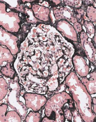
Most specimens taken to investigate hematuria look normal or close to normal on orthodox light microscopy, when the age of the person biopsied is taken into account, and provided that renal function is normal (Figs 5.1, 5.10, and 9.1).
236 |
9 Indication for Biopsy: Hematuria |
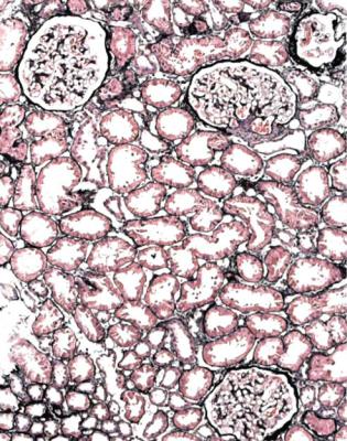
Fig. 9.1 Cortex in a renal biopsy specimen from a woman of 35 with hematuria and proteinuria. This is virtually normal on orthodox light microscopy, but shows IgA nephropathy on immunohistologic study. IgA nephropathy is sometimes called Berger’s disease. The name is almost always pronounced bur-ger, with a hard g, although the French pronunciation, ber-zhay, is perhaps more appropriate. In 1968, Jean Berger (born 1930), Professor of Pathology at the Necker Hospital in Paris, France, with his colleague Nicole Hinglais, who was an electron microscopist, reported the finding of IgA by immunofluorescence in 25 renal biopsy specimens. This was in a note of six short paragraphs, with no references (Berger J, Hinglais N. Les dépôts intercapillaires d’IgA-IgG. Journal d’Urologie et de Nephrologie 1968; 74: 694–695). This tiny paper was largely overlooked, and the significance of IgA deposition in glomeruli did not become widely appreciated until 1969, when Berger presented his findings at a meeting of the Transplantation Society (Berger J. IgA glomerular deposits in renal disease. Transplantation Proceedings 1969; 1: 939–944). The terms Berger’s disease and Berger’s nephropathy seem to have been first used in print in 1973. Berger was also the first to describe dense deposit disease, in 1963
Initial Assessment of Orthodox Light Microscopic Sections
The diagnosis in hematuria mainly depends on findings of immunohistology and electron microscopy. Examination of orthodox light microscopic sections is to assess the extent of irreversible damage, which alters the prognosis, and to ensure
Diagnosis of IgA Nephropathy |
237 |
that there are no unexpected findings. These include segmental glomerular changes of vasculitic type (Figs 7.12 and 7.18) and active interstitial nephritis (Fig. 7.34).
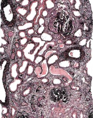
On orthodox light microscopic sections, there are often hints that the diagnosis may be IgA nephropathy or hereditary nephropathy of Alport type, rather than thin glomerular basement membrane disease. There may be more irreversible tubular damage than expected for the age, glomeruli may have definite mesangial expansion, and there may be segmental changes in glomeruli, particularly irregular areas of sclerosis at various sites in glomeruli (Fig. 8.1). Foamy cells in tissues outside glomeruli are often a striking feature in hereditary nephropathy of Alport type, even when there is little or no proteinuria (Fig. 9.2). Foamy cells can be seen in other conditions such as IgA nephropathy and membranous nephropathy, but generally when there is heavy proteinuria. Often the pathologist cannot easily determine if foamy cells are in tubules or in interstitial tissues.
The diagnosis can hardly ever be made in hematuria by examination of orthodox light microscopic sections alone.
Assessment of Immunohistologic Sections: Is There
IgA Nephropathy?
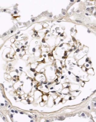
The next crucial investigation in hematuria is immunohistology. If this shows deposition of IgA in mesangium, the diagnosis is likely to be IgA nephropathy (Figs 8.2 and 9.3 – 9.6). Only one glomerulus is needed to give the diagnosis of IgA nephropathy (Fig. 4.8). The mesangial immunostaining begins sharply where the arterioles pass through Bowman’s capsule (Figs 8.2 and 9.4).
Often IgM and complement are also deposited in mesangium. Using an immunoperoxidase method, IgG is not found in typical IgA nephropathy, but may be seen using an immunofluorescence method. If IgG is found with IgA in mesangium by immunoperoxidase, this suggests lupus nephritis, which is almost the only other disorder in which IgA is found, if Henoch-Schönlein nephritis is regarded as part of IgA nephropathy, in its widest sense. C1q is more often found in lupus nephritis than in IgA nephropathy, and may help to discriminate between them if IgG is found on immunofluorescence. Lupus nephritis is usually diagnosed clinically or serologically before renal biopsy.
Diagnosis of IgA Nephropathy
The typical presentation of IgA nephropathy is with macroscopic hematuria at the time of an upper respiratory tract infection, characteristically without the delay between the infection and the urinary abnormality that occurs in acute postinfective glomerulonephritis. Hematuria or proteinuria, or both, may be detected in many other ways. At diagnosis, there may be chronic renal impairment or accelerated hypertension. IgA nephropathy hardly ever presents with the nephrotic syndrome.
238 |
9 Indication for Biopsy: Hematuria |
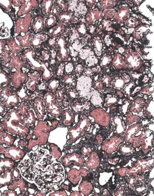
Fig. 9.2 Cortex in a renal biopsy specimen from a woman of 17 with hematuria and proteinuria. This is not normal on orthodox light microscopy, and has a little chronic damage with many foamy cells, probably both in tubules and in interstitial tissues. Electron microscopy shows hereditary nephropathy of Alport type.
Alport’s syndrome was suggested by R F Shaw and R A Glover in 1961 as a suitable name for the condition of familial renal disease with deafness, to honour Cecil Alport who described a family in 1927 (Alport AC. Hereditary familial congenital haemorrhagic nephritis. British Medical Journal 1927; 1: 504–506). The name is pronounced awl-port or al-port. Arthur Cecil Alport (1880–1959) was born in South Africa, and qualified in medicine in Edinburgh, United Kingdom. In 1922, he was appointed second assistant to the medical unit at St Mary’s Hospital Medical School in London, which he left in 1937 to become professor of medicine in Cairo, Egypt. The family he described had already been reported four times, by L G Guthrie in 1902, G Kendall and A F Hertz in 1912, Hertz again in 1923 but after a change of surname to Hurst, and J Eason, G L M Smith and G Buchanan in 1924. The observation had been made that males were more affected than females, but Alport was the first to mention deafness in the family. In 1968, M D Crawford and P J Toghill reported that they had traced the family described by Alport, and found that the disease had disappeared from it. The characteristic changes in glomerular basement membranes were first described in 1972. Electron microscopy was never done on glomeruli in Alport’s family
Diagnosis of IgA Nephropathy |
239 |
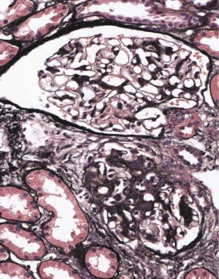
Fig. 9.3 Glomeruli in a renal biopsy specimen from a woman of 28 with hematuria and proteinuria. These have different amounts of mesangial expansion, and one has a segmental area of sclerosis. Immunohistologic findings are illustrated in Fig. 9.4, and show IgA nephropathy
IgA nephropathy usually has abnormalities of glomeruli visible on orthodox light microscopy, but sometimes can appear to have normal glomeruli (Fig. 9.5). The most common abnormality is mesangial expansion to different extents (Figs 8.1 and 9.3). Various classifications of the glomerular appearances have been made. In practice, these are of little clinical use, compared with the prognostic information given by assessment of the amount of chronic damage in the kidney, for instance, by measurement of the index of chronic damage (Fig. 5.6).
IgA nephropathy is probably a consequence of abnormal glycosylation of IgA1 molecules in plasma, which leads to their deposition in mesangium. This is likely to have genetic causes, and indeed IgA nephropathy occasionally runs in families. There is a high risk of progression to renal failure, although this may take a long time, meaning decades, if the kidney is close to normal at the time of diagnosis.
The pathologist cannot differentiate between the common type of IgA nephropathy and the type that is associated with various disorders, particularly chronic liver diseases, such as alcoholic cirrhosis, in which there may be reduced clearance
240 |
9 Indication for Biopsy: Hematuria |
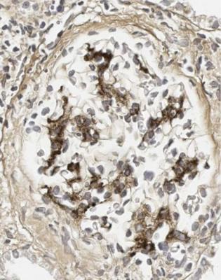
Fig. 9.4 Glomerulus in the renal biopsy specimen from the woman of 28, which is illustrated in Fig. 9.3. An immunoperoxidase method to detect IgA shows mesangial deposition, and gives the diagnosis of IgA nephropathy
of IgA1 from plasma. IgA nephropathy is one of several conditions seen in HIV infection, and may also be associated with dermatitis herpetiformis, celiac disease, inflammatory diseases of joints, and several other diseases in which the renal disorder may just be coincidental. Some people with IgA nephropathy have a raised concentration of IgA in the blood, but IgA nephropathy is not the only explanation of this change, and the pathologist should not be influenced by knowledge of the serum IgA concentration when a renal biopsy specimen is assessed.
Henoch-Schönlein Nephritis
Occasionally, there may be glomerular abnormalities of vasculitic type in IgA nephropathy, and these may occur with or without systemic features, such as a rash or joint symptoms (Figs 7.25–7.27, 9.7, and 9.8). Useful terms for this finding are vasculitic IgA nephropathy and Henoch-Schönlein nephritis, rather than
Henoch-Schönlein Nephritis |
241 |
Fig. 9.5 Glomerulus in a renal biopsy specimen from a man of 54 with microscopic hematuria, but no proteinuria. This is virtually normal on orthodox light microscopy, but shows IgA nephropathy on immunohistologic study, illustrated in Fig. 9.6
Henoch-Schönlein purpura, which is a clinical syndrome with various combinations of a rash, joint problems, gastrointestinal bleeding, acute renal failure, and hematuria. Biopsy of the skin or gastrointestinal tract in Henoch-Schönlein purpura shows a leukocytoclastic vasculitis with IgA in the walls of small blood vessels.
IgA nephropathy and Henoch-Schönlein nephritis are almost certainly the same condition. Henoch-Schönlein nephritis can be regarded as an acute vasculitic episode in IgA nephropathy, and is less common than IgA nephropathy not complicated by vasculitic glomerulonephritis. In adults, biopsy specimens showing Henoch-Schönlein nephritis are about one-sixth of the number that show uncomplicated IgA nephropathy.
The possible combinations of pathologic and clinical findings can be summarised in this way in conditions associated with IgA in glomeruli, which can be regarded as IgA nephropathy in its widest sense.
IgA nephropathy, without vasculitic glomerular lesions, may be found in a renal biopsy specimen when clinically there is apparently pure renal disease with
242 |
9 Indication for Biopsy: Hematuria |
Fig. 9.6 Glomerulus in the renal biopsy specimen from the man of 54, which is illustrated in Fig. 9.5. An immunoperoxidase method to detect IgA shows light mesangial deposition, and gives the diagnosis of IgA nephropathy. Definite deposition of IgA of any intensity in glomeruli is significant
hematuria and proteinuria, or when there is clinical Henoch-Schönlein purpura (Figs 4.8, 8.1, 8.2, 9.1, and 9.3–9.6). In adults, about nine-tenths with this finding do not have clinical Henoch-Schönlein purpura.
Henoch-Schönlein nephritis, or IgA nephropathy with vasculitic glomerular lesions, may be found in a renal biopsy specimen when there is clinical HenochSchönlein purpura, or when clinically there is apparently pure renal disease with hematuria and proteinuria (Figs 7.25–7.27, 9.7, and 9.8). In adults, about half with this finding have clinical Henoch-Schönlein purpura, and half do not.
Clinical Henoch-Schönlein purpura may be found in a renal biopsy specimen to have either Henoch-Schönlein nephritis or IgA nephropathy, without vasculitic glomerulonephritis (Fig. 9.7). In adults, there are approximately equal numbers of these findings.
IgA-associated disease apparently confined to the kidney, with hematuria and proteinuria, may be found in a renal biopsy specimen to have either IgA
Henoch-Schönlein Nephritis |
243 |
Fig. 9.7 Cortex in a renal biopsy specimen from a woman of 34 with hematuria, proteinuria, hypertension, clinically acute renal failure, and a rash. There is marked acute and chronic damage, and a surviving glomerulus shows mesangial expansion with healing segmental vasculitic lesions. There is IgA deposition in glomeruli on immunohistologic study. Features are those of late and active Henoch-Schönlein nephritis
nephropathy, without vasculitic glomerulonephritis, or Henoch-Schönlein nephritis (Figs 4.8, 7.25–7.27, 8.1, 8.2, 9.1, 9.3–9.6, and 9.8). In adults with these clinical features, most, about nine-tenths of them, do not have Henoch-Schönlein nephritis, but this means that Henoch-Schönlein nephritis is occasionally found in someone with no systemic features of Henoch-Schönlein purpura (Fig. 9.8).
Often children with typical clinical features of Henoch-Schönlein purpura do not have a renal biopsy, and are expected to get better. Biopsy may be done if there is heavy proteinuria at onset, or if proteinuria persists.
In the report, the pathologist should give the diagnosis as IgA nephropathy or Henoch-Schönlein nephritis, include the proportion of glomeruli with active vasculitic lesions, and give an assessment of the amount of irreversible damage in glomeruli and tubules.
244 |
9 Indication for Biopsy: Hematuria |
Fig. 9.8 Glomerulus in a renal biopsy specimen from a man of 26 with hematuria and proteinuria found on routine screening, without any other findings on medical examination. There is a large segmental vasculitic lesion with mild mesangial expansion in the rest of the tuft. Immunohistologic study shows IgA deposition in glomeruli. This is unexpected Henoch-Schönlein nephritis, without any systemic features of Henoch-Schönlein purpura
If IgA Nephropathy Has Been Excluded, Is There an Abnormality on Electron Microscopy?
If IgA nephropathy is excluded by immunohistology, and there is no other identifiable abnormality on light microscopy, the next investigation is electron microscopy. For pathologists without access to an electron microscope, specimens without IgA nephropathy, and with only changes attributable to age, can be reported to show kidney with no significant abnormality on light microscopy. The chance that a serious progressive disease has been missed is slight in adults, although there is a chance in children of early or mild hereditary nephropathy of Alport type.
The most likely finding on electron microscopy is thin glomerular basement membrane disease. This is one of the few disorders of any organ in which the name describes the structural findings.
Diagnosis of Thin Glomerular Basement Membrane Disease |
245 |
Diagnosis of Thin Glomerular Basement Membrane Disease
The reason for the renal biopsy is persistent microscopic hematuria in a person of either sex and of any age. There may be a family history of microscopic hematuria, or hematuria may be found by testing urine of relatives, but there should be no history of renal failure in the family. Macroscopic hematuria and proteinuria may occur, but are unusual. Thin glomerular basement membrane disease is common, and has been found in about one in 20 apparently normal people. The abnormality can occur with other disorders of the kidney.
If the diagnosis is correct, and if they have no other condition, such as hypertension, people with thin glomerular basement membrane disease should have normal renal function for the rest of their life. This outlook is different from that of people with either IgA nephropathy or hereditary nephropathy of Alport type, who may progress to renal failure.
The renal biopsy specimen looks normal on orthodox light microscopy, when the age of the person biopsied is taken into consideration (Figs 5.1, 5.7, and 5.10). This means that in middle aged and old people, there may be signs of chronic ischemic damage. There is no deposition of IgA in glomeruli on immunohistologic study. There may be deposition of IgM in mesangium, but if basement membranes are thin on electron microscopy, this immunohistologic finding can be ignored.
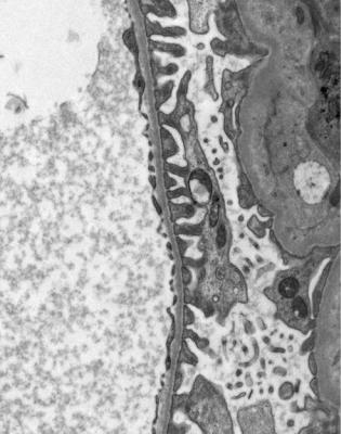
On electron microscopy, basement membranes appear strikingly thin. Usually measurement is not necessary, because there is such a definite difference from normal thickness, with which the pathologist should be familiar from examination of specimens with IgA nephropathy, for example (Figs 6.6 and 9.9). Membranes are uniform, without irregularities of the epithelial surface, and without signs of deposits. Usually a prominent lamina densa is seen in the centre of the membrane, although this is not seen in normal membranes. In children aged below 10 years, normal controls should be matched by age, because the glomerular basement membrane increases in thickness until then.
The pathologist should remember that glomerular capillary loops are threedimensional structures, and a random section can show basement membranes with any apparent thickness from the true minimum to any width at right angle to the minimum. Parts of the membrane where it appears to be sectioned perpendicularly are easily detected by the sharpness of the image of cytoplasmic membranes of endothelial and epithelial cells on each side, and these parts give the most accurate impression of the thickness. If necessary, the thickness can be measured here.
Measurement is difficult to do properly, because ideally this requires adequate numbers of random samples and suitable control measurements, all compared with measurements from calibration grids photographed at the same magnification on the electron microscope. This is tedious, is rarely done thoroughly, and is not usually necessary, because most examples of thin glomerular basement membrane disease can be diagnosed by simple inspection. Because the definition of thin glomerular basement membrane disease is arbitrary, and accurate measurement is difficult, the most important thing for the pathologist to do in equivocal specimens is to exclude other explanations of hematuria.
246 |
9 Indication for Biopsy: Hematuria |
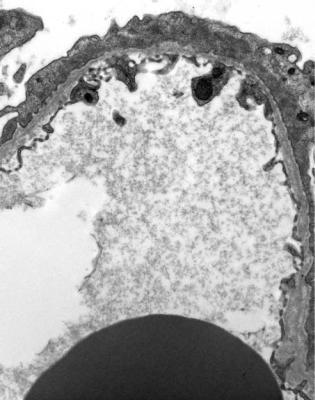
Fig. 9.9 Electron micrograph of part of the glomerular basement membrane in a renal biopsy specimen from a woman of 60 with persistent microscopic hematuria, but no proteinuria. This is uniformly thin, with a thickness of about 150 nm. Compared with normal basement membranes in Fig. 6.6, photographed at the same magnification, this membrane is not only thinner, but also has a more obvious lamina densa in the middle. Features are those of thin glomerular basement membrane disease
One problem is the specimen with basement membranes of different thicknesses, in places definitely thin but elsewhere apparently normal, without irregularity of the epithelial surface. This may be called patchy thin glomerular basement membrane disease.
If there is an irregular outer surface on basement membranes, the diagnosis is hereditary nephropathy of Alport type.
Diagnosis of Hereditary Nephropathy of Alport Type
This is not a single disease. The same pathologic appearances can be produced by many genetic abnormalities of type four collagen, which is found in glomerular basement membranes.
Diagnosis of Hereditary Nephropathy of Alport Type |
247 |
Most people with this condition have an abnormality on an X chromosome, which means that the disease is sex linked, carried by women who may appear unaffected, and phenotypically worse in males. There are other types of inheritance, autosomal recessive and autosomal dominant, and even in the sex linked type, the clinical features and severity vary between families. Often the disorder is found on investigation of a boy with hematuria, and there is a family history of deafness and renal failure in male relatives. There may be proteinuria, and occasionally this can be in the nephrotic range.
In the sex linked disorder, the alpha five chain of collagen type four is defective. In the other types, the alpha three chain or alpha four chain is defective. Lack of one of these chains leads to nonexpression of the others in glomerular basement membranes. In hereditary nephropathy of Alport type, glomerular basement membranes lack the Goodpasture antigen, which is in the noncollagenous domain of the alpha three chain of collagen type four, and is recognised by antibodies in Goodpasture’s disease (Figs 7.29 and 7.30). There is a likelihood that many people with thin glomerular basement membrane disease have a heterozygous defect in alpha three or alpha four chains of collagen type four, but such defects do not prevent expression of the Goodpasture antigen in glomerular basement membranes in thin glomerular basement membrane disease.
In hereditary nephropathy of Alport type, a renal biopsy specimen may range in orthodox light microscopic appearances from normal in a young person who has the disorder but is biopsied at an early stage, or in a woman who is a carrier of an abnormal X chromosome, to severely abnormal with extensive tubular atrophy, segmental and global sclerosis of glomeruli, and many foamy cells in the cortex (Fig. 9.2). Immunohistologic study shows no deposition of IgA in glomeruli.
Electron microscopy shows that basement membranes are not uniformly thin, and have an irregular epithelial surface (Figs 9.10 and 9.11). There are thick areas as well as thin, patches of complex splitting of the lamina densa, sometimes called basket weave pattern, and inclusions of electron dense particles in the basement membrane.
These features are often well developed in boys and young men, but may be slight in girls or women, in whom the pathologist may be in doubt whether the diagnosis is hereditary nephropathy. The changes usually become more marked with time, and a definite diagnosis may require another renal biopsy, a few years after the first. Each family with hereditary nephropathy of Alport type has a different genetic abnormality. There is no simple genetic screening test that at present helps to decide whether equivocal pathologic findings are those of hereditary nephropathy.
In hereditary nephropathy of Alport type, glomerular basement membranes lack the Goodpasture antigen, which can be identified by various antibodies. There are also antibodies that can be used immunohistologically to identify various chains of collagen type four, such as the alpha five chain. Such antibodies may be helpful in diagnosis, because they react with normal basement membranes, but may not react with basement membranes in hereditary nephropathy (Figs 9.12 and 9.13). Some antibodies may also be useful in skin biopsy specimens. One of the two X chromosomes is randomly inactivated in female cells, and this may give patchy
248 |
9 Indication for Biopsy: Hematuria |
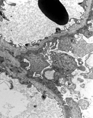
Fig. 9.10 Electron micrograph of glomerular basement membranes in a renal biopsy specimen from a woman of 28 with persistent microscopic hematuria and proteinuria. Her brother was on renal replacement therapy, but the cause of his renal failure was not known. The membranes have an irregular outer aspect, a range of thickness, and splitting of the lamina densa. These are features of hereditary nephropathy of Alport type. An image at higher magnification is shown in
Fig. 9.11
expression of collagen chains in glomeruli of women with a mutation giving the sex linked form of hereditary nephropathy of Alport type. This may cause difficulties with interpretation of immunohistologic findings. Antibodies to chains of collagen type four and the Goodpasture antigen should react normally in thin glomerular basement membrane disease (Fig. 9.12).
Other Findings in Hematuria
If orthodox light microscopy has shown only age-related changes, immunohistology has excluded IgA nephropathy, and electron microscopy has shown glomerular basement membranes of uniform normal thickness, the pathologist can give
Other Findings in Hematuria |
249 |
Fig. 9.11 Electron micrograph of part of a glomerular basement membrane in the renal biopsy specimen from the woman of 28 with hereditary nephropathy of Alport type, which is illustrated in Fig. 9.10. This is at the same magnification as the normal basement membrane in Fig. 6.6 and the thin basement membrane in Fig. 9.9. The basement membrane has an irregular outer aspect, variable width with thick and thin areas, splitting of the lamina densa, and small inclusions
the diagnosis as kidney with no detectable significant abnormality in the biopsy specimen. This does not mean that the kidney is normal because there may be an explanation of hematuria that has been missed, such as a vascular abnormality or a urologic disorder.
In both adults and children, most renal biopsy specimens taken for investigation of hematuria should be interpretable with this approach. The relatively few others may show such things as diabetic glomerulopathy, lupus nephritis, and segmental sclerosing conditions. Often, the clinical information given to the pathologist on the request form in these is incomplete or wrong. For example, the underlying disease may not be mentioned, or there may be chronic renal failure, or the main nephrologic problem may be proteinuria in the nephrotic range rather than hematuria.
250 |
9 Indication for Biopsy: Hematuria |
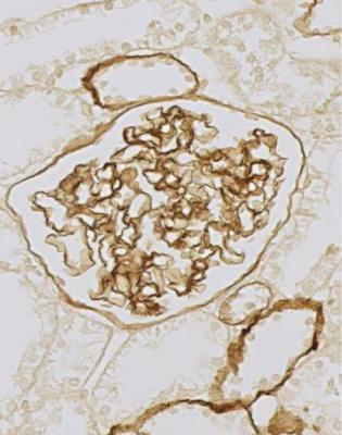
Fig. 9.12 Cortex in a renal biopsy specimen from a woman of 40 with thin glomerular basement membrane disease. This is stained by an immunoperoxidase method with an antibody to the Goodpasture antigen. The antigen is detected in basement membranes of the glomerular tuft, Bowman’s capsule, and a few tubules. This distribution is normal, unlike the findings in hereditary nephropathy of Alport type, illustrated in Fig. 9.13
Summary: Hematuria
A renal biopsy is not done on every person with hematuria, and is only done after appropriate investigations to exclude other causes of hematuria.
Most specimens taken to investigate hematuria look normal or close to normal on orthodox light microscopy when the age of the person biopsied is taken into account.
In children, most specimens in hematuria show one of three disorders: thin glomerular basement membrane disease, IgA nephropathy, or hereditary nephropathy of Alport type.
In adults, most specimens in hematuria show either IgA nephropathy or thin glomerular basement membrane disease.
Further Reading: Hematuria |
251 |
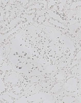
Fig. 9.13 Cortex in the renal biopsy specimen from the woman of 17 with hereditary nephropathy of Alport type that is illustrated in Fig. 9.2. This is stained by an immunoperoxidase method with the antibody to the Goodpasture antigen that is illustrated in Fig. 9.12. No Goodpasture antigen is found in either glomerular or tubular basement membranes. Similar findings are expected with antibodies against alpha three and five chains of collagen type four
Further Reading: Hematuria
D’Agati VD, Jennette JC, Silva FG. Non-Neoplastic Kidney Diseases. Atlas of Nontumor Pathology, first series, fascicle 4. Washington, DC: American Registry of Pathology and Armed Forces Institute of Pathology, 2005. Chapters 4, 12.
Jennette JC, Olson JL, Schwartz MM, Silva FG. Heptinstall’s Pathology of the Kidney. Sixth ed. Philadelphia: Lippincott Williams and Wilkins, 2007. Chapters 10, 11.