

Chapter 8
Indication for Biopsy: Chronic Renal Failure
Introduction to Chronic Renal Failure
This is permanently reduced renal excretory function, meaning a reduced glomerular filtration rate, due to a reduced number of nephrons. This contrasts with acute renal failure, in which the number of nephrons may be normal, and renal function may recover. Chronic renal failure, at least in adults, usually appears to have been present for months or years, rather than days or weeks, unlike acute renal failure. Chronic renal impairment can be used with the same meaning.
For many decades, the term Bright’s disease was used as a diagnosis in clinical disorders of the kidney, particularly in chronic renal failure (Figs 6.34 and 6.50).
The separation from acute renal failure is not always straightforward. Renal failure that clinically appears chronic may have acute changes in a biopsy specimen, and vice versa. There may also be acute disease superimposed on chronic disease. There may be hematuria, proteinuria that may be heavy enough to produce the nephrotic syndrome, and hypertension, which clinically may be in the accelerated phase. If the nephrotic syndrome is present, the biopsy specimen should be assessed in the way suggested in Chapter 6.
People with chronic renal failure come to medical attention in many ways, and may have no symptoms or only nonspecific symptoms, such as lassitude, nausea, and anorexia. Symptoms related to urination, such as pain on passing urine or difficulty of urination usually indicate a disorder of the lower urinary tract, not of the kidneys. Renal function is assessed by chemical tests, and chronic renal failure is often detected almost as an incidental finding.
After an amount of chronic damage that is different for each person, chronic renal failure becomes relentlessly progressive, even if the original cause is no longer active, and even if hypertension is controlled. One reason is that loss of some nephrons leads to increased functional activity and structural enlargement in remaining nephrons, which lead to irreversible damage in some of these, which in turn imposes an increased work load on surviving nephrons, and so on. This progresses to end-stage renal failure, when renal function is not enough to support life. Renal replacement therapy is then required, which means hemodialysis or peritoneal dialysis or renal transplantation.
A. J. Howie, Handbook of Renal Biopsy Pathology. |
203 |
C Springer 2008 |
|
204 |
8 Indication for Biopsy: Chronic Renal Failure |
Not every person with chronic renal failure is investigated by renal biopsy. Several causes can be diagnosed clinically or radiologically, without the need for biopsy. In some centres, biopsy is rarely used to establish the diagnosis in chronic renal failure, if there are small kidneys with no clues to the cause, while in others, biopsy is used commonly.
Because the kidneys are often small in chronic renal failure, percutaneous needle biopsy is more difficult than in conditions with kidneys of normal size, and the specimen may not contain cortex. Provided that the specimen is suitable for analysis, there are always abnormalities in chronic renal failure. The pathologist should almost always be able to give information of help to nephrologists, and can often suggest the diagnosis.
Assessment of Chronic Renal Failure
Nephrologists usually rely on measurement of serum creatinine concentration as an indicator of renal excretory function. This is crude and has a nonlinear relation with glomerular filtration rate, but is much cheaper and simpler than accurate measures of glomerular filtration rate. There may be a reduction of glomerular filtration rate to half the normal value while the serum creatinine concentration is in the normal range. In practice, the pathologist can understand a high serum creatinine concentration to be a sign of definite renal impairment, while a concentration in the normal range may still occur in the presence of a surprising amount of renal damage in a renal biopsy specimen (Fig. 5.5).
Accurate determination of glomerular filtration rate requires measurement of clearance, the amount of plasma that is cleared of a substance by excretion in the urine in an interval of time. The ideal substance for clearance measurements is completely filtered by glomeruli, and neither reabsorbed nor secreted by tubules. The pathologist should be aware that measurement of creatinine clearance can be unreliable as an indication of glomerular filtration rate, partly because creatinine is secreted by tubules, but mainly because this investigation requires collection of urine in a timed period. The collection is often incomplete, and this is the largest reason for error in calculations, giving an underestimate of creatinine excretion. The unreliability of reported creatinine clearances explains why a pathologist may see virtually normal kidney in a specimen from someone said to have renal impairment based on this investigation, although if the serum creatinine concentration is high, or if accurate tests of glomerular filtration rate show renal impairment, there will always be structural changes in the kidney. Glomerular filtration rate is often estimated rather than measured, using various formulas based on serum creatinine concentration and other factors such as age and body size.
Stages of Chronic Kidney Disease
Because chronic renal failure is not all or none, meaning that the definition is arbitrary, chronic kidney disease is divided into stages to help clinical management. This classification is not so helpful to pathologists, but is given here to explain
Causes of Chronic Renal Failure |
205 |
the meaning of stages if numbers are written on request forms. These stages are based on measurements or estimates of the glomerular filtration rate, standardised to a body surface area of 1.73 m2. Body surface area is calculated from a formula based on height and weight, and the reference area of 1.73 m2 was derived from supposedly average adult height and weight. Standardisation allows the stages to be applied irrespective of age or body size, including in children.
1.Stage one of chronic kidney disease means a normal glomerular filtration rate, defined as over 90 ml/min/1.73 m2, but with at least one of these features of renal disease: persistent proteinuria, persistent hematuria after exclusion of other causes, structural abnormality of the kidney shown by ultrasound examination, or a glomerular disorder shown by biopsy. Stage one is not renal failure, as conventionally defined.
2.Stage two is mild renal impairment, defined as a glomerular filtration rate of 60–90 ml/min/1.73 m2, with at least one of the other features of renal disease.
3.Stage three is moderate renal impairment, with a glomerular filtration rate of 30–59 ml/min/1.73 m2.
4.Stage four is severe impairment, with a glomerular filtration rate of 15–29 ml/min/1.73 m2.
5.Stage five is established renal failure, meaning either the glomerular filtration rate is under 15 ml/min/1.73 m2 or the person is on dialysis.
Causes of Chronic Renal Failure
There are many causes of chronic renal failure, and these are different between the sexes, at different ages, and in different parts of the world. More males than females have chronic renal failure.
In children, the main causes are developmental abnormalities of the kidney, with malformation of the urinary tract. Abnormalities include renal agenesis, which means complete failure of development of a kidney, hypoplasia, which means that a kidney is small but otherwise normally formed, and dysplasia, which means abnormal, incomplete differentiation of part or all of a kidney. Dysplasia is almost always associated with other disorders of the urinary tract, such as duplex ureters, prenatal vesicoureteric reflux, and obstruction of the urinary tract, particularly by posterior urethral valves in boys. Among other causes of chronic renal failure in children are glomerular disorders, reflux nephropathy, metabolic conditions such as cystinosis and hyperoxaluria, and cystic disorders, particularly juvenile nephronophthisis, which is pronounced nefro-no-ff-thigh-siss or nefro-no-thigh-siss, and is a group of disorders similar to medullary cystic disease. Phthisis comes from a Greek word meaning wasting away or decay.
In adults, the commonest cause is diabetes mellitus. Hypertensive renal damage or hypertensive nephrosclerosis is often used as a diagnosis in a person with hypertension, small symmetrical kidneys, chronic renal failure, and no other obvious cause. Many other conditions can cause chronic renal failure, such as nondiabetic
206 |
8 Indication for Biopsy: Chronic Renal Failure |
glomerular disorders, reflux nephropathy, autosomal dominant polycystic kidney disease, ischemia from atherosclerosis of the aorta and renal arteries, collectively called renovascular disease, and obstruction of the urinary tract. In some countries, analgesic nephropathy and tuberculosis of the urinary tract are common, and there are parts of the world where there are characteristic disorders associated with chronic renal failure, such as the endemic nephropathy of the Balkans.
Value of a Renal Biopsy in Chronic Renal Failure
The loss of nephrons is irreversible, and in theory every person with chronic renal failure, if they live long enough, will eventually need renal replacement therapy. There is no treatment at present that will regenerate nephrons after atrophy, although there are ways to slow down the rate of progression of chronic renal failure, such as by control of hypertension. Accordingly, some nephrologists think that a biopsy will give little or no information of any use, and that this does not justify the difficulties associated with the procedure.
Renal biopsy may be justified in chronic renal failure for these reasons:
1.The specimen may show a reversible and treatable condition, with predominantly acute tubular damage, rather than tubular atrophy. A combination of acute and chronic changes may also be seen, which means that there could be an improvement in renal function.
2.Even though the specimen usually confirms established chronic damage in the kidney, knowledge of the diagnosis may help in clinical decisions, such as need for further investigations, study of relatives, and selection of renal replacement therapy. Some conditions commonly recur in renal allografts, and the knowledge that there is such a condition may influence management.
3.The specimen may allow the pathologist to give an indication of the prognosis, meaning how long the kidney will function without the need for replacement therapy. This is because there is a statistical relation between the amount of chronic damage at biopsy and the length of survival of renal function.
Findings in Renal Biopsy Specimens in Chronic Renal Failure
Although there are many explanations of chronic renal failure, only a few conditions are seen commonly in biopsy specimens.
In adults, many specimens show evidence of chronic renal damage due to something other than a glomerular disorder. Most of the rest have IgA nephropathy, various segmental sclerosing glomerular conditions, or diabetic glomerulopathy.
In biopsy specimens in children, many have evidence of a nonglomerular disorder, often familial.
Assessment of Renal Biopsy Specimens in Chronic Renal Failure |
207 |
Assessment of Renal Biopsy Specimens in Chronic Renal Failure
Chronic renal failure is more likely than other indications for renal biopsy to have more than one disease process or type of abnormality in the kidney.
The general rule is that renal function is more closely correlated with structural changes in tubules than with any other changes. Chronic renal failure is associated with chronic abnormalities in tubules.
The pathologist should first determine that most or all of the tubular damage is chronic, which means that the clinical problem is indeed chronic renal failure, rather than acute. This is important because chronic damage will not get better, and its associated renal impairment will not reverse to give normal function, but acute damage and acute renal impairment may recover to normal.
Chronic damage is detected as various combinations of reduced number of tubules, shrunken tubules, thickening of tubular basement membranes, thyroidization, which means more marked dilatation of tubules and more marked flattening of tubular epithelium than seen in acute tubular damage, and cysts, although the distinction between dilated tubules and cysts is arbitrary. These tubular changes may be accompanied by interstitial fibrosis, an interstitial infiltrate of chronic inflammatory cells, global sclerosis of glomeruli, and chronic intimal thickening in small arteries. Surviving tubules may be larger than normal, and may have acute damage if there is an acute deterioration of renal function in someone with chronic renal failure (Figs 5.2, 5.3, 5.12, and 5.14).
If the number of tubules without atrophy appears appropriate for the age of the person biopsied, the problem is acute rather than chronic, and the specimen should be analysed in the way suggested in Chapter 7.
The next stage of assessment is to see if there is evidence of a glomerular disorder. This is unlikely if there is no proteinuria and no hematuria. Proteinuria below the nephrotic range and hematuria may occur in disorders that are not primarily of glomeruli, but proteinuria in the nephrotic range almost always indicates a glomerular disorder that is the explanation of the chronic renal failure.
Is There a Glomerular Disorder?
Two common glomerular disorders in chronic renal failure are straightforward to diagnose. These are IgA nephropathy and diabetic glomerulopathy.
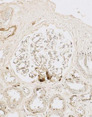
IgA nephropathy has mesangial increase that may differ in extent between glomeruli (Fig. 8.1). There is IgA deposition on immunohistologic study, and IgA may still be seen in glomeruli with global sclerosis (Fig. 8.2).
Diabetic glomerulopathy has a history of diabetes mellitus given on the request form, and several features such as mesangial increase with Kimmelstiel-Wilson nodules and thickened glomerular basement membranes (Figs 6.34–6.44).
Segmental sclerosing glomerular conditions are usually easy to see in renal biopsy specimens in chronic renal failure, but interpretation of their significance
208 |
8 Indication for Biopsy: Chronic Renal Failure |
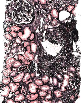
Fig. 8.1 Cortex in a renal biopsy specimen from a man of 62 with chronic renal failure, hematuria, proteinuria, and hypertension. There is patchy tubular atrophy. A glomerulus has mesangial increase. Immunohistologic study shows mesangial deposition of IgA, and gives the diagnosis of IgA nephropathy
is difficult. These are often considered to be one condition called focal segmental glomerulosclerosis, but this is not helpful to nephrologists because the term is applied to several different disorders. The pathologist may be able to give a more precise diagnosis of more use in prognosis and management.
Late stages of nearly every glomerular disorder and nonglomerular disorder may be complicated by the development of segmental areas of sclerosis in glomeruli. If the glomerular disorder can be recognised, this should be given as the diagnosis. Some texts say that focal segmental glomerulosclerosis can occur in IgA nephropathy, membranous nephropathy, diabetic glomerulopathy, and so on. This is an example of the lack of precision of the term focal segmental glomerulosclerosis. A better approach is to give the diagnosis as late IgA nephropathy or late membranous nephropathy, or a late stage of whatever other condition can be recognised.
Segmental abnormalities are often automatically considered focal and segmental, although they may not be focal but diffuse, which means present in every
Differentiation of Types of Segmental Sclerosing Glomerular Disorders |
209 |
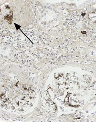
Fig. 8.2 Cortex in a renal biopsy specimen from a man of 49 with chronic renal failure, hematuria, proteinuria, and hypertension. An immunoperoxidase method to detect IgA shows deposition of IgA in the mesangium of surviving glomeruli, and also in a glomerulus with global sclerosis (arrowed). The diagnosis is IgA nephropathy
glomerulus, even though this may not be apparent on a single section or on a few random sections. A value of serial sections is that they help the pathologist to find segmental lesions, because just one lesion in a specimen may make a significant difference to the diagnosis, and because the proportion of glomeruli that contain segmental lesions may also influence the diagnosis.
Differentiation of Types of Segmental Sclerosing
Glomerular Disorders
In a biopsy specimen in chronic renal failure, a segmental sclerosing glomerular condition may be either a consequence of reduced number of glomeruli or a marker of an underlying glomerular disorder. Differentiation between these possibilities may be difficult, but should be attempted.
210 |
8 Indication for Biopsy: Chronic Renal Failure |
Reduced number of glomeruli is an equivalent term to reduced renal mass or reduced nephron number. Reduced renal mass can be understood to mean that the number of nephrons is ultimately inadequate to maintain a glomerular filtration rate appropriate for the size of the body, although there may be increases in size and function of surviving nephrons to compensate for a time.
The reduction could be congenital, in which too few nephrons were formed in the fetal kidney, or acquired, following any cause of loss of nephrons. A congenitally small kidney with compensatory enlargement of glomeruli is sometimes said to show oligomeganephronia, or oligomeganephronic hypoplasia, in which the components oligo, mega, hypo, and plasia come from Greek words meaning few, big, under, and forming or moulding. Only the most marked examples of this are likely to be recognised, and lesser degrees of inadequate formation of nephrons are usually too subtle to be detected. Severe obesity can be considered a cause of relatively reduced renal mass, which is disproportionately small compared with the body mass.
Effects of reduced renal mass are called glomerular overload, or hyperfiltration, or hyperperfusion. Surviving glomeruli enlarge, which may stretch and damage visceral epithelial cells. This may lead to proteinuria, loss of visceral epithelial cells, adhesion of the glomerular tuft to Bowman’s capsule, development of areas of segmental sclerosis, and eventual global sclerosis of glomeruli with atrophy of their tubules.
There are clues that a segmental sclerosing condition may be explained by reduced glomerular number. There may be more extensive global sclerosis of glomeruli and tubular atrophy than expected for the age of the person biopsied, usually with ischemic shrinkage of the tuft and wrinkled basement membranes in some surviving glomeruli, the worst affected of which have thickening of the inner aspect of Bowman’s capsule. Other glomeruli appear large (Fig. 5.11). Surviving tubules are also enlarged. The sclerosed areas in glomeruli are often at the hilum of glomeruli, although why they usually have this distribution is unclear (Figs 5.13 and 8.3). Sometimes, the pathologist can suggest that there are overload effects even when there are no segmental lesions, but the other features are well marked.
Usually, the segmental sclerosis in overload changes is genuinely or truly focal, meaning that not every glomerulus is affected. This is stressed, because some conditions called focal segmental glomerulosclerosis are actually diffuse disorders, affecting every glomerulus.
Segmental lesions at the hilum correspond with the perihilar variant of focal segmental glomerulosclerosis in the Columbia classification.
If the pathologist suspects that there are changes consistent with reduced glomerular number, there may be clinical evidence of reduced renal mass, possibly even a single kidney or relatively reduced renal mass in obesity. As well as chronic renal failure, there should be proteinuria, which occasionally is in the nephrotic range, although without the other features of the nephrotic syndrome. Glomerular overload effects are not considered an adequate explanation of the nephrotic syndrome, and if this is found, there should be another diagnosis, most likely a late stage of classic segmental sclerosing glomerulonephritis (Figs 6.27 and 6.28).
Differentiation of Types of Segmental Sclerosing Glomerular Disorders |
211 |
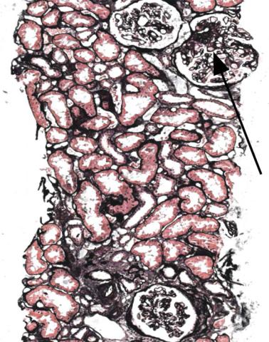
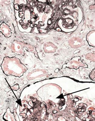
Fig. 8.3 Cortex in a renal biopsy specimen from a man of 38 with chronic renal failure, proteinuria, and asymmetrical kidneys. There had been denervation of the bladder in a road traffic collision, several years before the biopsy. This specimen from the larger kidney shows patchy tubular atrophy, glomeruli with ischemic shrinkage, enlargement of tubules and another glomerulus, and an area of sclerosis at the hilum of the large glomerulus (arrowed). These changes are interpreted to be effects of reduced glomerular numbers, following chronic damage from drainage problems, which particularly affected the other kidney
Even if there are apparently two kidneys of normal size on clinical investigation, the finding of the typical changes of overload effects should be reported as consistent with reduced nephron number. Many examples of segmental sclerosing glomerular lesions in so-called hypertensive nephrosclerosis are probably explained by reduced renal mass (Fig. 8.4).
212 |
8 Indication for Biopsy: Chronic Renal Failure |
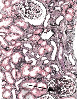
Fig. 8.4 Cortex in a renal biopsy specimen from a woman of 67 with apparently normal renal function, proteinuria, and hypertension. There are early atrophic changes in tubules, which suggest there is renal impairment. One glomerulus has an area of sclerosis (arrowed). The diagnosis could be given as a truly focal segmental sclerosing disorder, possibly even hypertensive nephrosclerosis, but glomeruli and tubules appear large, and there is a likelihood that this is due to reduced renal mass
A practical problem is the finding of a segmental sclerosing lesion that is not at the glomerular hilum, in a biopsy specimen in which there is no identifiable glomerular disorder such as IgA nephropathy, glomeruli are not markedly enlarged, and there is little evidence of chronic damage in the form of global sclerosis of glomeruli and tubular atrophy. The separation of this type of abnormality from overload changes is arbitrary, and probably many if not all examples of this are early or mild forms of overload changes.
This condition may be called a truly or genuinely focal segmental sclerosing disorder to avoid the ambiguity of the term focal segmental glomerulosclerosis (Figs 8.4 and 8.5). This is a condition in which only one segmental lesion has to be found to suggest the diagnosis. Proteinuria below the nephrotic range is usually a feature of a truly focal segmental sclerosing disorder.
Differentiation of Types of Segmental Sclerosing Glomerular Disorders |
213 |
Fig. 8.5 Glomerulus in a renal biopsy specimen from a woman of 37 with proteinuria. An immunoperoxidase method to detect C9 shows deposition in a segmental lesion. This glomerulus and the rest appear slightly large, but otherwise normal. The diagnosis can be given as a truly focal segmental sclerosing disorder
If every glomerulus or nearly every glomerulus has segmental sclerosis, the condition is not focal but diffuse. In such circumstances there is likely to be a story of the nephrotic syndrome at some time, and the diagnosis can be given as a late classic segmental sclerosing condition (Figs 6.27 and 6.28). This is important to recognise because this type of disorder may recur in a renal allograft, although the segmental sclerosing disorder associated with reduced glomerular number does not recur, nor do truly focal segmental sclerosing disorders.
Occasionally, there is evidence of a segmental sclerosing condition, but the specimen is so small or the changes are so late that the pathologist can only report a segmental sclerosing glomerular condition of undetermined significance. Even this is more helpful to nephrologists than the vague term focal segmental glomerulosclerosis.
214 |
8 Indication for Biopsy: Chronic Renal Failure |
Differential Diagnosis of Overload Changes in Glomeruli
Strikingly large glomeruli are rarely seen except when there is reduced glomerular number. One condition that can be associated with uniformly large glomeruli is chronic hypoxia. The pathologist is given the information on the request form that the person biopsied has a congenital heart disorder with cyanosis due to a right to left shunt, or a chronic lung disease with hypoxia, or sleep apnea (Fig. 8.6). There is also mesangial expansion that is most marked at the glomerular hilum. The uniformity of the enlargement and relatively little tubular atrophy support the diagnosis of hypoxic changes in glomeruli, and are against a diagnosis of overload effects.
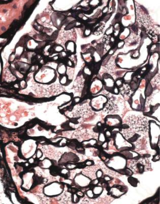
Fig. 8.6 Cortex in a renal biopsy specimen from a man of 34 with chronic renal failure, proteinuria, and sleep apnea. All glomeruli appear large with expanded mesangium, particularly at the hilum. These are hypoxic changes in glomeruli
Is There Evidence of Another Glomerular Disorder? |
215 |
Is There Evidence of a Glomerular Disorder Other than IgA Nephropathy, Diabetic Glomerulopathy, or a Segmental Sclerosing Condition?
Renal biopsy specimens in chronic renal failure may have evidence of late stages of glomerular abnormalities, such as vasculitic glomerulonephritis (Figs 7.15 and 7.16), amyloid (Fig. 6.49), subendothelial membranoproliferative glomerulonephritis (Fig. 6.80), membranous nephropathy (Fig. 6.2), or lupus nephritis (Figs 6.52 and 6.57).
Interpretation is made simpler if there was an earlier biopsy specimen from the same person that had established the diagnosis. A later specimen may show another disease, or a coincidental disorder, or a complication of treatment, rather than just a later stage of the initial condition.
If there is no previous specimen, the pathologist should examine glomeruli without global sclerosis and without much segmental sclerosis to see if there are any features of identifiable disorders.
Irradiation damages the kidney and leads to chronic renal failure. There are glomerular abnormalities that resemble subendothelial membranoproliferative glomerulonephritis with doubled basement membranes, although the mesangium may show disruption that is called mesangiolysis, rather than expansion, and few or no immune deposits are found on immunohistologic study (Fig. 8.7). Generally, tubules have severe atrophy. Blood vessels have chronic changes, and they may also show a small vessel vasculopathy, also called thrombotic microangiopathy.
A few rare inherited disorders may give chronic renal failure, and some have characteristic changes in glomeruli. An example is Fabry’s disease, in which there is lack of a lysosomal enzyme alpha galactosidase A. This has distinctive features in the kidney (Fig. 8.8). Lecithin cholesterol acyl transferase deficiency is a condition in which lipid deposits accumulate in glomerular basement membranes to give an irregularly vacuolated appearance. Another inherited disorder is hereditary nephropathy of Alport type, which requires electron microscopy to make the diagnosis. This is considered in Chapter 9, because the typical presentation is with hematuria.
Is There No Evidence of a Glomerular Disorder?
If glomeruli appear normal or have only ischemic changes, there is likely to be a nonglomerular disorder, or a combination of such disorders, to explain chronic renal failure. These types of disorder should also underlie glomerular changes ascribed to effects of reduced glomerular number.
Such disorders include these:
1.Chronic ischemic damage, including effects of hypertension.
2.Problems with urinary drainage, meaning obstruction of the urinary tract and reflux of urine into the kidney.
216 |
8 Indication for Biopsy: Chronic Renal Failure |
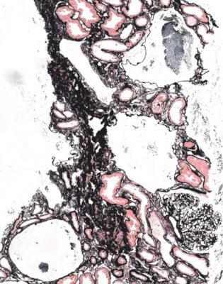
Fig. 8.7 Glomeruli in a renal biopsy specimen from a man of 42 with chronic renal failure, hematuria, proteinuria, and hypertension, and a history of abdominal irradiation for Hodgkin’s disease, 17 years before the renal biopsy. In one glomerulus, basement membranes are doubled, and in the other, the mesangium appears to have shrunk or disappeared in several areas (arrowed). These are features of irradiation nephropathy
3.Problems in the renal medulla, such as gout, papillary necrosis, and the inherited disorders juvenile nephronophthisis and medullary cystic disease. These can be regarded as intrarenal causes of drainage problems.
4.Other cystic diseases.
5.Tubular disorders, such as damage from toxins, paraproteins, or crystals.
6.Late stages of interstitial nephritis, including granulomatous disorders.
Few specimens have features characteristic of only one condition, but many have features suggestive of either a likely explanation of chronic renal failure, or a likely combination of explanations.
Frequently, all late renal damage is labelled chronic tubulo-interstitial nephritis or chronic interstitial nephritis. These names are not helpful because nearly all diseases lead eventually to similar appearances in the kidney. This was recognised by Ellis in 1942: “Chronic interstitial nephritis is merely the end stage of various forms of
Is There Evidence of Chronic Ischemic Damage? |
217 |
Fig. 8.8 Glomerulus in a renal biopsy specimen from a man of 28 with chronic renal failure, hematuria, and proteinuria. His mother was on hemodialysis. Visceral epithelial cells have finely vacuolated cytoplasm characteristic of Fabry’s disease. Johannes Fabry (1860–1930), pronounced fah-bree, was a German dermatologist who described the skin changes of this condition in 1898. Sometimes, the name Anderson Fabry disease is used. William Anderson (1842–1900), pronounced in the usual way, independently described the skin changes also in 1898, when he was a dermatologist in London, United Kingdom
renal disorder, and for a proper understanding of Bright’s disease this term should be discarded” (Fig. 6.34).
If possible, the pathologist should try to find clues to the explanation of late damage. Sometimes, the diagnosis can only be given as late nonglomerulonephritic renal damage, but even that is helpful to nephrologists (Fig. 8.9). The various possible explanations of such damage may be considered in turn.
Is There Evidence of Chronic Ischemic Damage?
Chronic ischemic damage, including hypertensive damage, as the explanation of chronic renal failure, is suggested by changes in glomeruli, tubules, and blood
218 |
8 Indication for Biopsy: Chronic Renal Failure |
Fig. 8.9 Cortex in a renal biopsy specimen from a woman of 37 with chronic renal failure, small symmetrical kidneys, hypertension for a few years, and recent urinary tract infection. There is severe chronic damage, without evidence of a glomerulonephritic disorder. Otherwise, no clue to the cause is seen
vessels, and by a lack of other identifiable abnormalities. Almost all renal biopsy specimens from middle aged and old people have evidence of chronic ischemic damage, and this may not be the only explanation of chronic renal failure. Clinical clues that there may be more extensive chronic ischemic damage than expected from the age of the person biopsied include evidence of hypertension, renal artery stenosis, asymmetrical kidneys, ischemia of the legs called peripheral vascular disease, and other features of atherosclerosis, such as myocardial infarction or stroke.
Glomeruli show either ischemic changes only, or features consistent with effects of reduced glomerular number, including segmentally sclerosed areas (Figs 5.9, 5.12, and 8.4). Tubules show atrophy with fibrosis and a light infiltrate of lymphocytes (Figs 5.5, 7.1, and 7.48). Blood vessels may show only the changes likely to be seen in most specimens in chronic renal failure with intimal fibroelastic thickening in arteries and hyaline arteriolosclerosis (Figs 5.14 and 5.15).
Is There Evidence of a Medullary Disorder |
219 |
These changes are seen in so-called hypertensive nephrosclerosis. Problems with this diagnosis are discussed in Chapter 5, and are summarised here. Undoubtedly, hypertension causes ischemia of the kidney, and produces chronic ischemic changes. What is unclear is the cause of essential hypertension, and whether such hypertension damages a previously normal kidney, or develops as a consequence of an underlying renal disorder, such as a congenitally reduced number of nephrons. Almost all conditions that affect the kidney produce hypertension, and hypertension causes chronic renal damage, which perpetuates the hypertension. This is a mechanism that contributes to the progression of chronic renal failure.
Other changes seen in blood vessels may be loose, concentric, mucoid intimal thickening in small arteries and fibrinoid necrosis of arterioles with the appearance of a small vessel vasculopathy, also called thrombotic microangiopathy (Figs 7.49 and 7.51). There may be evidence of atherosclerotic or thrombotic embolism in arteries with cholesterol clefts in vessels, where fat has been removed during processing of the specimen (Fig. 7.10).
These features can be associated with signs of other things. Changes of small vessel vasculopathy, also called thrombotic microangiopathy, consistent with accelerated hypertension, may occur in IgA nephropathy. Identical vascular changes are found in scleroderma and related conditions, and in some examples of hemolytic uremic syndrome. Atherosclerotic embolism is common in association with diabetic glomerulopathy.
Is There Evidence of a Disorder of Urinary Drainage?
Problems with urinary drainage can be impossible to diagnose on a biopsy specimen, but there may be clues.
Pus in tubules seen as clusters of living and dead neutrophil polymorphs in the lumen, often with similar cells in tubular epithelium and around tubules, is a strong sign of ascending infection, also called acute pyelonephritis (Fig. 7.36). This sign may be seen in any renal condition, and the pathologist should suggest that there is ascending infection as well as the other condition.
Sometimes, clusters of various inflammatory cells are found within tubules when there is an interstitial inflammatory infiltrate, as in acute interstitial nephritis (Figs 7.34 and 7.35). If the pathologist cannot be sure if these clusters are pus or not, a sensible course is to suggest the possibility of ascending infection. Renal biopsy is not usually done if there is clinical evidence of ascending infection, but sometimes this may be impossible to exclude, for instance if there is no urine output.
Reflux nephropathy, also called chronic pyelonephritis, is suggested by coarse areas of chronic damage to the kidney, with a dense infiltrate of lymphocytes, thyroidization of tubules, sometimes the presence of lymphoid polyps in tubules, and signs of ascending infection (Figs 5.3 and 8.10). Not all these features may be present, and there may not be enough evidence of them to allow the pathologist to make a confident diagnosis. Granulomas containing giant cells in a specimen with these other features suggest tuberculous pyelonephritis (Fig. 8.11).
220 |
8 Indication for Biopsy: Chronic Renal Failure |
Fig. 8.10 Cortex in a renal biopsy specimen from a man of 31 with chronic renal failure, proteinuria, and HIV infection. A tubule contains a lymphoid polyp consistent with reflux nephropathy
Obstruction of drainage of the kidney will produce hydronephrosis if present for long enough. Obstruction is usually diagnosed by methods other than biopsy, and may be difficult to diagnose with certainty on a biopsy specimen unless the biopsy specimen includes medulla showing thinning (Figs 5.2 and 8.12).
Is There Evidence of a Medullary Disorder?
Hundreds of nephrons drain into only a few terminal collecting ducts. This means that the medulla is a funnel into which the cortex drains. A small amount of damage in the medulla can have extensive effects in the cortex.
Features of late nonglomerulonephritic damage in the cortex may be due to a medullary lesion (Fig. 8.13). This is rarely confirmed or excluded by a biopsy specimen, but by chance a specimen may contain evidence of a medullary lesion.
Urate deposition is seen as spaces from which crystals have been removed, surrounded by chronic inflammatory cells (Fig. 4.7). Urate deposition could be a sign of gouty nephropathy, but is more likely to be coincidental in another condition.
Is There Evidence of a Medullary Disorder |
221 |
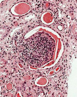
Fig. 8.11 Cortex in a renal biopsy specimen from a man of 41 with chronic renal failure, hematuria, proteinuria, and hypertension. There is chronic damage with extensive thyroidization of tubules. In the chronic inflammatory infiltrate, there are granulomas that contain giant cells. These features suggest tuberculous pyelonephritis
Papillary necrosis is seen as ghostly areas that are poorly stained, often with a rim of inflammatory cells at the junction with viable tissues (Fig. 8.14). Papillary necrosis is a powerful feature because it gives the diagnosis. One cause is use of analgesic drugs when the condition is called analgesic nephropathy. Other causes are sickle cell disease, arteritis, and severe acute pyelonephritis, especially if there is diabetes mellitus.
Changes apart from papillary necrosis in sickle cell disease include the pattern of subendothelial membranoproliferative glomerulonephritis without immune deposits, segmental sclerosing glomerular lesions, which may be due to reduced nephron numbers, tubular atrophy, and iron deposition in tubules as a result of hemolysis (Fig. 8.15). Sickle cell trait can give similar but milder changes.
Cysts may be seen at the corticomedullary junction and in the medulla, and could indicate juvenile nephronophthisis or medullary cystic disease (Fig. 8.16). These are
222 |
8 Indication for Biopsy: Chronic Renal Failure |
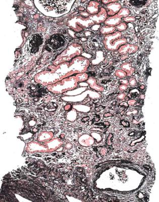
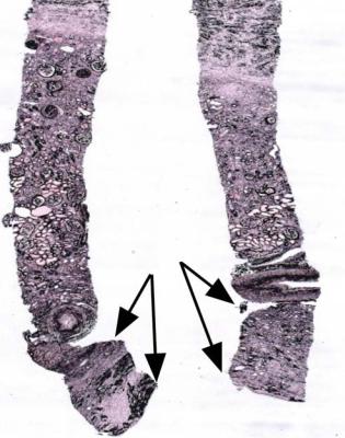
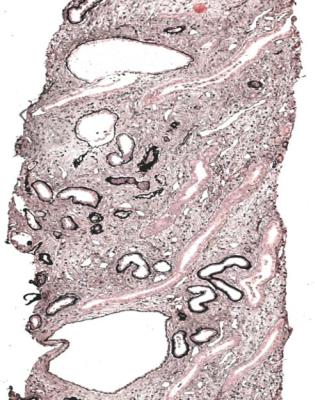
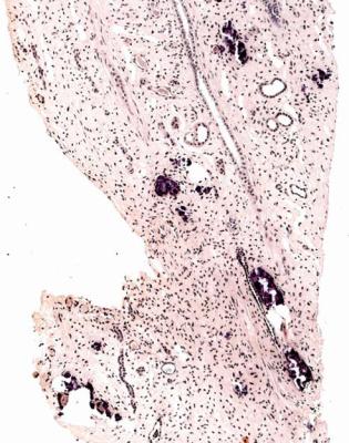
Fig. 8.12 A renal biopsy specimen from a woman of 66 with chronic renal failure, progressive fibrosis around the aorta, and fibrosis in the pleura, peritoneum, and elsewhere. The other kidney was known to have blockage of the ureter, but the biopsied kidney appeared nearly normal on imaging. The two pieces include perirenal tissues and full thickness of the cortex and medulla. There is retroperitoneal fibrosis with thinned cortex, which has areas of chronic damage. The medulla, between both sets of arrows, is severely thinned. This shows there is hydronephrosis, presumed to be due to ureteric obstruction from retroperitoneal fibrosis
inherited disorders with similar structural findings, but with several different genetic abnormalities.
Nephronophthisis is a group of autosomal recessive disorders of nephrocystin proteins, and the renal disease is often associated with abnormalities of other organs. Although nephronophthisis is traditionally considered a cystic disorder, there may be few or no cysts. Tubular basement membrane abnormalities are described in nephronophthisis, including thickening, thinning, and splitting to give a laminated appearance, but similar changes may be seen in areas of tubular atrophy in many other conditions.
Medullary cystic disease has autosomal dominant inheritance, and presents later in life than nephronophthisis, usually in young adults. One form of this condition may be the same as familial juvenile hyperuricemic nephropathy, which is
Is There Evidence of a Cystic Disorder in the Cortex? |
223 |
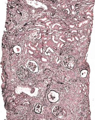
Fig. 8.13 Cortex in a renal biopsy specimen from a woman of 64 with chronic renal failure. There is severe chronic damage without evidence of a glomerular disorder, but otherwise without clues to the cause. There was no medulla in this specimen. A renal biopsy specimen taken in another country a few months before this specimen was reviewed. This contained medulla, which showed acute papillary necrosis, later attributed to nonsteroidal anti-inflammatory drugs. The cortical damage seen in this figure is a late consequence of papillary necrosis
associated with mutations in the gene for Tamm-Horsfall protein, also called uromodulin in this context. Medullary cystic disease is not the same as medullary sponge kidney, in which there are dilated collecting ducts towards the tip of papillae. If there are no complications of these dilated ducts, which predispose to formation of stones, the kidney should be otherwise normal in medullary sponge kidney.
Is There Evidence of a Cystic Disorder in the Cortex?
Cysts may be seen in the cortex (Fig. 8.17). Their significance is often difficult to determine. Any late renal disease may have cysts, sometimes called simple, although their pathogenesis is likely to be complicated rather than simple.
224 |
8 Indication for Biopsy: Chronic Renal Failure |
Fig. 8.14 Medulla in a renal biopsy specimen from a man of 68 with chronic renal failure that rapidly worsened over a short time, insulin resistant or type two diabetes mellitus for 20 years, and recent urinary tract infection. There is acute papillary necrosis
Autosomal dominant polycystic kidney disease rarely requires a renal biopsy to be diagnosed, because the disease is usually apparent on other investigations, but biopsy may be done in a young person before the diagnosis is made in other ways. This condition shows cysts of various sizes in all parts of the nephron, including glomeruli. Disorders of more than one gene give similar clinical features, and a polycystin protein is abnormal in each.
Autosomal recessive infantile polycystic kidney disease may be seen in a biopsy specimen as dilated collecting ducts that appear tubular, rather than rounded and truly cystic. The abnormal protein is fibrocystin. This, like polycystins and several other proteins that are abnormal in inherited cystic diseases of the kidney, is important in the structure and function of the centrosome and cilium in tubular cells.
Glomerular cysts may indicate familial glomerulocystic disorders.
Is There Evidence of a Tubular Disorder? |
225 |
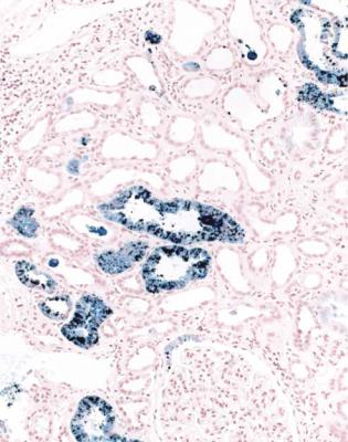
Fig. 8.15 Cortex in a renal biopsy specimen from a man of 19 with chronic renal failure, the nephrotic syndrome, and sickle cell disease. Perls’ Prussian blue stain shows deposition of iron in tubular cells. The glomerular disorder that accounts for the nephrotic syndrome has the pattern of subendothelial membranoproliferative glomerulonephritis. These findings are complications of sickle cell disease
Is There Evidence of a Tubular Disorder?
Paraproteinemias may have several effects on the kidney, although they may have none at all, because paraproteins are common in the elderly, and may be coincidental with any other renal disorder.
The commonest effect they have is tubular damage as a result of consequences of filtered light chains, which is called myeloma kidney or light chain cast nephropathy or similar names. This is easy to detect if there are characteristic casts, meaning deposits within the lumen of tubules that appear dry, cracked, and more palely stained than usual casts. Myeloma casts are surrounded by giant cells (Figs 7.40 and 7.41). Usual casts are seen in virtually any kidney, especially if there is tubular damage, and appear moist, intact, and without a cellular response around them, although there may be red blood cells or leucocytes mixed in with them. In
226 |
8 Indication for Biopsy: Chronic Renal Failure |
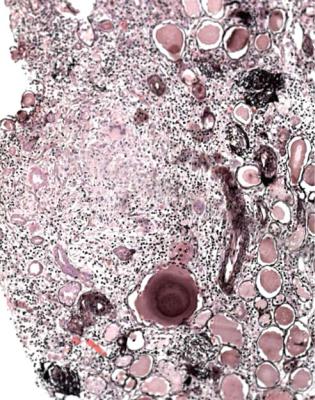
Fig. 8.16 Medulla in a renal biopsy specimen from a man of 30 with chronic renal failure and small, symmetrical kidneys. There is chronic damage with cysts. No cysts were seen in the cortex. These findings suggest medullary cystic disease
myeloma, the typical casts may be sparse or undetectable in a biopsy specimen, and the diagnosis of myeloma kidney may be given by immunohistologic study to detect kappa and lambda light chains, which shows deposition of one type in tubular epithelium (Figs 7.42 and 7.43).
Myeloma kidney should be considered by the pathologist as a possible diagnosis in any specimen from an old person with otherwise unexplained renal failure, either acute or chronic.
Other effects of abnormal immunoglobulins on the kidney include amyloid of AL type (Figs 6.49, 6.63–6.67, and 6.74–6.76). This usually presents with the nephrotic syndrome, and the renal biopsy specimen may give the first hint of a disorder of immunoglobulins. Another effect may be seen if the abnormal immunoglobulins are cryoglobulins, which means that they can precipitate in the plasma in certain conditions (Figs 7.31–7.33). Cryoglobulinemic effects on the kidney usually present with acute renal failure. Another effect of abnormal immunoglobulins is the nodular glomerulosclerosis of light chain glomerulopathy, which resembles
Is There Evidence of a Tubular Disorder? |
227 |
Fig. 8.17 Cortex in a renal biopsy specimen from a man of 20 with chronic renal failure. There are cysts about the size of glomeruli, and elsewhere remnants of tufts are seen in cysts. This appears a glomerulocystic disorder, rather than an early form of autosomal dominant polycystic kidney disease
diabetic glomerulopathy, and usually presents with proteinuria and chronic renal failure (Figs 6.45–6.48).
Deposits of various chemicals may be seen in biopsy specimens. A few calcified deposits within tubules are common, but calcification of tubular basement membranes or interstitial tissues is uncommon, and suggests hypercalcemia (Fig. 8.18). This may be coincidental, or may itself be the cause of renal damage. Extensive calcification may be seen in some disorders of tubular function (Fig. 8.19). Oxalate crystals are within tubules and are birefringent, which means they appear bright when the specimen is examined between crossed polariser and analyser (Figs 7.46 and 7.47). Such crystals may indicate poisoning by antifreeze, or oxalosis, which may be the result of a genetic disorder, or the consequence of increased oxalate absorption from the intestines. More commonly, oxalate deposition is a complication of other renal disorders that cause acute tubular damage. In cystinosis, cystine crystals are also birefringent, but, unlike oxalate crystals, are within cells (Fig. 8.20).
228 |
8 Indication for Biopsy: Chronic Renal Failure |
Fig. 8.18 Cortex in a renal biopsy specimen from a woman of 37 with chronic renal failure, sarcoid, and hypercalcemia. There are calcified deposits in a band of atrophic tubules (arrowed)
Tubular atrophy may be due to drugs or toxins, such as lithium or lead, but the pathologist is unlikely to be able to identify the cause even if there is a history of exposure to a relevant drug or toxin. Chinese herb nephropathy appears due to a toxin aristolochic acid. Balkan endemic nephropathy may also be from the effects of tubular toxins from plants or fungi.
Fanconi syndrome is sometimes seen when there is damage to proximal tubules, with failure to reabsorb some substances from the glomerular filtrate, such as glucose, phosphate, amino acids, and small proteins, which appear in the urine. There is usually renal failure as well. Inherited diseases can produce this, but most are rare. The commonest is cystinosis, although mitochondrial disorders and many others may be found. Acquired disorders can also give Fanconi syndrome, but few have
Is There Evidence of an Interstitial Nephritis? |
229 |
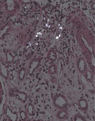
Fig. 8.19 Medulla in a renal biopsy specimen from a woman of 45 with chronic renal failure, medullary calcification on imaging, normal serum calcium concentration, and distal renal tubular acidosis. Intratubular and interstitial deposits of calcified material are seen in this section, which is stained by Congo red with hematoxylin counterstain
characteristic structural features in tubules. These include the nephrotic syndrome and paraproteinemias occasionally, and effects of drugs, particularly anti retroviral drugs used to treat HIV infection, and chemotherapeutic agents used in cancer (Fig. 8.21).
Is There Evidence of an Interstitial Nephritis?
Acute interstitial nephritis with active damage to tubules may be seen when the clinical problem is acute or acute on chronic renal failure, rather than truly chronic renal failure (Figs 7.34 and 7.35).
Granulomas may be seen in renal biopsy specimens. These are well-defined microscopic collections of macrophages with or without necrosis, giant cells, and other cells such as lymphocytes. Granulomatous interstitial nephritis may be seen
230 |
8 Indication for Biopsy: Chronic Renal Failure |
Fig. 8.20 Cortex in a biopsy specimen of a renal allograft present for nearly 2 years in a woman of 18, who had had chronic renal failure due to cystinosis. Examination of the section between polariser and analyser that are nearly crossed shows birefringent crystals of cystine within macrophages that are infiltrating the graft
in sarcoid or tuberculosis when the tubular damage is mostly chronic (Figs 7.38 and 8.11), and as a response to drugs, such as penicillins, and in immunodeficiency states when the tubular damage is mostly acute (Fig. 7.37). True granulomas are not seen in the kidney in Wegener’s granulomatosis, despite the name.
The term late interstitial nephritis may be used when the pathologist is confident that there is no other explanation of late renal damage. In practice, this should be a rare diagnosis, because to be sure of it there must have been an earlier biopsy specimen which showed acute interstitial nephritis. The pathologist should make it clear that the term is not used in the same sense as the discredited term chronic interstitial nephritis, often applied to any late changes in the kidney. Sjögren’s syndrome of dry mouth and eyes and other clinical features may be associated with interstitial nephritis, and usually the renal damage is late and inactive at the time of diagnosis (Fig. 7.39).
Summary: Chronic Renal Failure |
231 |
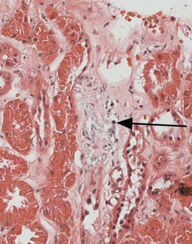
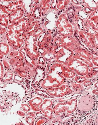
Fig. 8.21 Cortex in a renal biopsy specimen from a man of 54 with chronic renal failure, slight proteinuria, and clinical evidence of Fanconi syndrome. He had been treated with chemotherapy for a lymphoma. There is patchy chronic damage with acute tubular damage seen as irregularity of cell size and a few abnormal nuclei, but there are no structural clues to the cause of the damage. Guido Fanconi (1882–1979), pronounced fan-ko-nee, was a Swiss pediatrician. His name is given to several conditions as well as the tubular disorder
Summary: Chronic Renal Failure
Most people with chronic renal failure do not have a renal biopsy.
Chronic renal failure is associated with chronic abnormalities in tubules, the most important of which is atrophy.
In adults, many renal biopsy specimens show evidence of chronic renal damage due to something other than a glomerular disorder. Findings include chronic ischemic damage, problems with urinary drainage, problems in the renal medulla, cystic diseases, tubular disorders, and late stages of interstitial nephritis, including granulomatous disorders.
232 |
8 Indication for Biopsy: Chronic Renal Failure |
Most of the rest have IgA nephropathy, diabetic glomerulopathy, or segmental sclerosing glomerular conditions, which are often a complication of reduced renal mass.
In biopsy specimens in children, many have evidence of a nonglomerular disorder.
Further Reading: Chronic Renal Failure
D’Agati VD, Jennette JC, Silva FG. Non-Neoplastic Kidney Diseases. Atlas of Nontumor Pathology, first series, fascicle 4. Washington, DC: American Registry of Pathology and Armed Forces Institute of Pathology, 2005. Chapters 2, 4, 16, 17, 20, 22.
Howie AJ. Nephrogenesis and developmental and genetic disorders of the urogenital tract. In: Burge DM, Griffiths DM, Steinbrecher HA, Wheeler RA, editors. Paediatric Surgery, second edition. London: Hodder Arnold, 2005: pp 415–421.
Jennette JC, Olson JL, Schwartz MM, Silva FG. Heptinstall’s Pathology of the Kidney. Sixth ed. Philadelphia: Lippincott Williams and Wilkins, 2007. Chapters 10, 18, 19, 21, 22, 26.