

Chapter 11
Indication for Biopsy: Renal Allograft
Introduction to Renal Allograft Biopsy Specimens
Most renal allograft biopsies are taken because of impaired excretory function called graft dysfunction. This could be called acute or chronic renal failure, as appropriate, although these terms are rarely used following transplantation. The extreme cases are when the graft has not functioned at all or has completely lost function.
Allograft means a graft from a human donor who is not genetically identical with the recipient. Allois from the Greek for other. Graft comes from the Greek for a stylus or style, an instrument used for writing on wax tablets, from the resemblance between its shape and that of shoots of plants inserted to grow in other plants, which is how grafting began. Virtually all transplanted kidneys are allografts, and that is the word used in this text.
An isograft is much rarer. Isois from the Greek for equal. This graft is from a donor genetically identical with the recipient, which means that the pair are monozygotic or identical twins. The recipient has no immune response against the graft, and immunosuppression is not necessary. A successful graft between identical twins in 1954 in Boston, Massachusetts, had a great effect, because it showed that kidney transplantation was feasible, even though it was not the first human transplant, as is sometimes said. A xenograft, from the Greek for foreign, is a graft from a donor of a different species to the recipient. Xenografts are experimental at the moment, but had been tried in people before 1954, as had allografts. An autograft, from the Greek for self, is removal of a person’s own kidney and replacement in the body, but this is not a procedure that will cure chronic renal failure.
Transplanted kidneys are from people whose brainstem has died, called cadaveric donors, or from living people. Cadaveric kidneys are removed either while the donor is on a ventilator, and the kidneys are still perfused with oxygenated blood, or after circulation has stopped, in a non heart beating donor. Potential live donors are usually investigated thoroughly, to make sure they have no condition of the kidneys or other organs that would be a contraindication to transplantation. This may reveal an abnormality that requires investigation by renal biopsy, analysed as in the appropriate chapter, depending on the indication for biopsy.
A. J. Howie, Handbook of Renal Biopsy Pathology. |
263 |
C Springer 2008 |
|
264 |
11 Indication for Biopsy: Renal Allograft |
Reasons for Biopsy of an Allograft Other than Dysfunction
There are a few other reasons for biopsy of an allograft or potential allograft.
1.Sometimes a kidney is sampled after surgical removal, to see if it is suitable to be transplanted.
Renal transplantation is rarely an emergency. After removal, a kidney is perfused with a suitable solution to remove blood and is kept cold in ice. The interval between perfusion and preparation of the kidney for transplantation is the cold ischemic time, which is ideally under 24 h but if necessary can be longer, and some kidneys have been grafted more than 48 h after removal. Outcome of the transplant is slightly worse if the cold ischemic time is over 24 h. The warm ischemic time is the interval between removal of the kidney from ice, and opening of the vascular clamps to perfuse the graft with blood. The shorter this time is, the less damage the kidney is likely to have.
If a pre-transplant sample is taken, the pathologist usually has time to have sections prepared of the specimen embedded in paraffin wax rather than frozen sections, which are ready more quickly than paraffin sections but are less satisfactory for detailed microscopy. The kidney may be from a donor known or suspected to have significant renal disease, and the pathologist may be able to diagnose this and give an assessment of whether the disease and the amount of chronic damage make transplantation undesirable. This is a matter of opinion rather than of established rules, but the amount of chronic damage that has to be present before the outcome is affected is much more than is usually seen in transplanted kidneys (Fig. 11.1).
Sections of frozen material may be necessary in an emergency. The surgeon may have noted a lesion when the kidney is finally prepared for transplantation, and the potential recipient is under a general anesthetic. The pathologist may diagnose a neoplasm or an early stage of adult polycystic kidney disease, which are usually contraindications to transplantation.
2.A sample of a kidney is often taken by the surgeon immediately after the kidney has been grafted, either as a wedge of the capsular surface or as a needle biopsy.
This is called an implantation biopsy, or post perfusion biopsy, or 30 min biopsy, or other terms. Such a specimen is useful, because the pathologist can determine the condition of the transplanted kidney, particularly the extent of chronic damage and state of the blood vessels, which may have evidence of chronic intimal thickening, atherosclerotic embolism, or hyalinosis (Figs 5.15, 11.2, and 11.3). Knowledge of this pre-existing damage helps the pathologist to interpret changes in later specimens. The specimen is also useful to show who has received a graft and when they received it. Ideally, the pathologist should be informed about the original renal disease in the recipient, although the surgeon may not know this.
Sometimes, the surgeon gives details of the donor, such as age, sex, and cause of death if it was a cadaveric donor, or whether it was a living donor. These specimens often show globules of cytoplasm in the lumen of tubules,
Reasons for Biopsy of an Allograft Other than Dysfunction |
265 |
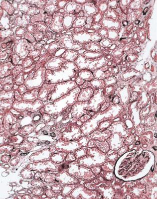
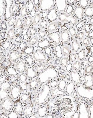
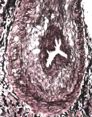
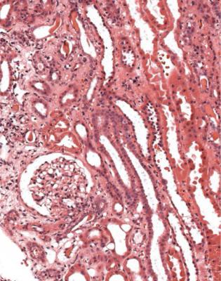
Fig. 11.1 Cortex in a wedge biopsy specimen of kidney removed after death from a man of 34, who had a head injury, severe loss of blood, and progressive renal impairment over a few hours before death. The biopsy was to see if the kidney could be transplanted. There is a little tubular atrophy, and most tubules show acute damage with flattened epithelium. Tubules also have fine vacuolation, which is consistent with use of mannitol. The pathologist recommended that the kidney should be used. The kidney was transplanted into a man of 48. Although there was no function for 10 days, the graft worked well afterwards
particularly proximal tubules, that may reflect autolysis, but seem to have no significance related to graft function. This is sometimes called preservation injury, or ischemia/reperfusion injury. Proximal tubules may have fine vacuolation of their cytoplasm if the donor had had cerebral edema treated with the osmotic diuretic mannitol, but this change also seems to have no significance related to graft function (Fig. 11.1). Despite these and other changes, the pathologist may be able to identify features of genuine acute tubular damage, which must have been present in the donor (Figs 7.3 and 7.4). The significance is that a graft with this damage is likely to take longer to function than one without the damage.
There may be an unsuspected glomerular disorder, or something else (Fig. 4.9). If every implantation biopsy specimen is investigated thoroughly, thin glomerular basement membrane disease may be found in about one in 20
266 |
11 Indication for Biopsy: Renal Allograft |
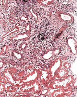
Fig. 11.2 Cortex in a wedge biopsy specimen of kidney taken at the time of transplantation into a woman of 24 from a boy of 16, who died of a subarachnoid hemorrhage. The kidney appears normal, and the graft worked well
samples, while other conditions, such as IgA nephropathy or diabetic glomerulopathy, will be found less commonly. Identification of such conditions is helpful in interpretation of later specimens, but whether pre-existing glomerular disorders have a significant effect on outcome of a graft by themselves is doubtful. The amount of chronic damage associated with those disorders, or arising from other factors such as ischemia, is more important in prognosis. Even the amount of chronic damage is less important in outcome than immunologic, vascular, ureteric, and other problems that develop after transplantation. Some conditions should get better after transplantation. For instance, IgA should disappear from glomeruli in a kidney from a donor with IgA nephropathy, provided that the recipient does not also have IgA nephropathy.
3.Occasionally, if a surgeon is operating on a graft for reasons such as ureteric or vascular problems, a biopsy is taken just because the surgeon is there.
4.Some centres take biopsies at set intervals after transplantation, irrespective of renal function. These are called protocol biopsy specimens.
Value of Biopsy of a Renal Allograft |
267 |
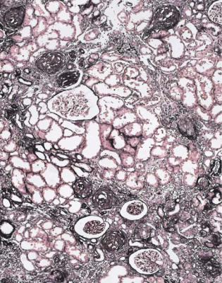
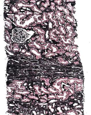
Fig. 11.3 Cortex in a wedge biopsy specimen of kidney taken at the time of transplantation into a woman of 46 from a woman of 57, who died of a brainstem infarct. There is extensive chronic ischemic damage. Neither this kidney nor the other one from the same donor worked well. An index of chronic damage (Fig. 5.6) of 40 % or more indicates that graft survival is likely to be shorter than average. In this specimen, the index of chronic damage is 46 %
5.Allografted people may have a renal problem other than abnormal excretory function, or as well as abnormal excretory function, such as the nephrotic syndrome, and this may be the reason for the biopsy.
Value of Biopsy of a Renal Allograft
The most common reason for surgeons or nephrologists to take an allograft biopsy specimen is to determine whether there is evidence of a significant immunologic reaction by the recipient against the graft, which is traditionally called rejection. This is one of the few circumstances in which a pathologist’s report can have a decisive immediate influence on clinical management, and may help to save a graft that otherwise would have been lost, or help to avoid unnecessary treatment, or indicate
268 |
11 Indication for Biopsy: Renal Allograft |
the need for further investigations. Biopsy is much better done before treatment of rejection, rather than afterwards, when the pathologist may not be able to say whether or not there had been significant rejection.
Technical Handling of Renal Allograft Biopsy Specimens
Specimens taken for investigation of graft dysfunction should be considered urgent, and processed and sectioned with the shortest possible delay. The same set of sections that is prepared for other specimens can be cut, and the pathologist should be given the sections stained by hematoxylin and eosin. Most conditions of immediate clinical significance can be assessed on sections stained by hematoxylin and eosin, even if only provisionally.
Most specimens need only orthodox light microscopic stains for adequate assessment. The full set should be prepared, to allow the pathologist to see many sections, and to make use of the stains in interpretation, for instance in judgment of the extent of tubular atrophy. Immunohistologic study or electron microscopy may occasionally be required, to investigate such things as possible viral infection, or possible recurrence of the original renal disease in the graft.
Cortex is required to allow the pathologist to assess allograft biopsy specimens. How much is required is a matter of judgment. The Banff group, so called because their meeting at Banff in Canada in 1991 was the first of a regular series at various sites, suggested in 1997 that an adequate specimen had at least ten glomeruli and at least two arteries. The minimum requirement was seven glomeruli and one artery, with at least two cores of cortex or two areas of cortex on the same core, studied with multiple sequential sections, 3–4 μm thick, on seven slides, three stained with hematoxylin and eosin, three with either periodic acid Schiff or periodic acidmethenamine silver, and one with a connective tissue stain.
Approach to the Diagnosis in Renal Allograft Biopsy Specimens
The single most important piece of information that the pathologist needs on the request form with a biopsy specimen from a renal allograft is how long the kidney has been in the recipient.
The two common decisions that a pathologist should make are whether there is significantly active, acute rejection that may respond to treatment, and whether there is chronic damage that will not respond to treatment. Generally, the first problem arises in specimens taken soon after transplantation, meaning in the first few weeks, and the second problem in specimens taken long after transplantation.
Another problem for the pathologist is to decide whether there is any other identifiable reason for graft dysfunction.
Most specimens can be given a satisfactory diagnosis by a pathologist. An important rule is that there may be more than one condition in a specimen.
Antibody-mediated Rejection |
269 |
Traditionally, rejection is divided into acute and chronic, although the pathogenesis of chronic rejection is not understood, and this may not be an immunologic process at all.
Is There Evidence of Acute Rejection?
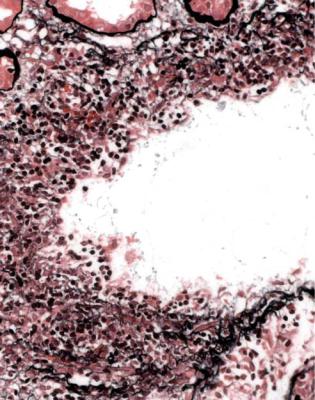
Before satisfactory identification of blood group and histocompatibility antigens of the potential donor and recipient, and cross-matching procedures between cells from the donor, such as lymphocytes, and serum from the recipient, there were examples of hyperacute rejection caused by an immediate response of antibodies in the recipient against the endothelium in the kidney. Soon after the vascular anastomoses were completed and blood was allowed to perfuse the graft, this became discolored and never worked, and the pathologist saw thrombus in glomerular capillaries, either in an implantation specimen or in a specimen taken within a few days of transplantation. In theory, hyperacute rejection should not occur these days, but may still be seen if there have been technical problems with matching donor and recipient (Fig. 11.4).
Antibody-mediated Rejection
Hyperacute rejection is the most dramatic type of antibody-mediated rejection, which may be seen in a form that can be called delayed hyperacute or accelerated acute rejection. This may be due to antibodies in the recipient against HLA or non-HLA antigens in the graft. Pre-existing antibodies could have been induced in a recipient by blood transfusion, pregnancy, or previous transplantation. If antibodymediated rejection occurs, these antibodies either were not evident on the crossmatching procedures, or were detected but considered insignificant. New antibodies against the graft may also develop, despite immunosuppression.
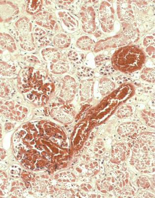
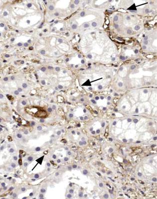
This type of rejection often occurs in the first few weeks after transplantation, and a biopsy specimen shows many neutrophils adherent to the endothelium of capillaries between tubules, and often in glomeruli as well (Fig. 11.5). A marker that there has been a reaction between antibodies and endothelium is C4d, which is an inactivated form of the complement component C4. C4d detected by an immunohistologic method on the endothelium of intertubular capillaries is sometimes considered evidence of antibody-mediated rejection, especially if C4d is widespread (Fig. 11.6).
Interpretation of the clinical significance of C4d deposition is not straightforward. For example, extensive deposition is almost always found in recipients who have been apparently depleted of antibodies to A or B blood groups or HLA antigens to allow them to receive an ABO or HLA incompatible graft, and yet many have an uncomplicated outcome after transplantation. C4d may also persist for several days after rejection has been treated (Fig. 11.7).
270 |
11 Indication for Biopsy: Renal Allograft |
Fig. 11.4 Cortex in a nephrectomy specimen removed 7 days after transplantation into a man of 63. The donor and recipient had a close match on HLA typing, but could not be directly tested for histocompatibility, because no lymphocytes were available from the donor. The graft never worked. There is thrombosis in glomeruli and small vessels with the appearances of hyperacute rejection
Although the Banff group recommended that every renal allograft biopsy specimen should be examined immunohistologically using an antibody to C4d, there is contradictory evidence whether this is justified in routine practice. The test is probably only useful when there are clinical or pathologic suggestions that there may be antibody-mediated rejection.
Diagnosis of antibody-mediated rejection should not be made on the finding of C4d alone. There should also be changes in a biopsy specimen such as neutrophils in intertubular capillaries and thrombosis in glomeruli, and the recipient should be shown by immunologists to have antibodies against donor antigens. The clinical importance of this diagnosis is that treatment may be required that is different from treatment of conventional rejection.

Conventional Acute Rejection |
271 |
Fig. 11.5 Cortex in a needle biopsy specimen of kidney taken 4 days after transplantation into a man of 25. There was no graft function. Tubules show severe acute damage, with a heavy infiltrate of neutrophils in intertubular capillaries. Immunoperoxidase staining for C4d is shown in Fig. 11.6, and helped to suggest the diagnosis of antibody mediated rejection
Conventional Acute Rejection
Conventional acute rejection takes two forms that can occur independently or together, and can be seen with antibody-mediated rejection. The forms are acute cellular rejection, sometimes called acute tubular and interstitial rejection or acute tubulo-interstitial rejection, and acute vascular rejection. These days, acute vascular rejection is uncommon, particularly in its severe form. Acute rejection is commonest soon after transplantation, within the first few weeks, but can occur at any time after transplantation, for instance if there is a change in dose or type of immunosuppressive drugs. About one quarter of transplant recipients have at least one episode of acute rejection.
272 |
11 Indication for Biopsy: Renal Allograft |
Fig. 11.6 Cortex in the biopsy specimen from the man of 25 shown in Fig. 11.5, stained by an immunoperoxidase method for C4d. This is seen on the endothelium of many intertubular capillaries, some of which are arrowed. Before the transplant, there was a close HLA match between donor and recipient, and recipient serum had no cytotoxic effect on donor lymphocytes. When the biopsy findings suggested antibody mediated rejection, further immunologic investigation showed that the recipient had an antibody to an HLA class two antigen, found on donor B cells, but not included in routine HLA typing. The man had had a previous renal transplant, which had the same class two antigen as the new donor
Diagnosis of Acute Cellular Rejection, also Called Acute Tubulo-interstitial Rejection
Acute cellular rejection, in its more severe stages, is easy to diagnose. Sometimes, sections appear dark even before the pathologist looks at them with a microscope. At low magnification, there is an infiltrate of inflammatory cells, patchily or uniformly throughout the cortex (Fig. 11.8). At high power, the cells are mostly lymphocytes, and there is acute damage to tubules, which are separated by the inflammatory infiltrate and edema (Fig. 11.9). Inflammatory infiltrates are not considered significant if they are in areas of fibrosis, such as in the subcapsular cortex if there is chronic ischemic damage or in connective tissues around arcuate vessels (Fig. 11.10).
Diagnosis of Acute Cellular Rejection, also Called Acute Tubulo-interstitial Rejection |
273 |
Fig. 11.7 Cortex in a needle biopsy specimen of kidney taken 2 weeks after transplantation into a woman of 43. Vascular access to allow hemodialysis was becoming so difficult that a cadaveric kidney was accepted for transplantation, even though the recipient had antibodies to HLA class two antigens in the donor. With extra immunosuppression at transplantation, graft function was satisfactory until 9 days after transplantation, when there was a swollen, tender kidney with oliguria and a marked rise in serum creatinine concentration. Biopsy was not done at that time, because there was a fear of rupture of the graft. Intensive treatment for presumed antibody-mediated rejection produced clinical improvement in symptoms, but not in renal function. The biopsy specimen is stained by an immunohistologic method for C4d, and shows extensive deposition in the endothelium of intertubular capillaries, but without any other evidence of rejection. The graft recovered function without additional treatment
Acute cellular rejection is a reaction against antigens on tubular cells by T lymphocytes, particularly CD8 positive cytotoxic cells. The feature that is the strongest indicator of this type of rejection is infiltration of lymphocytes across tubular basement membranes to lie inside the tubular epithelium (Fig. 11.9). This is often called tubulitis. A lymphocytic infiltrate is disregarded in atrophic tubules, which are recognised by reduction in size of the tubules, with thickening of their basement membrane.
274 |
11 Indication for Biopsy: Renal Allograft |
Fig. 11.8 Cortex in a needle biopsy specimen of kidney taken 6 days after transplantation into a man of 35. Initial function was satisfactory, but then deteriorated. There is a lymphocytic infiltrate with edema. Tubules are acutely damaged, and there is an infiltrate of lymphocytes into them. Features are those of significant acute cellular rejection
Diagnosis of Significant Acute Cellular Rejection
The problem is that acute cellular rejection is not all or none. There is a gradation from no cellular infiltrate to widespread infiltration of the cortex. The extent of lymphocytic infiltration of tubules varies widely, even in the same specimen. A difficulty for the pathologist is to decide when the features are those of acute cellular rejection of sufficient intensity to be clinically significant. This means that the immunologic response is producing enough damage to give renal dysfunction and that treatment of the response is likely to allow the kidney to recover.
The distinction between significant acute cellular rejection and a clinically insignificant cellular infiltrate is arbitrary. Grading schemes have been devised to try to standardise interpretation of changes in renal allograft biopsy specimens. At the moment, these schemes largely depend upon guesswork and the opinion of the pathologist rather than upon measurement or strictly objective criteria.

Diagnosis of Significant Acute Cellular Rejection |
275 |
Fig. 11.9 Cortex in a needle biopsy specimen of kidney taken 2 months after transplantation into a man of 24. Function was satisfactory, but suddenly deteriorated. There is a lymphocytic infiltrate with edema. Tubules are acutely damaged, and there is an infiltrate of lymphocytes into them, obscuring the epithelium in several tubules. Features are those of significant acute cellular rejection
In the scheme suggested by the Cooperative Clinical Trials in Transplantation (CCTT), the threshold for diagnosis of clinically significant acute cellular rejection is that the mononuclear cell inflammatory infiltrate occupies at least 5 % of the cortical area, by guesswork, and that in the most heavily infiltrated areas there are at least three tubules with an infiltrate of lymphocytes in ten serial microscopic fields with a ×40 objective. The original scheme stated that there must also be at least two of these three additional features, edema, tubular degeneration or injury, and reactive lymphoblasts, although these features are not necessary when the biopsy is taken for graft dysfunction, but may be useful in interpretation of protocol biopsy specimens.
In the scheme suggested by the Banff group in 1997, the threshold for diagnosis of clinically significant acute cellular rejection is that at least 25 % of the cortical area, by guesswork, has an inflammatory infiltrate, and there are at least four lymphocytes within a tubule in more than one area. In the Banff scheme, there is no
276 |
11 Indication for Biopsy: Renal Allograft |
Fig. 11.10 Cortex in a needle biopsy specimen of kidney taken 8 days after transplantation into a man of 17. There was no function. There are dense clusters of lymphocytes at the corticomedullary junction around arcuate vessels, but these are disregarded in assessment of rejection. There is no evidence of significant rejection in the cortex. A few tubules contain crystals (arrowed), a feature of acute tubular damage
requirement for edema, tubular degeneration or injury, and reactive lymphoblasts. Also, in the Banff scheme, changes between roughly the CCTT threshold and the Banff threshold for clinically significant acute cellular rejection are called borderline or suspicious for rejection, except that the lower limit for the Banff scheme is that the inflammatory infiltrate occupies at least 10 % of the cortical area, by guesswork.
The borderline category is not helpful to surgeons or nephrologists who are looking for guidance to help their decision whether to treat rejection. For this reason, the pathologist should take the responsibility and say whether the changes are significant or not. The Banff category of borderline or suspicious for rejection usually indicates clinically significant acute cellular rejection in renal allograft biopsy specimens, other than those from grafts that have been treated for rejection shortly before the biopsy, or those that are protocol biopsy specimens. The pathologist should know whether either of those conditions applies to a particular specimen, and can report accordingly, for instance that there seems to have been significant acute cellular rejection with an apparent response to treatment.
Summary of Acute Rejection |
277 |
Acute cellular rejection is identical in appearance to acute interstitial nephritis (Figs 7.34 and 7.35). In theory, an allograft could have an acute interstitial nephritis as an allergic response to a drug, but no pathologist with the tests routinely available would be able to differentiate this with certainty from acute cellular rejection.
Diagnosis of Acute Vascular Rejection
Acute vascular rejection is easy to diagnose. Any features of it that the pathologist is confident are present are enough to indicate clinically significant rejection, even if only one artery is affected.
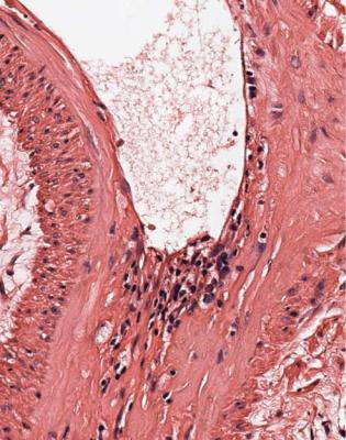
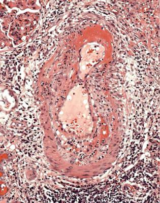
These features are widening of the space between the endothelium and the underlying tissues in arteries of any size, with infiltration of lymphocytes into this space (Figs 11.11 and 11.12). This abnormality is sometimes called intimal arteritis, intimal vasculitis, endarteritis, or arterial endothelialitis. The infiltrating cells are mostly T lymphocytes of both CD4 and CD8 types. Changes in the intima of venules and veins are not considered significant (Fig. 11.13).
Acute vascular rejection is an easier diagnosis to make than acute cellular rejection, because any definite features of acute vascular rejection are significant, no matter how small or scarce. Not even the full circumference of an artery has to be affected (Fig. 11.11). Compared with acute cellular rejection, acute vascular rejection is generally more difficult to treat, less likely to return to normal, and more likely to leave significant chronic damage, and no definite features of it should be ignored or considered insignificant.
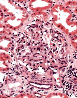
Another feature that may be seen in acute rejection is a glomerular disorder called acute allograft glomerulopathy. Glomeruli appear hypercellular, with swollen endocapillary cells and an infiltrate of lymphocytes (Fig. 11.14). This is uncommon, usually occurs shortly after transplantation, and is often associated with signs of significant rejection, particularly acute vascular rejection.
Acute vascular rejection is considered severe when affected arteries have damage in the media as well as in the intima. Specifically, there is fibrinoid necrosis of the media, with or without infiltration of inflammatory cells into the media. There may be thrombosis in these vessels. Severe acute vascular rejection is often accompanied by interstitial hemorrhage, and sometimes by necrosis of parts of the kidney (Fig. 11.15). This type of rejection is particularly likely to cause irreversible damage to a graft, but is rare, and in practice, virtually only seen when immunosuppression is withdrawn.
Summary of Acute Rejection
The pathologist should use one of five diagnoses related to acute rejection. These are antibody-mediated rejection, significant acute cellular rejection, acute vascular rejection, severe acute vascular rejection, and no evidence of significant acute rejection.
The three diagnoses of rejection, other than the antibody-mediated type, correspond to types one, two, and three, respectively, in the CCTT and Banff systems,
278 |
11 Indication for Biopsy: Renal Allograft |
Fig. 11.11 Cortex in a needle biopsy specimen of kidney taken 13 days after transplantation into a woman of 43 from a woman of 50, who died of a subarachnoid hemorrhage. Initial function was satisfactory, but then deteriorated. An arcuate artery has pre-existing chronic intimal thickening from the donor. There are lymphocytes under the endothelium in places, a sign of acute vascular rejection
although these have different criteria to separate significant and insignificant acute cellular rejection.
More elaborate schemes, such as those suggested by the Banff group, are ways of recording and grading the great variety of changes that can be seen in allografts. In general, these are poorly reproducible between pathologists, and of little or no clinical use.
Is There Any Other Acute Abnormality Instead of Acute Rejection, or in Addition to It?
If an allograft is not working, there is a structural disorder of tubules, as there is in any kidney with acute renal failure.

Other Acute Abnormalities |
279 |
Fig. 11.12 Cortex in a needle biopsy specimen of kidney taken 10 days after transplantation into a man of 33. There was no function. An artery has loose intimal swelling with an infiltrate of lymphocytes. Features are those of acute vascular rejection
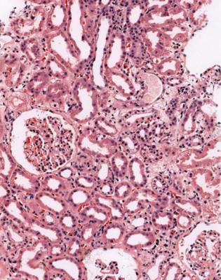
In the early stages after transplantation, a common clinical problem is delayed graft function. The main differential diagnosis is between significant acute rejection as a cause of acute tubular damage, and acute tubular damage without significant rejection, almost always called acute tubular necrosis by nephrologists and surgeons.
Acute tubular damage is inevitable to some extent after transplantation, although it may be so clinically insignificant that the graft works immediately. Acute tubular damage is likely to be more severe and longer lasting in grafts from cadaveric donors rather than living donors, in grafts from cadaveric donors who had extensive trauma or prolonged hypotension or whose heart was not beating at the time of removal of the kidney, and in grafts with particularly long cold or warm ischemic times (Fig. 11.16).
Both acute rejection and pure acute tubular damage can occur at any time after transplantation, and usually there is an explanation in the clinical history. An episode of acute rejection long after transplantation may be due to a change in immunosuppression, such as failure of the recipient to take drugs. An episode of pure acute
280 |
11 Indication for Biopsy: Renal Allograft |
Fig. 11.13 Cortex in the needle biopsy specimen of kidney taken 2 months after transplantation into the man of 24 with significant acute cellular rejection, which is illustrated in Fig. 11.9. A vein has an infiltrate under its endothelium, but this is not a sign of acute vascular rejection
tubular damage may be due to an event such as a myocardial infarct or an episode of sepsis with hypotension and underperfusion of the graft.
Other conditions that may occur especially in the early stages after transplantation include infarction and infection.
Infarction may be due to thrombosis in large arteries or veins, although this is now rare, or to severe acute vascular rejection, which is also rare, and should have characteristic changes in arteries (Fig. 11.15). Thrombus may be seen in small vessels as a complication of acute or chronic rejection, often without infarction (Fig. 11.17). Infarction may occur in part of the graft if one pole has its own artery separate from the main artery, and the accessory artery has not been anastomosed to a recipient artery, or if there is thrombosis only of some vessels. The affected part undergoes infarction (Fig. 11.18). The pathologist should report only that the tissue in the biopsy specimen is infarcted, not that the whole graft is infarcted. This is an example of the general rule that a biopsy specimen may not be representative of the whole kidney.
Other Acute Abnormalities |
281 |
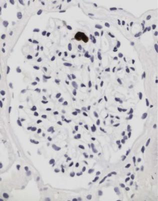
Fig. 11.14 Glomerulus in a needle biopsy specimen of kidney taken 6 months after transplantation into a woman of 41, whose immunosuppressive drugs had been reduced. The glomerulus appears hypercellular with endothelial swelling, an infiltrate of lymphocytes, some of which are clustered and appear to run together, and prolapse into the tubular opening. Features are those of acute allograft glomerulopathy. There is evidence of acute cellular rejection elsewhere in the specimen
Infection of a graft may be ascending infection with pus in tubules, sometimes with evidence of bacteria or fungi (Fig. 7.36). Purulent infections and tuberculosis can also be carried to the graft in the blood, and there may be purulent infection around a graft (Fig. 4.3).
Cytomegalovirus infection is common as a systemic infection, but is now unusual in the graft itself. Large, infected cells with intranuclear inclusions may be seen in glomeruli or tubules, and can be confirmed to contain cytomegalovirus by immunohistologic study (Figs 11.19 and 11.20).
Another viral infection is with BK virus, a polyomavirus related to simian virus (SV) 40, and which can be detected immunohistologically using antibodies to SV40. BK virus initially infects transitional epithelium of the urinary tract and cells of the collecting duct, to produce large, distorted nuclei, containing indistinct inclusions
282 |
11 Indication for Biopsy: Renal Allograft |
Fig. 11.15 Cortex in a nephrectomy specimen from a woman of 22 removed 19 months after transplantation, and after immunosuppression had been withdrawn. An artery has fibrinoid necrosis, a sign of severe acute vascular rejection
(Figs 11.21 and 11.22). There is usually a chronic inflammatory response around infected tubules.
Is There Evidence of Chronic Rejection?
Gradual loss of graft function is still a common problem, despite advances in control of acute rejection. The median survival of function is under 10 years, although the aim should be to have adequate function until the recipient dies of a cause unrelated to the kidney. Decline in function may be a sign of so-called chronic rejection, one feature of which is a disorder of blood vessels that can be called chronic vascular rejection. Although this is usually seen years after transplantation, changes may begin to appear within a few weeks of grafting.
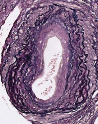
This is a condition in which arteries develop concentric intimal thickening, without an inflammatory infiltrate in the intima (Fig. 11.23). The intimal changes do
Is There Evidence of Chronic Rejection? |
283 |
Fig. 11.16 Cortex in a needle biopsy specimen of kidney taken 11 days after transplantation into a woman of 43. There was a period of hypotension, and there was no renal function. The graft is viable, and shows acute tubular damage without evidence of significant rejection
not have concentric rings of elastin fibres, and in this way can be differentiated from the chronic intimal thickening caused by age and hypertension (Fig. 11.24). Sometimes, there is an appearance of a new artery inside the old, with muscular media and internal elastic lamina (Fig. 11.25). There may be intimal foamy cells.
Usually, the affected arteries are larger than those included in biopsy specimens. Because of this, the pathologist may see no direct evidence of the arterial changes, and the diagnosis is suspected from changes in tubules, which show atrophy as a result of ischemia, and glomeruli, which may be shrunken, also from ischemia. Large renal veins may have similar changes to those in arteries, but these are unlikely to be seen in a biopsy specimen. Intertubular capillaries may have thickening and multilayering of their basement membrane, and deposition of C4d in their wall.
Glomeruli may also show chronic allograft glomerulopathy. There is mesangial expansion and doubled basement membranes, not always globally, often with areas of segmental sclerosis (Fig. 11.26). On immunohistologic study, there is slight
284 |
11 Indication for Biopsy: Renal Allograft |
Fig. 11.17 Cortex in a needle biopsy specimen of kidney taken 3 months after transplantation into a man of 24. There is evidence of significant acute cellular rejection with a thrombus in a small vein
deposition of IgM and complement in mesangium and subendothelial areas. On electron microscopy, glomeruli show subendothelial areas widened by material that lacks immune deposits (Fig. 11.27). This glomerulopathy may be accompanied by proteinuria, which is occasionally heavy enough to be in the nephrotic range.
In chronic rejection, there may be collections of lymphocytes, but these are in atrophic areas, and there is no sign of the active tubular damage that indicates significant acute cellular rejection. A few grafts have heavy infiltrates of plasma cells. Although this is sometimes called plasma cell-rich acute rejection, the infiltrates are mostly in areas with tubular atrophy rather than acute tubular damage, and there is usually little or no response to conventional treatment for acute cellular rejection.
Acute rejection can coexist with chronic rejection. In practice, a pathologist may have difficulty in deciding whether a lymphocytic infiltrate, in a graft with chronic damage, is a sign of significantly active cellular rejection. A feature which suggests significant acute cellular rejection is the presence of acute tubular damage in areas of lymphocytic infiltration, with lymphocytes inside tubules that are not atrophic (Fig. 11.28).

Differential Diagnosis of Late Damage in a Renal Allograft |
285 |
Fig. 11.18 Cortex in a needle biopsy specimen of kidney taken 1 month after transplantation into a man of 37. There is hemorrhagic necrosis without evidence of acute vascular rejection. Only part of the kidney had venous thrombosis on radiographic investigation. The graft recovered function
Differential Diagnosis of Late Damage in a Renal Allograft
Late damage in an allograft, meaning tubular atrophy in particular, does not only result from chronic rejection (Fig. 11.29). All late changes were grouped in the Banff system in 1991 under the term chronic allograft nephropathy. This was meant to be just a description, but the term was often taken to mean a specific diagnosis, synonymous with chronic rejection. For this reason, in 2005, the Banff group suggested that chronic allograft nephropathy should be no longer used.
Possible explanations of late damage in addition to chronic rejection are these.
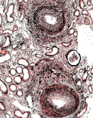
1.Damage may have been present in the kidney before transplantation. The implantation biopsy specimen may help the pathologist to decide how much damage was pre-existing (Figs 5.15 and 11.3).
2.Calcineurin inhibitors, used as immunosuppressants, have an ischemic effect on the kidney. These drugs are tacrolimus and cyclosporine, whose
286 |
11 Indication for Biopsy: Renal Allograft |
Fig. 11.19 Glomerulus in a needle biopsy specimen of kidney taken 7 weeks after transplantation into a man of 53. There was acute graft dysfunction. Cytomegalovirus was detected in the blood. The biopsy specimen shows no evidence of significant rejection, but the glomerulus appears hypercellular, and contains large cells with atypical nuclei. Immunoperoxidase findings are shown in
Fig. 11.20
Recommended International Non Proprietary Name is ciclosporin in the European Union. These may cause reversible acute tubular damage in the short term or irreversible tubular atrophy in the long term. These drugs may also produce fine vacuolation of tubular cells, often called isometric vacuolation, but similar changes may be seen in acute tubular damage from any cause, and as a result of mannitol treatment (Figs 5.9, 7.4, and 11.1). Calcineurin inhibitors may rarely cause a small vessel vasculopathy, also called thrombotic microangiopathy, of the type seen in the hemolytic uremic syndrome (Fig. 7.9). A pathologist cannot often diagnose acute effects of calcineurin inhibitors with confidence, and can only suggest toxicity as a possibility if other things, such as significant acute rejection, have been excluded (Fig. 11.30).
Arteriolar hyalinosis is a late complication of these drugs. This finding may be consistent with drug toxicity in children’s grafts, but is often little help in adult kidneys, in which hyalinosis is common (Fig. 5.15). The hyalinosis produced by calcineurin inhibitors is sometimes claimed to be characteristic, because this bulges into the tissues around an arteriole, but this feature can be seen in people not treated with these drugs (Fig. 11.31). The tubular atrophy produced by calcineurin inhibitors is often said to be accompanied by striped fibrosis, running
Differential Diagnosis of Late Damage in a Renal Allograft |
287 |
Fig. 11.20 Glomerulus in the biopsy specimen from the man of 53 shown in Fig. 11.19. An immunoperoxidase method to detect cytomegalovirus shows a large, infected cell
radially through the cortex in the position of medullary rays. This may be seen in other conditions, and the drugs may give more widespread atrophy (Fig. 11.32). Glomeruli may have segmental sclerosis, which, again, can be seen in other conditions.
Although the late changes said to be characteristic of effects of calcineurin inhibitors may be seen in kidneys of people without a renal graft, treated with these drugs for another reason, such as after transplantation of a heart or liver, whether the changes are only produced by calcineurin inhibitors is unlikely (Fig. 2.1). There does not appear to have been a formal comparison between renal grafts examined at several years after transplantation, some treated with these drugs, and a control group treated only with other immunosuppressants. If the various late changes are seen, they are best reported to be consistent with effects of calcineurin inhibitors.
3.There may be permanent damage to the graft from attacks of acute rejection. This is more likely after acute vascular rejection than acute cellular rejection. Previous infection of the graft may also have produced chronic damage.
288 |
11 Indication for Biopsy: Renal Allograft |
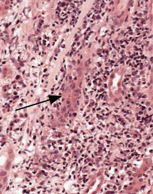
Fig. 11.21 Cortex in a needle biopsy specimen of kidney taken 4 months after transplantation into a man of 62. There is a heavy chronic inflammatory infiltrate with abnormal nuclei in tubules, particularly the one arrowed. Immunoperoxidase findings are shown in Fig. 11.22
4.The graft may have a disorder of its blood supply, such as effects of hypertension, stenosis of the renal artery, or thrombosis of veins. Vascular problems cause ischemia, with acute and chronic effects, but there may be no indication in a biopsy specimen of the precise cause or causes of ischemia.
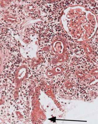
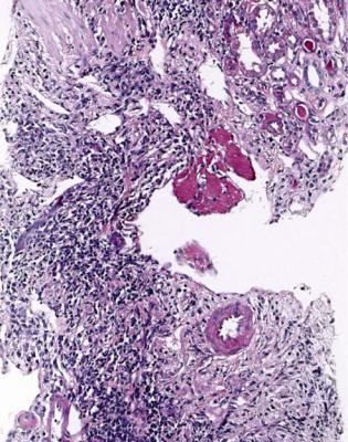
5.The graft may have a disorder of its drainage, such as urinary reflux or stenosis of the ureter. A clue to a drainage problem is the presence of pus in tubules (Fig. 7.36). Another clue is solid masses in interstitial tissues or veins of material that stains with periodic acid Schiff and contains few cells (Fig. 11.33). This material is precipitated Tamm-Horsfall protein (Fig. 11.34). These extratubular deposits, which include tubulo-venous ruptures, are seen in obstruction of the urinary tract, but can also be complications of acute rejection. In severe, acute obstruction, Tamm-Horsfall protein may reflux within tubules to precipitate in Bowman’s space.
6.Effects of reduced nephron number, with hyperfiltration and overload changes, are likely, after any other process has caused loss of nephrons. These

Differential Diagnosis of Late Damage in a Renal Allograft |
289 |
Fig. 11.22 Cortex in the biopsy specimen from the man of 62 shown in Fig. 11.21. An immunoperoxidase method to detect polyomaviruses using an antibody to SV40 shows several infected tubular cells. This finding is consistent with BK virus infection. B and K were the initials of the first person, a renal allograft recipient, from whom the virus was isolated in 1971
effects in turn damage the remaining nephrons, and cause further loss, with progressive and irreversible decline in graft function.
Discrimination between explanations of chronic damage may be impossible for the pathologist. An important point to remember is that more than one process may be occurring, and this is probably the case in most long-standing grafts. With no clues, the pathologist should report that there is chronic damage to the graft, without evidence of significantly active rejection.
Is There a Glomerular Disorder?
Glomerular disorders may be seen in an allograft. There are several possible explanations.
290 |
11 Indication for Biopsy: Renal Allograft |
Fig. 11.23 Cortex in a needle biopsy specimen of kidney taken 3 months after transplantation into a woman of 47. There is concentric intimal thickening in arteries with the appearances of chronic vascular rejection
1.Pre-existing disease in the donor. The implantation biopsy specimen should allow the pathologist to determine if the transplanted kidney had a glomerular disorder, such as IgA nephropathy. This can be assessed retrospectively if necessary.
2.Acute allograft glomerulopathy (Fig. 11.14).
3.Chronic allograft glomerulopathy (Figs 11.26 and 11.27).
4.Infection, such as by cytomegalovirus (Figs 11.19 and 11.20).
5.Recurrence of a glomerular disorder, known to be present in the recipient before transplantation. Many glomerular disorders are consequences of diseases of the immune system. Immunosuppression, used to prevent rejection, does not always protect the grafted kidney from the effects of the underlying immune disease.
Disorders that virtually always recur are dense deposit disease, also called membranoproliferative glomerulonephritis type two (Figs 6.16–6.18), fibrillary/
Is There a Glomerular Disorder? |
291 |
Fig. 11.24 Artery in a nephrectomy specimen of kidney from a man of 69 removed 10 years after transplantation. An elastin hematoxylin van Gieson stain shows that the intima has two types of thickening, an outer ring of concentric elastin fibres, representing changes present before transplantation, and an inner ring of fibrous tissue without elastin fibres, representing chronic vascular rejection
immunotactoid glomerulopathy (Figs 6.77 and 6.78), and diabetic glomerulopathy (Figs 6.34–6.44). These rarely cause the graft to fail, at least in the short term. Some forms of atypical hemolytic uremic syndrome almost always recur, and almost always cause loss of the graft (Fig. 7.53).
Other disorders that frequently recur are IgA nephropathy and its associated condition, Henoch-Schönlein nephritis (Figs 7.25–7.27, 8.1, 8.2, 9.1, and 9.3–9.8), the classic type of focal segmental glomerulosclerosis associated with the nephrotic syndrome (Figs 6.25–6.28), vasculitic glomerulonephritis (Figs 7.12–7.24), amyloid (Figs 6.64–6.76), nodular light chain glomerulopathy (Figs 6.45– 6.48), and subendothelial membranoproliferative glomerulonephritis, in which heavy deposition of IgG, IgM, and complement in a subendothelial distribution allows discrimination from chronic allograft glomerulopathy (Figs 6.80 and 6.82). Electron microscopy
292 |
11 Indication for Biopsy: Renal Allograft |
Fig. 11.25 Artery in a needle biopsy specimen of kidney taken 10 months after transplantation into a man of 60. There is the appearance of a new muscular media around the lumen, separated from the original media by loose, poorly cellular tissue and the original internal elastic lamina. This is a feature of chronic vascular rejection
also differentiates these conditions and shows deposits in a subendothelial distribution in membranoproliferative glomerulonephritis (Fig. 6.84).
Lupus nephritis and membranous nephropathy do not often recur, and rarely cause loss of the graft. Goodpasture’s disease only recurs if transplantation is done when the recipient still has antibodies to glomerular basement membranes (Figs 7.29 and 7.30).
In a similar way to glomerular disorders, metabolic diseases may recur. Genetic oxalosis recurs in grafts, unless the recipient has also had liver transplantation to treat the metabolic problem (Figs 7.46 and 7.47). Cystinosis does not recur, but cystine crystals may be carried into a graft by macrophages (Fig. 8.20). Fabry’s disease probably does not recur. Cystic diseases do not recur.
6.Development of a new glomerular disorder, also called de novo glomerular disorder. This usually appears long after transplantation, and rarely causes graft loss by itself.
Is There a Glomerular Disorder? |
293 |
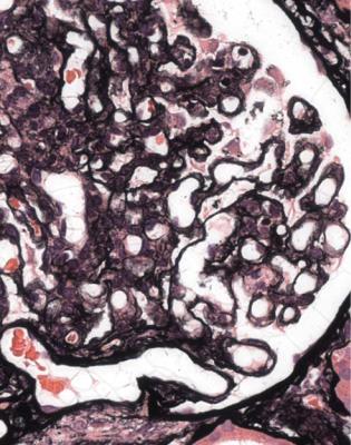
Fig. 11.26 Glomerulus in a needle biopsy specimen of kidney from a man of 27 taken 17 years after transplantation. There is mesangial expansion, and several capillary loops have doubled basement membranes. These are features of chronic allograft glomerulopathy
Diagnosis requires knowledge of the recipient’s original renal disease and recognition that the glomerular disorder is neither a recurrence nor chronic allograft glomerulopathy. The commonest is membranous nephropathy (Figs 6.8– 6.14). Hereditary nephropathy of Alport type does not recur in grafts, but recipients develop antibodies to part of the type four collagen molecule that they lack, which is the antigen detected by antibodies in Goodpasture’s disease (Fig. 9.13). Grafts in recipients with hereditary nephropathy of Alport type can sometimes be shown to have linear deposition of IgG in glomerular basement membranes, but they hardly ever develop the vasculitic glomerulonephritis of Goodpasture’s syndrome.
In a similar way, children with the Finnish type of congenital nephrotic syndrome, who do not express nephrin in their glomeruli, may develop antibodies against nephrin in the graft. These antibodies can produce the nephrotic syndrome.
A glomerular disorder that may develop long after transplantation is the type associated with hyperperfusion effects, or reduced glomerular numbers, with enlargement, segmental sclerosis often at the glomerular hilum, progressive loss of
294 |
11 Indication for Biopsy: Renal Allograft |
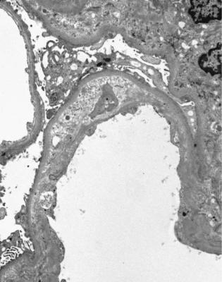
Fig. 11.27 Electron micrograph of part of a glomerulus in a needle biopsy specimen of kidney from a woman of 31 taken 5 years after transplantation. In one capillary loop, there is loose material on the inside of the original basement membrane with formation of a new basement membrane next to the capillary lumen. This corresponds with the doubled basement membranes seen on light microscopy in chronic allograft glomerulopathy as in Fig. 11.26
glomeruli by global sclerosis, and proteinuria (Fig. 8.3). This may be the explanation of the segmental lesions reported as an effect of calcineurin inhibitors.
Is There Evidence of a Neoplastic Disorder, or Any Other Disorder?
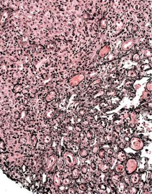
Lymphomas are rare in grafted kidneys. These are often called post transplant lymphoproliferative disorders, but are now classified in the same way as other lymphomas. They are usually a complication of Epstein Barr virus infection. This virus persists in B lymphocytes, and its replication is kept in check by T lymphocytes. Immunosuppression may release the virus from this control, and can lead to proliferation of B cell clones. There is an infiltrate of lymphoid cells, usually large and all the same (Fig. 11.35). These are B lymphocytes that express CD20 on immunohistologic study, with expression of markers of Epstein Barr virus infection (Fig. 11.36).
Summary: Renal Allografts |
295 |
Fig. 11.28 Cortex in a needle biopsy specimen of kidney from a woman of 18 taken 2 years after transplantation. There is chronic damage, but there is also evidence of significant acute cellular rejection
Grafts may develop other neoplasms, such as Kaposi’s sarcoma or renal call carcinoma, and can show abnormalities that can occur in any kidney, such as cysts or amyloid (Fig. 6.63). Often, a thick fibrous capsule develops around a graft, and this makes biopsy difficult. A capsule may contain crystals or sutures from surgical procedures, surrounded by giant cells. There may be evidence around a graft of pre-existing disease in the recipient, such as amyloid derived from beta two microglobulin related to hemodialysis.
Summary: Renal Allografts
Most renal allograft biopsies are done because of impaired excretory function, called graft dysfunction.
The two common problems for a pathologist are to decide whether there is significantly active, acute rejection that may respond to treatment, and whether there is chronic damage that will not respond to treatment.
296 |
11 Indication for Biopsy: Renal Allograft |
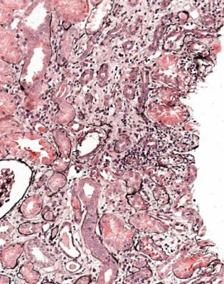
Fig. 11.29 Cortex in a needle biopsy specimen of kidney taken 3 months after transplantation into a man of 40. There is chronic damage with atrophic tubules that contrast with surviving enlarged tubules, but there are no clues to the cause
There are five diagnoses related to acute rejection. These are antibody-mediated rejection, significant acute cellular rejection, acute vascular rejection, severe acute vascular rejection, and no evidence of significant acute rejection.
Late damage in an allograft, meaning tubular atrophy in particular, may result from chronic rejection, but there are several other possible explanations.
Other disorders that may occur in grafts include glomerular abnormalities that may be due to a variety of causes, vascular and ureteric problems, infections, and neoplasms.
Summary: Renal Allografts |
297 |
Fig. 11.30 Cortex in a needle biopsy specimen of kidney taken 2 months after transplantation into a man of 44. There is a little chronic damage, but there is no evidence of significant acute rejection. The clinical diagnosis was cyclosporine toxicity. Function improved when the dose of cyclosporine was reduced

298 |
11 Indication for Biopsy: Renal Allograft |
Fig. 11.31 Arteriole in a needle biopsy specimen of kidney from a woman of 46 with chronic renal failure from a nonglomerulonephritic cause. Nodules of hyalinosis bulge into tissues around the arteriole. This is sometimes claimed to be a feature of effects of calcineurin inhibitors, but this woman had never been treated with these drugs
Summary: Renal Allografts |
299 |
Fig. 11.32 Cortex in a needle biopsy specimen of kidney from a woman of 67 with chronic renal failure from a nonglomerulonephritic cause. There are streaks of chronic damage running radially through the cortex in the position of medullary rays. This is sometimes called striped fibrosis, which is said to be characteristic of effects of calcineurin inhibitors. This woman had never been treated with these drugs
300 |
11 Indication for Biopsy: Renal Allograft |
Fig. 11.33 Cortex in a needle biopsy specimen of kidney taken 4 months after transplantation into a man of 51. On this section stained by periodic acid Schiff, there is material that protrudes into a vein. Tubulo-venous ruptures like this often suggest obstruction of the urinary tract, and previously there had been a temporary blockage of a urinary catheter in this man. The nature of the material is described in the legend to Fig. 11.34

Summary: Renal Allografts |
301 |
Fig. 11.34 Cortex in a needle biopsy specimen of kidney taken 16 days after transplantation into a man of 53. An immunoalkaline phosphatase method to detect Tamm–Horsfall protein shows a deposit of the protein in a vein, suggestive of urinary obstruction with tubulo-venous rupture. There was ultrasonographic evidence of hydronephrosis. Tamm–Horsfall protein is the most abundant protein in normal urine, and is produced by the thick limb of the loop of Henle. Tamm, pronounced as it looks, and Horsfall, pronounced horse-fawl or horse-fal, described a protein purified from urine that reacted with viruses, because of its content of carbohydrates (Tamm I, Horsfall FL. A mucoprotein derived from human urine which reacts with influenza, mumps, and Newcastle disease viruses. Journal of Experimental Medicine 1952; 95: 71–97). Igor Tamm (born 1922) and Frank Lappin Horsfall (1906–1971) were virologists at the Rockefeller Institute for Medical Research, New York, where Tamm later became a Professor of Virology. Horsfall became President and Director of the Sloan-Kettering Institute for Cancer Research. Friedrich Gustav Jacob Henle (1809–1885), pronounced hen-lee, was a German anatomist who reported loops in the medulla in 1862
302 |
11 Indication for Biopsy: Renal Allograft |
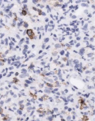
Fig. 11.35 Medulla in a needle biopsy of kidney taken 12 weeks after transplantation into a man of 21. There is an infiltrate in half the field of large lymphoid cells, shown on immunoperoxidase staining to be B lymphocytes. Features are those of a lymphoma. Investigation of Epstein Barr virus is shown in Fig. 11.36
Summary: Renal Allografts |
303 |
Fig. 11.36 The lymphoid infiltrate shown in Fig. 11.35, examined by an immunoperoxidase method for a late membrane protein of Epstein Barr virus. Many cells express the viral antigen
304 |
11 Indication for Biopsy: Renal Allograft |
Further Reading: Renal Allografts
Colvin RB, Cohen AH, Saiontz C, et al. Evaluation of pathologic criteria for acute renal allograft rejection: reproducibilty, sensitivity, and clinical correlation. Journal of the American Society of Nephrology 1997; 8: 1930–1941. This gives details of the system suggested by the Cooperative Clinical Trials in Transplantation.
D’Agati VD, Jennette JC, Silva FG. Non-Neoplastic Kidney Diseases. Atlas of Nontumor Pathology, first series, fascicle 4. Washington, DC: American Registry of Pathology and Armed Forces Institute of Pathology, 2005. Chapter 26.
Jennette JC, Olson JL, Schwartz MM, Silva FG. Heptinstall’s Pathology of the Kidney, Sixth ed. Philadelphia: Lippincott Williams and Wilkins, 2007. Chapter 28.
Racusen LC, Solez K, Colvin RB, et al. The Banff 97 working classification of renal allograft pathology. Kidney International 1999; 55: 713–723.