

Chapter 6
Indication for Biopsy: Nephrotic Syndrome
Introduction to the Nephrotic Syndrome
This means heavy proteinuria accompanied by hypoalbuminemia and edema, often seen as swelling of the ankles and higher up the lower limbs, which is usually the clinical feature that makes people seek the help of a physician. Occasionally, proteinuria is in the nephrotic range without the other features of the syndrome. Renal excretory function may be normal or abnormal. There may be hematuria or hypertension. There is hyperlipidemia as part of the syndrome.
Edema and hypertension are due to increased retention of sodium and water by renal tubules. Hyperlipidemia is due to increased synthesis of low density lipoproteins, and decreased catabolism of cholesterol.
The nephrotic syndrome is commoner in boys than in girls, and in men than in women.
The nephrotic syndrome is dangerous because it is associated with a risk of thrombosis, susceptibility to infection, and wasting of muscles. Thrombosis is due to increased concentration of coagulation factors in the blood, with reduced concentration of thrombolytic and antithrombotic factors. Susceptibility to infection is partly due to loss of immunoglobulins in the urine, and to increased catabolism of immunoglobulins. Muscle wasting is due to increased protein catabolism.
Syndrome, in this context, means a set of clinical features produced by different diseases. The conditions that produce the nephrotic syndrome have something in common, because they all increase glomerular permeability to protein. The nephrotic syndrome always indicates a glomerular disorder. Protein can be found in the urine for reasons other than increased glomerular permeability, but nothing else can produce heavy enough loss of protein to give the nephrotic syndrome. In glomerular proteinuria, the main protein in the urine is albumin. The selectivity of the proteinuria, or relative concentrations of albumin or similarly sized proteins, and larger proteins, was considered clinically useful in the past, but is now rarely measured.
A. J. Howie, Handbook of Renal Biopsy Pathology. |
49 |
C Springer 2008 |
|
50 |
6 Indication for Biopsy: Nephrotic Syndrome |
Detection and Measurement of Proteinuria
Understanding the detection and measurement of proteinuria may help the pathologist in interpretation of renal biopsy specimens. In particular, knowledge of potential difficulties and inaccuracies in measurement may be useful.
Proteinuria is detected by the use of reagent strips, called dipsticks, that are dipped in urine, and change colour if protein is present. These strips detect other substances as well, such as blood, and are the way in which microscopic hematuria is discovered. This is called urinalysis. Proteinuria can only be assessed crudely by this method.
Usually, if proteinuria is detected, this is measured. Often, this is expressed as output per 24 h, which is sometimes loosely called output per day, although that expression is ambiguous, because there is an implication that the night is ignored. Output per 24 h is measured, because the protein content of the urine varies over 24 h. Total protein output is often assessed. In normal adults, the total is under 150 mg per 24 h, and this includes Tamm–Horsfall protein and others derived from the urinary tract, as well as tiny amounts of protein from the blood. There are various chemical methods in use, some sensitive to interference by materials other than proteins. A specific protein, albumin, is sometimes measured by immunologic methods. Urinary albumin excretion is a more sensitive test of significant proteinuria than total protein output, and in normal adults is under 30 mg per 24 h.
Heavy proteinuria in adults is defined arbitrarily as at least 3 g total protein output in the urine per 24 h, although some definitions say 3.5 g.
All measurements that rely on a timed urine collection are suspect, because this has the risk of inaccuracy arising from incomplete collection. For this reason, proteinuria is often assessed in other ways. Measurement of the concentration of either total protein or albumin in a urine sample is relatively straightforward, but is not usually used on its own. The state of dilution or concentration of the urine affects this measurement, but this can be standardised by simultaneous measurement of the concentration of creatinine in the sample. These days, proteinuria is often expressed as the ratio of concentrations of either total protein or albumin to creatinine in a single sample of urine, in various units. Clinical chemical laboratories usually have their own reference range for these ratios.
In children, timed urine collections are hardly ever done and if they are, body size has to be considered when proteinuria is measured as total output. Use of protein/creatinine or albumin/creatinine concentration ratios removes the need to calculate body surface area or other indicators of body size.
Value of a Renal Biopsy in the Nephrotic Syndrome
Most or all adults with the nephrotic syndrome have a renal biopsy, but most children with the nephrotic syndrome do not. This is because they are assumed to have minimal change nephropathy, and are managed accordingly. A biopsy is done in a child if there are unusual clinical features, such as presentation in late childhood or
Approach to the Diagnosis in the Nephrotic Syndrome |
51 |
with an insidious onset. Minimal change nephropathy typically gives an abrupt onset of edema, and is particularly common in children under the age of five, especially boys. A biopsy is also done if the response to treatment is not typical of minimal change nephropathy.
The diagnosis in the nephrotic syndrome is a great help to nephrologists, to make clinical decisions about management. For instance, minimal change nephropathy should respond to appropriate treatment and should not progress to chronic renal failure, although membranous nephropathy may not respond to any treatment and may progress.
The biopsy specimen may give clues that there are complications of the nephrotic syndrome, particularly renal vein thrombosis.
The amount of chronic damage in the specimen is a guide to the long-term outlook for the kidney.
Approach to the Diagnosis in the Nephrotic Syndrome
Renal biopsy specimens in the nephrotic syndrome are usually straightforward and satisfying for the pathologist, because a definite diagnosis can almost always be given.
The pathologist should examine the renal biopsy specimen with the rule in mind that common things are common. The prevalence of various conditions in specimens depends upon the age of people biopsied, the part of the world, and whether nephrologists biopsy everyone with the nephrotic syndrome, or are selective in their biopsy practice. The syndrome is commoner in developing countries than elsewhere.
In adults with the nephrotic syndrome, three conditions are found in the majority of specimens. These are membranous nephropathy, various segmental sclerosing disorders often called focal segmental glomerulosclerosis, and minimal change nephropathy.
The pathologist has a high expectation of finding one of these three conditions before the specimen is examined. The proportion of specimens that have these conditions varies depending upon factors such as the willingness of nephrologists to biopsy people with the nephrotic syndrome associated with other disorders, particularly diabetes mellitus and systemic lupus erythematosus. Where nearly all adults with the nephrotic syndrome are biopsied, membranous nephropathy, segmental sclerosing disorders, and minimal change nephropathy together account for about two-thirds of specimens in descending order of frequency, and these conditions with diabetic glomerulopathy, lupus nephritis, and amyloid account for nearly nine-tenths of specimens. Various glomerular conditions are found in the relatively few other adults with the nephrotic syndrome.
In the relatively few children with the nephrotic syndrome who have a renal biopsy, most have minimal change nephropathy, and about three-quarters have this condition or a segmental sclerosing disorder.
Various other findings occur in the rest of the children, with different frequencies from adults. Membranous nephropathy is rare in children in higher income
52 |
6 Indication for Biopsy: Nephrotic Syndrome |
countries, and diabetic glomerulopathy and amyloid are hardly ever seen in children. There is a group of disorders causing congenital or infantile nephrotic syndrome, some of which are only found in young children.
Initial Inspection of a Renal Biopsy Specimen in the Nephrotic
Syndrome
Although glomeruli have to be examined in detail in the nephrotic syndrome, renal biopsy specimens should first be inspected at low power on the microscope, to see whether tubules look normal or not. If they are not normal, the next question is whether there is acute damage, or chronic damage, or a mixture. In acute tubular damage, there is an apparently normal number of tubules in the renal cortex, when the age of the person biopsied is taken into consideration. Acutely damaged tubules
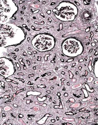
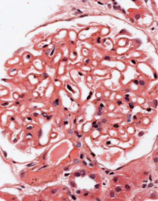
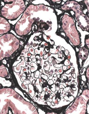
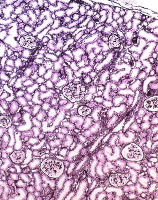
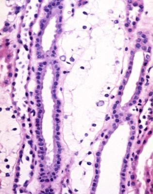
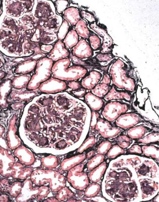
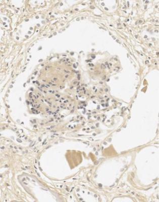
Fig. 6.1 Cortex in a renal biopsy specimen from a woman of 49 with the nephrotic syndrome and mild acute renal impairment. The nephrotic syndrome is explained by minimal change nephropathy. There is extensive acute damage to tubules, which are dilated, with irregular epithelium. This appearance in the nephrotic syndrome suggests the possibility of renal vein thrombosis, which the woman was shown to have on radiologic investigation
Initial Inspection of a Renal Biopsy Specimen in the Nephrotic Syndrome |
53 |
have any of a variety of changes including irregular flattening of the epithelium, vacuolation, loss of the brush border, and accumulation of material in the lumen. If acute tubular damage is easily seen, there is likely to be acute renal failure as well as the nephrotic syndrome.
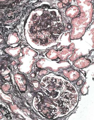
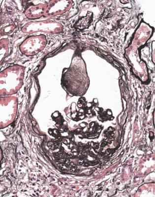
Acute tubular damage, especially if widespread and uniform, may indicate renal vein thrombosis, a complication of the nephrotic syndrome arising from the hypercoagulable state. Renal vein thrombosis is difficult to diagnose with certainty on a renal biopsy specimen. Thrombus is unlikely to be seen in veins included in a biopsy specimen, but if the pathologist sees extensive tubular damage, renal vein thrombosis should be reported as a possibility (Fig. 6.1). This damages tubules by impairing blood flow through the kidney, and causing ischemia. The danger is that if there is renal vein thrombosis that is not treated, there may be irreversible damage to tubules (Fig. 6.2). This is not the only cause of acute tubular damage in
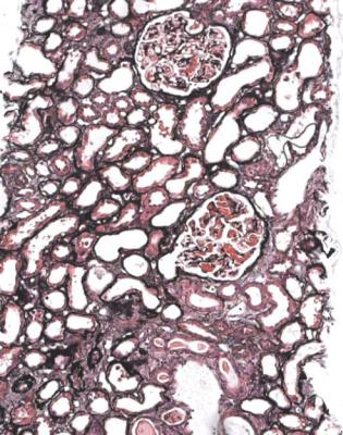
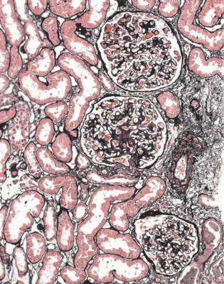
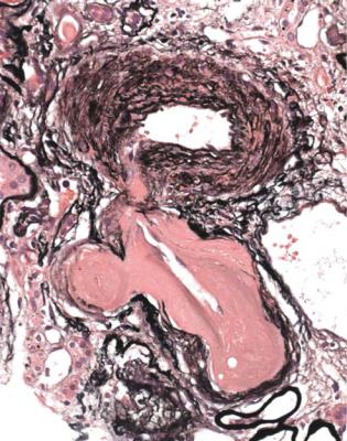
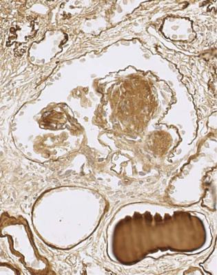
Fig. 6.2 Cortex in a renal biopsy specimen from a man of 38 known to have membranous nephropathy, shown by a biopsy nearly 2 years before this one. The nephrotic syndrome had persisted since the first biopsy, and acute renal failure had developed 1 month before this biopsy. There is now widespread uniform tubular atrophy, which suggests longstanding renal vein thrombosis. This was confirmed by radiologic investigation. Renal function did not recover
54 |
6 Indication for Biopsy: Nephrotic Syndrome |
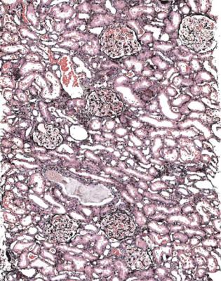
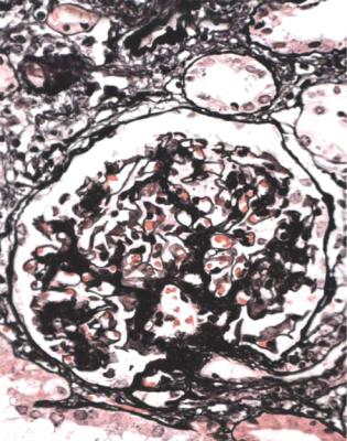
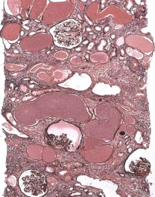
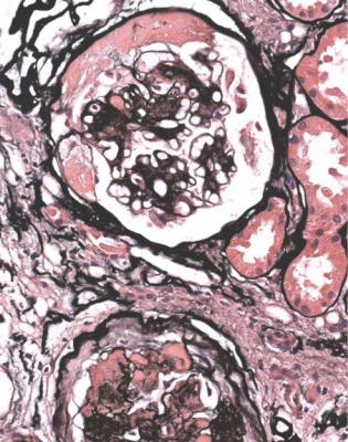
Fig. 6.3 Cortex in a renal biopsy specimen from a man of 56 with the nephrotic syndrome and acute renal failure. The nephrotic syndrome is explained by an early form of classic segmental sclerosing glomerulonephritis. There is acute tubular damage, resembling the appearances in Fig. 6.1. Renal vein thrombosis was suggested as a possible explanation, but was excluded by appropriate investigations. Later, the renal failure recovered almost to normal, but the cause was not identified
the nephrotic syndrome. Other explanations include toxic effects of luminal protein on tubules, and intravascular fluid depletion, for instance from effects of powerful diuretics, or movement of fluid out of vessels as a consequence of the nephrotic state, particularly from osmotic consequences of the low plasma albumin concentration (Fig. 6.3).
Chronic damage means changes such as global sclerosis of glomeruli, atrophy of tubules, and interstitial fibrosis. An amount of chronic damage that seems disproportionately large for age has prognostic significance, indicates that there is a longstanding disorder, and means that renal function is never likely to recover to be completely normal.
Diagnosis of Minimal Change Nephropathy |
55 |
Do Glomeruli Look Normal? Is the Diagnosis Minimal Change
Nephropathy?
Minimal change nephropathy is a diagnosis that has immense prognostic significance, since the person with it should respond to appropriate treatment and never progress to renal failure, even though there may be relapses of the nephrotic syndrome. The condition can occur at any age. Children respond more quickly than adults, but even without treatment, the condition should eventually resolve, provided there are no complications of the nephrotic state.
In most people, the explanation of minimal change nephropathy is not yet known, but a likely cause is that lymphocytes produce a circulating factor that increases the permeability of glomerular basement membranes to protein. Rare causes of the condition are nonsteroidal anti-inflammatory drugs and lymphomas, but the pathologist is usually not able to detect these on a renal biopsy specimen.
The diagnosis of minimal change nephropathy should only be used when there is the nephrotic syndrome, but is sometimes misused when applied by pathologists to specimens taken for hematuria or other indications, and which seem to show no abnormality.
Minimal change nephropathy is a difficult diagnosis to make. The pathologist should use the term with care. Because it is largely a diagnosis of exclusion of other things, the fewer the glomeruli in a specimen, the less certain a pathologist can be about the diagnosis.
Diagnosis of Minimal Change Nephropathy
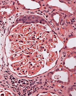
In minimal change nephropathy, glomeruli look normal on light microscopy. There is no increase in cellularity, nor are there segmental abnormalities, nor enlargement, although a pathologist may find difficult in certainty about these features (Figs 6.4 and 6.5). Only one definite segmental abnormality has to be seen to exclude this diagnosis.
Immunohistology shows no deposition of immunoproteins on glomerular basement membranes, and either no deposition of immunoproteins or only IgM in mesangium. Mesangial deposition of IgM does not exclude minimal change nephropathy, if all other features support this diagnosis.
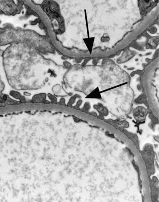
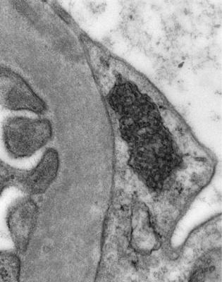
Electron microscopy can be done, but is not necessary for the diagnosis of minimal change nephropathy. This will show loss of epithelial foot processes on the outside of glomerular basement membranes, and replacement by sheets of epithelial cell cytoplasm, which is due to swelling of foot processes, rather than their fusion, as it is sometimes called (Figs 6.6 and 6.7). Effacement is also used to describe this change, which seems a response to proteinuria, found in all specimens in the nephrotic syndrome. Electron microscopy will exclude immune deposits, for instance on glomerular capillary loops, but satisfactory immunohistologic staining excludes these anyway.
56 |
6 Indication for Biopsy: Nephrotic Syndrome |
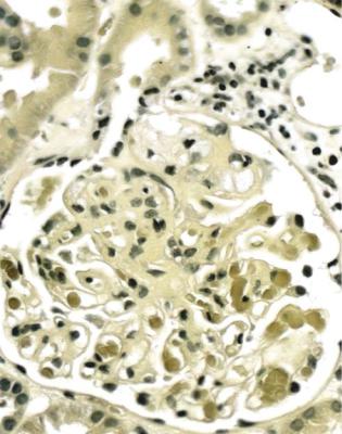
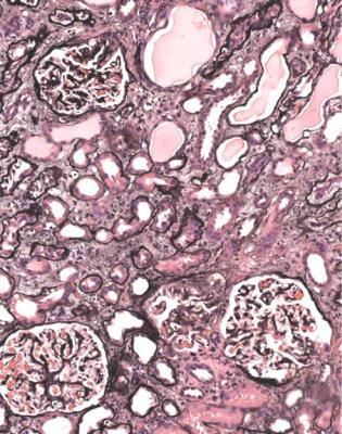
Fig. 6.4 Cortex in a renal biopsy specimen from a man of 62 who was spontaneously beginning to recover from the nephrotic syndrome. Glomeruli appear of normal size and cellularity, have no segmental lesions on any of the full set of sections, and show no significant deposition of immunoproteins on immunohistologic study. The diagnosis is minimal change nephropathy
Differential Diagnosis of Minimal Change Nephropathy
On initial sections, without a full set of stains and without immunohistology, the pathologist may see no glomerular abnormality. If pressed for a diagnosis by nephrologists, the pathologist should be cautious, and offer a provisional diagnosis that minimal change nephropathy is a possibility. Early membranous nephropathy may show little or no abnormality on orthodox staining, and can be easily mistaken for minimal change nephropathy, but will be immediately apparent on immunohistology. The full set of sections may show a segmental abnormality, which excludes minimal change nephropathy. The more glomeruli that are examined, the more confident the pathologist can be that there is no segmental disorder. Small amounts of amyloid in glomeruli may be missed, if a Congo red stain is not done or is technically unsatisfactory, or if the mesangium is not inspected thoroughly.
Differential Diagnosis of Minimal Change Nephropathy |
57 |
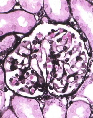
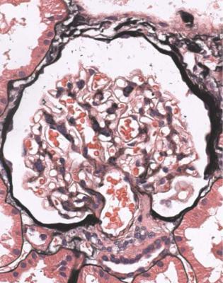
Fig. 6.5 Glomerulus in a renal biopsy specimen from a girl aged 21 months with the nephrotic syndrome for 2 weeks. This glomerulus and all others appear normal, and there is no significant deposition of immunoproteins on immunohistologic study. The diagnosis is minimal change nephropathy
Are Glomeruli Abnormal? Is There Membranous Nephropathy?
Membranous nephropathy is almost always an easy diagnosis to make. Because the condition affects the whole of every glomerulus, only one glomerulus is needed to make the diagnosis.
Membranous nephropathy is the commonest finding in the nephrotic syndrome in adults in several parts of the world, but is generally rare in children. This disorder is also common in cats and dogs with the nephrotic syndrome.
In most people, the cause is not yet apparent. Sometimes, in the absence of known causes or associations, the word “idiopathic” is used. This appears a respectable term, but simply means that the cause is not known. The pathogenesis of membranous nephropathy may be related to the fact that the main immunoglobulin subclass found in glomeruli in this condition is IgG4. This is associated with antibodies of low affinity for antigen, which do not activate the classic pathway of the complement
58 |
6 Indication for Biopsy: Nephrotic Syndrome |
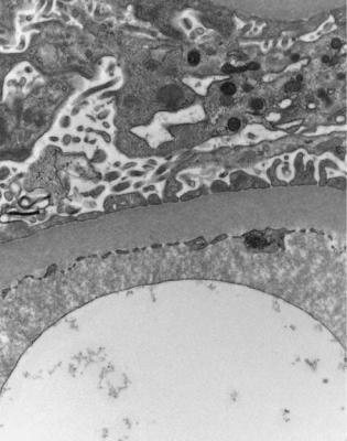
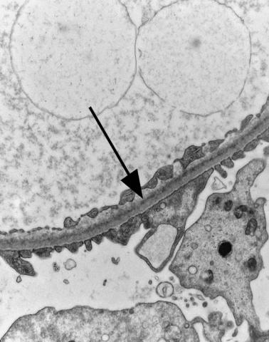
Fig. 6.6 Electron micrograph of a renal biopsy specimen from a man of 47 with mild acute tubular damage, but no glomerular abnormality. Glomerular basement membranes have the normal adult appearance and thickness, from 270 to 340 nm. On the outside of the membranes, there are foot processes of epithelial cells (arrowed), separated by slit pores
system. Immune complexes containing IgG4 may not be cleared from the circulation, but settle in glomeruli, where they dissociate on the inside of basement membranes and reform on the outside.
A few people have an underlying explanation of membranous nephropathy. Recognised factors include drugs such as gold and penicillamine, infections such as with hepatitis B and hepatitis C viruses, neoplasms such as carcinoma of the bronchus, and use of skin creams containing mercury. The pathologist is unlikely to be able to identify an underlying explanation of membranous nephropathy from appearances in a biopsy specimen, although there may be clues, such as inclusions in lysosomes of proximal tubular cells after treatment with gold. Systemic lupus erythematosus can produce nearly every glomerular disorder including membranous nephropathy, but the diagnosis should be given as lupus nephritis, suitably qualified, rather than membranous nephropathy, if this is an abnormality found in a person with lupus.
Diagnosis of Membranous Nephropathy |
59 |
Fig. 6.7 Electron micrograph of a renal biopsy specimen from a man of 38 with the nephrotic syndrome and minimal change nephropathy. Glomerular basement membranes are covered on the outside by a sheet of epithelial cell cytoplasm
Diagnosis of Membranous Nephropathy
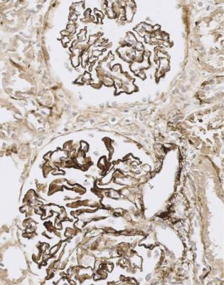
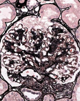
Orthodox light microscopy often suggests the diagnosis, because even on sections stained by hematoxylin and eosin, glomerular basement membranes appear uniformly thickened and more rigid than normal. The earliest stages may not show this, and may be mistaken for minimal change nephropathy if immunohistology is not done (Figs 6.8 and 6.9). Later stages are easy to identify on sections stained by periodic acid-methenamine silver, on which there are uniform spikes on the outside of glomerular basement membranes, which progress to give the appearance of chain links as the tips of the spikes join together (Fig. 6.10). These days, spikes are usually not well developed at first presentation of membranous nephropathy, and because they are not essential for the diagnosis, there is no need for the pathologist to search hard for them, either on especially thin sections or with an oil immersion objective lens on the microscope. Generally, the cellularity of glomeruli in membranous nephropathy appears normal, or close to normal.
60 |
6 Indication for Biopsy: Nephrotic Syndrome |
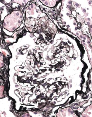
Fig. 6.8 Glomerulus in a renal biopsy specimen from a man of 73 with the nephrotic syndrome. On hematoxylin and eosin staining, there is uniform thickening of basement membranes, which makes them appear rigid. These features suggest membranous nephropathy, but further investigations, particularly immunohistologic study, are necessary to allow the diagnosis to be made
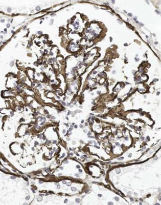
Immunohistology gives the diagnosis, because there is strikingly uniform, granular deposition of IgG and complement on the outer surface of basement membranes, in every loop of every glomerulus (Figs 6.11 and 6.12). These granules may be so close that they give the impression of linear deposition on basement membranes, but inspection of thin parts of sections or tangential cuts of basement membranes will show the granularity. IgM is often seen in mesangium, but not other immunoproteins. Additional findings of mesangial deposition of IgG, and IgA on basement membranes, indicate that the membranous nephropathy is a sign of lupus nephritis.
If immunohistology is done properly, electron microscopy is not necessary for the diagnosis of membranous nephropathy, but will confirm regular deposits on the outer aspect of glomerular basement membranes (Fig. 6.13).
There may be segmental changes of various types in membranous nephropathy, which do not usually change the diagnosis, unless there are clinical or pathologic
Diagnosis of Membranous Nephropathy |
61 |
Fig. 6.9 Glomerulus stained by periodic acid-methenamine silver in a renal biopsy specimen from a man of 80 with the nephrotic syndrome. There is a hint of thickening of basement membranes, but there are no spikes. Immunohistology illustrated in Figs 6.11 and 6.12 shows that this is membranous nephropathy. The lack of spikes indicates an early stage
indications of lupus nephritis, when that should be given as the diagnosis. Almost always, the pathologist is given the information on the request form that the person biopsied has systemic lupus erythematosus, but if not, this should always be kept in mind as a possible diagnosis in any young woman with the nephrotic syndrome. Clues to lupus nephritis are the presence of other immunoglobulins as well as IgG on basement membranes, and the finding of other glomerular changes such as mesangial hypercellularity, mesangial deposition of immunoproteins especially IgG and IgA, and segmental abnormalities of vasculitic type.
A segmental disorder that is common in membranous nephropathy, and should not change the diagnosis, is adhesion of the glomerular tuft to Bowman’s capsule next to the opening of the proximal tubule. This may be seen at all stages of membranous nephropathy (Figs 6.10 and 6.14). In late stages, there may be areas of segmental sclerosis at various sites in glomeruli, but the diagnosis should still be membranous nephropathy.
62 |
6 Indication for Biopsy: Nephrotic Syndrome |
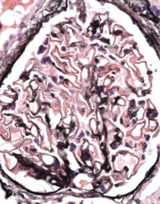
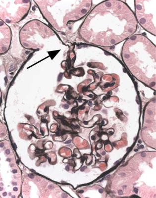
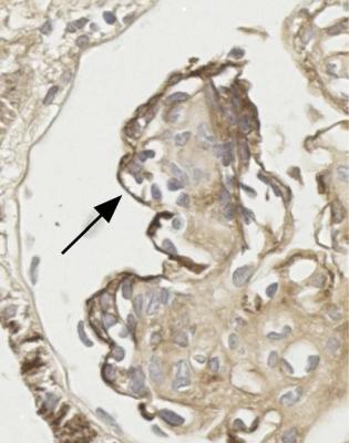
Fig. 6.10 Glomerulus stained by periodic acid-methenamine silver in the renal biopsy specimen, which is illustrated in Fig. 6.2, from the man of 38 known to have membranous nephropathy for 2 years. There are uniform spikes on the outside of capillary loops. These indicate a later stage of membranous nephropathy than in Fig. 6.9. The arrow indicates adhesion of the glomerular tuft to Bowman’s capsule, next to the tubular opening, opposite the hilum
Rarely, in people who have no evidence of systemic lupus erythematosus, membranous nephropathy is seen with vasculitic glomerular lesions, and this combination should be given as the diagnosis (Fig. 6.15). When these two conditions occur together, there is generally a more aggressive clinical course than is usual in membranous nephropathy. Goodpasture’s disease, in which there are antibodies to glomerular basement membranes, and glomeruli show global and diffuse vasculitic lesions, has occasionally been seen in combination with membranous nephropathy.
There is a system of staging membranous nephropathy based on changes in glomerular basement membranes, but this is less useful than assessment of the extent of chronic damage to tubules in prognosis.
Proteinuria is lost in many children with membranous nephropathy as a complication of hepatitis B viral infection. At 10 years after diagnosis of membranous
Differential Diagnosis of Membranous Nephropathy |
63 |
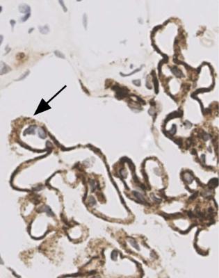
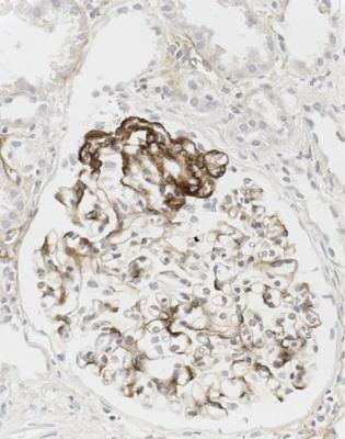
Fig. 6.11 Cortex in the renal biopsy specimen from the man of 80 that is illustrated in Fig. 6.9, stained by an immunoperoxidase method to detect complement component C9. On microscopy at low magnification, there seems linear staining of every glomerular capillary loop, but the staining is finely granular. This indicates membranous nephropathy
nephropathy, about one-third of adults have progressed to renal failure or have died, about one-third have recovered, and about one-third have persistent proteinuria.
Differential Diagnosis of Membranous Nephropathy
A disorder that may be confused with membranous nephropathy is dense deposit disease, also called membranoproliferative or mesangiocapillary glomerulonephritis type two, although these names are misleading because there is little resemblance to membranoproliferative or mesangiocapillary glomerulonephritis type one, also called subendothelial membranoproliferative glomerulonephritis. This use of the same name for different conditions perpetuates a historical mistake.
Dense deposit disease is much less common than membranous nephropathy, but may present with the nephrotic syndrome, and can give the appearance of thickened glomerular basement membranes.
64 |
6 Indication for Biopsy: Nephrotic Syndrome |
Fig. 6.12 Glomerulus in the renal biopsy specimen from the man of 80, which is illustrated in Figs 6.9 and 6.11, stained by an immunoperoxidase method to detect C9. Especially where basement membranes are seen tangentially, there are uniform fine granules, arrowed. This finding indicates membranous nephropathy
Dense deposit disease is usually diagnosed in children or young adults, who often have the nephrotic syndrome, but may have various clinical features as well, such as hematuria, renal failure, and partial lipodystrophy, which means a lack of subcutaneous fat. Complement component C3 has a persistently low concentration in serum. Often, there is an antibody in serum to one of the components of the alternate pathway of complement activation, C3 convertase. The antibody, called C3 nephritic factor, prevents inactivation of this component by its inhibitor, factor H, and allows persistent generation of active C3. Generally, the disorder is unremitting, and progresses to renal failure.
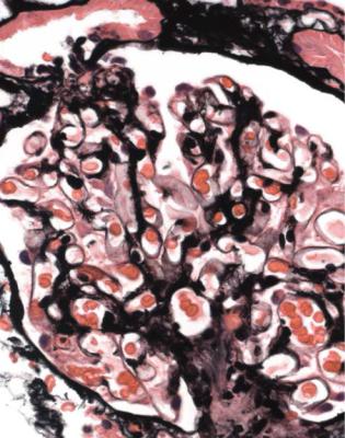
In dense deposit disease, glomeruli can appear nearly normal, or have various amounts of mesangial hypercellularity. Clues to the diagnosis are that there are no spikes, basement membranes are not uniformly thickened, and often the membranes have a brown colour, rather than black, on sections stained by periodic acid-methenamine silver (Fig. 6.16). On immunohistology, there are coarse
Differential Diagnosis of Membranous Nephropathy |
65 |
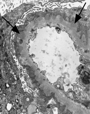
Fig. 6.13 Electron micrograph of a renal biopsy specimen from a man of 80 with the nephrotic syndrome. There are immune deposits (arrowed) on the outer aspect of the glomerular basement membrane. These indicate early membranous nephropathy
granules of complement in mesangium and glomerular basement membranes, generally without immunoglobulins (Fig. 6.17). If this diagnosis is suspected, electron microscopy should be done. This shows large, irregular dense deposits within glomerular basement membranes and mesangium, rather than regular deposits on the outside of membranes that would indicate membranous nephropathy (Fig. 6.18). Similar immunohistologic and electron microscopic findings to those in glomeruli may be seen in Bowman’s capsule and tubular basement membranes.
Are Glomeruli Abnormal? Is There a Segmental Sclerosing
Disorder?
Segmental sclerosing glomerular disorders are difficult. The term focal segmental glomerulosclerosis is widespread, but those who use it accept that it is applied to many different conditions, and that it has no specific meaning. This problem has
66 |
6 Indication for Biopsy: Nephrotic Syndrome |
Fig. 6.14 Glomerulus stained by periodic acid-methenamine silver in a renal biopsy specimen from a man of 52 with the nephrotic syndrome. Immunohistology shows that this is membranous nephropathy, early because there are no spikes. There is adhesion of the tuft to Bowman’s capsule at the opening of the tubule, with hyaline material in capillary loops at this site
arisen through uncritical use of the term during the development of renal pathology as a specialty, when the same label was given to different entities, and the label is now difficult to remove. A pathologist can often refine the diagnosis to give more useful information to nephrologists. Whenever possible, the pathologist should try to avoid use of the unqualified term focal segmental glomerulosclerosis as a diagnosis.
Although the adjectives focal and segmental almost always appear together, the important one is segmental, meaning affecting only part of the glomerulus. Segmental lesions may be diffuse, meaning present in every glomerulus, rather than focal, but this will not be apparent on a single section or on a few random sections.
In the nephrotic syndrome, if glomeruli have segmental areas of foamy cells, or sclerosed material, or hyaline material, or a mixture of these, such as in Fig. 6.19, there is a temptation to use the diagnosis focal segmental glomerulosclerosis. The pathologist should first consider two questions.
Segmental Abnormalities at the Tubular Opening |
67 |
Fig. 6.15 Glomerulus in a renal biopsy specimen from a man of 36 with the nephrotic syndrome and acute renal failure, taken 5 months after a biopsy that had shown early membranous nephropathy. There is now a segmental glomerular lesion of vasculitic type, in addition to membranous nephropathy
1.Is there evidence of an underlying condition, such as membranous nephropathy? If so, the underlying condition should be given as the diagnosis.
2.What is the site of the segmental abnormalities in glomeruli?
Segmental Abnormalities at the Tubular Opening
In the nephrotic syndrome, the first site in a glomerulus at which segmental abnormalities appear is next to the opening of the tubule, which can also be called the tubular origin (Fig. 6.14). This part of the glomerulus should always be examined closely in a biopsy specimen. Serial sections give the pathologist the best chance to see tubular origins. Only if there are no structural changes at this site, in a specimen that at first glance has no segmental abnormalities, can a segmental sclerosing condition be said to be unlikely. The fewer the glomeruli seen in a biopsy specimen,
68 |
6 Indication for Biopsy: Nephrotic Syndrome |
Fig. 6.16 Glomerulus stained by periodic acid-methenamine silver in a renal biopsy specimen from a woman of 24 with persistent proteinuria postpartum. There is mesangial increase and basement membranes appear thickened, but not so uniformly as in membranous nephropathy. This is dense deposit disease, confirmed by further investigations that include immunohistology, illustrated in Fig. 6.17
the less confident the pathologist can be about exclusion of a segmental sclerosing condition.
If structural changes are seen next to the opening of the tubule, interpretation depends on the condition of the rest of the glomerular tuft. This is because these changes, that can be called tip changes, are not a disease in themselves, but are always a complication of a glomerular disorder.
Tip changes develop as a consequence of temporary prolapse of the glomerular tuft into the opening of the tubule, because the tuft swells acutely in various disorders (Fig. 6.20). The earliest recognisable changes are adhesion of the basement membrane of the tuft to the basement membrane of Bowman’s capsule in the vicinity of the tubular opening. There are various abnormalities in the affected part of the tuft, including accumulation of foamy macrophages in capillary loops, and sometimes vacuolation and granularity of the cytoplasm of epithelial cells, with flattening
Segmental Abnormalities at the Tubular Opening |
69 |
Fig. 6.17 Glomerulus in the renal biopsy specimen from the woman of 24 with dense deposit disease that is illustrated in Fig. 6.16. Immunoperoxidase staining to detect C9 shows heavy deposition in glomerular basement membranes, but not so regularly or completely as in membranous nephropathy, illustrated in Figs 6.11 and 6.12
of the first proximal tubular cells (Figs 6.20 and 6.21). Later the foamy cells disappear, and are replaced by sclerosed material stained by periodic acid-methenamine silver, or hyaline material stained by eosin (Figs 6.14 and 6.19), or the only evidence of tip changes may be persistence of a thin adhesion or strand of basement membrane material, linking the tuft and Bowman’s capsule (Figs 6.10 and 6.22). Immunohistologic study shows IgM and complement in tip changes (Fig. 6.23).
The rest of the tuft in a specimen with tip changes can appear normal or abnormal.
If the rest of the tuft appears normal in a person with the nephrotic syndrome, the diagnosis would have been minimal change nephropathy, if tip changes had not been present (Figs 6.21, 6.22, and 6.24). This corresponds to the glomerular tip lesion, as it was originally defined. The clinical course should be that of minimal change nephropathy, without progression to renal failure, which supports the idea that this is indeed minimal change nephropathy, complicated by tip changes. Although only
70 |
6 Indication for Biopsy: Nephrotic Syndrome |
Fig. 6.18 Electron micrograph of a glomerulus in a renal biopsy specimen from a girl of 13 with proteinuria, progressive renal impairment, a low serum concentration of C3, and C3 nephritic factor in serum. Basement membranes have irregular thickening by material typical of dense deposit disease
one tip change has to be found to alter the diagnosis from minimal change nephropathy, in the glomerular tip lesion, as originally defined, tip changes are usually present in every glomerulus, although this is not apparent without rigorous search of many serial sections.
A problem is that the term glomerular tip lesion has been used with meanings other than the original one. A common usage is to apply the term to all tip changes, whether or not there is the nephrotic syndrome, and irrespective of the condition of the glomerular tuft away from the tubular origin.
If the rest of the tuft is abnormal, the diagnosis depends on the features away from the tip changes. Membranous nephropathy is commonly associated with tip changes, but these should not alter the diagnosis of membranous nephropathy (Figs 6.10 and 6.14).
Another common appearance in the nephrotic syndrome is of tip changes in glomeruli that are abnormal and seem large, with mesangial increase, although the tip changes may be overlooked unless specifically sought. The tip changes
Segmental Abnormalities at the Tubular Opening |
71 |
Fig. 6.19 Glomerulus in a renal biopsy specimen from a man of 37 with the nephrotic syndrome. Next to the tubular opening, adherent to Bowman’s capsule, there is a large segmental area of solid material with traces of foamy cells. Many pathologists would call this focal segmental glomerulosclerosis, but this diagnosis can be refined by study of the distribution of abnormalities in glomeruli, and of other features in the kidney
themselves may be large, occupying one-third or more of the glomerulus, and there may be marked acute tubular damage (Figs 6.19, 6.23, 6.25, and 6.26). This condition may evolve into one with more obvious segmental areas of sclerosis, and can be called the early form or early stage of classic segmental sclerosing glomerulonephritis. In this name, the adjective classic refers to the traditional clinical usage of the term focal segmental glomerulosclerosis in association with the nephrotic syndrome. To make this more understandable to nephrologists, the terms early classic focal segmental glomerulosclerosis, or early focal segmental glomerulosclerosis, could be used.
This condition is less likely to do well clinically than minimal change nephropathy or the glomerular tip lesion as originally defined, but is more likely to do better than expected from the traditional view of focal segmental glomerulosclerosis associated with the nephrotic syndrome, which is that there is almost always progression to renal failure, with no response to treatment.
72 |
6 Indication for Biopsy: Nephrotic Syndrome |
Fig. 6.20 Glomerulus in a renal biopsy specimen from a man of 28 with acute renal failure, from which he recovered spontaneously shortly after the biopsy. The glomerulus has many neutrophils in capillary loops, and other features of acute postinfective glomerulonephritis. The tuft is prolapsed into the tubular opening. This is an early stage of development of tip changes
Occasionally, in specimens with few glomeruli or with only a few sections, the pathologist may not detect any segmental abnormalities, but may see definite mesangial expansion, usually with glomerular enlargement and IgM deposition in mesangium on immunohistologic study. This should not be called minimal change nephropathy, but there is no satisfactory name. Such specimens will often progress to ones with segmental sclerosing lesions, and may be called an early mesangial proliferative form of focal segmental glomerulosclerosis, or diffuse mesangial hypercellularity, or similar terms.
There may be difficulty for a pathologist to determine whether glomeruli appear normal away from tip changes, or are enlarged, or have mesangial increase, because these are subjective decisions, and the distinction between definitely normal and definitely abnormal is arbitrary. There are some specimens in which the pathologist is in doubt. In these, the best course is to describe the dilemma, and suggest to nephrologists that the clinical course cannot be confidently predicted. Many people will do well, but some may not, with persistence of the nephrotic syndrome and
Segmental Abnormalities at the Tubular Opening |
73 |
Fig. 6.21 Glomerulus in a renal biopsy specimen from a woman of 64 with the nephrotic syndrome, from which she recovered spontaneously. Within the tubular opening is a part of the tuft that contains swollen cells with foamy cytoplasm. This is a tip change at an early stage, slightly more advanced than seen in Fig. 6.20. The rest of the tuft appears normal, and the diagnosis is glomerular tip lesion
progression to renal failure. Features that suggest there may be a poorer prognosis are any of the following.
1.Large tip changes affecting at least one-third of the tuft.
2.Definite mesangial increase.
3.Acute tubular damage affecting at least one-quarter of proximal tubules.
The more of these features that are seen, the less likely is a good outcome.
One classification of focal segmental glomerulosclerosis, often called the Columbia classification after Columbia University in New York, uses the term tip variant of focal segmental glomerulosclerosis, which corresponds to both the glomerular tip lesion as originally defined, and early classic segmental sclerosing glomerulonephritis. This term may be helpful when the pathologist sees tip changes, but no other segmental abnormalities, and cannot be sure that glomeruli are otherwise normal. Some people have used the term glomerular tip lesion with
74 |
6 Indication for Biopsy: Nephrotic Syndrome |
Fig. 6.22 Glomerulus in a renal biopsy specimen from a man of 27 with the nephrotic syndrome, which had remitted and relapsed a few times over the previous 13 years. There is a thin adhesion, arrowed, between the tuft and Bowman’s capsule next to the tubular opening, where the basement membranes of the two have fused. This is one of the late stages of a tip change. Glomeruli otherwise appear normal away from tubular openings, and the diagnosis is glomerular tip lesion
the same meaning as the tip variant of focal segmental glomerulosclerosis, which is different from the original definition of the glomerular tip lesion.
Segmental Abnormalities Not Only at the Tubular Opening
In the nephrotic syndrome, segmental abnormalities away from the tubular opening develop after those at the opening, and are a sign of a later or more advanced condition. If segmental abnormalities are at various sites in glomeruli, including at the tubular origin, whether the sites are multiple within the same glomerulus or differ between glomeruli, the diagnosis is best given as the late form or the late stage of classic segmental sclerosing glomerulonephritis (Figs 6.27 and 6.28). To make this more understandable to nephrologists, the terms late classic focal segmental glomerulosclerosis, or late focal segmental glomerulosclerosis, could
Segmental Abnormalities Not Only at the Tubular Opening |
75 |
Fig. 6.23 Glomerulus in a renal biopsy specimen from a woman of 37 with the nephrotic syndrome, examined by an immunoperoxidase method to detect IgM. An early tip change shows heavy deposition of IgM. Otherwise there is mesangial increase with light deposition of IgM. The diagnosis is early classic segmental sclerosing glomerulonephritis, which can also be called early classic focal segmental glomerulosclerosis
be used. Often, the segmental abnormalities are not focal, if sections of enough glomeruli are studied, which is why the segmental feature of the abnormalities is more important to stress.
This type of disorder most closely corresponds with the typical textbook description of focal segmental glomerulosclerosis associated with the nephrotic syndrome. What is not often realised is that this description applies to a condition late in its course. This can be confirmed by the high proportion of glomeruli with global sclerosis, and the large amount of tubular atrophy, usually seen in biopsy specimens given the conventional diagnosis. Because the diagnosis is made late, the clinical course usually accords with the conventional view of a condition that does not respond to treatment and progresses to renal failure.
In the Columbia classification, these changes are called focal segmental glomerulosclerosis, not otherwise specified.
76 |
6 Indication for Biopsy: Nephrotic Syndrome |
Fig. 6.24 Glomerulus in a renal biopsy specimen from a woman of 60 with the nephrotic syndrome, from which she recovered without treatment. There is a tip change, and the rest of the tuft appears normal. The diagnosis is glomerular tip lesion
The classic type of segmental sclerosing glomerulonephritis associated with the nephrotic syndrome appears due in some people to a circulating factor, possibly from lymphocytes, that has a toxic effect on glomerular visceral epithelial cells. Part of the evidence for this is that the nephrotic syndrome may recur if someone with this condition receives a renal allograft. Other causes include genetic disorders of various proteins found in glomerular epithelial cells, including podocin and alpha actinin four. Use of the drug heroin may produce this type of segmental sclerosing abnormality.
In adults, classic segmental sclerosing glomerulonephritis probably affects all glomeruli, although in its late stage there may be more global sclerosis of glomeruli in the deep cortex. In children, the glomeruli containing segmental abnormalities may be only in the deep cortex, and may be missed if the corticomedullary junction is not included in the biopsy specimen (Figs 6.29 and 6.30).
Minimal change nephropathy should not progress to a segmental sclerosing glomerular disorder. If this appears to have happened, the diagnosis of minimal
Human Immunodeficiency Viral Infection and its Effects in the Kidney |
77 |
Fig. 6.25 Cortex in the renal biopsy specimen from the man of 37 that is illustrated in Fig. 6.19, which shows a glomerulus with a large tip change. Glomeruli appear large with mesangial increase, but have no segmental abnormalities in this field, because there are no tubular openings. There is acute tubular damage. The diagnosis is early classic segmental sclerosing glomerulonephritis, which can also be called early classic focal segmental glomerulosclerosis
change nephropathy was probably wrong, or there is another explanation, such as effects of long-term use of calcineurin inhibitor drugs.
Clinically, the term malignant focal segmental glomerulosclerosis was used in the past when a segmental sclerosing glomerular disorder progressed rapidly, but a more accurate diagnosis is usually possible now.
Human Immunodeficiency Viral Infection and its Effects in the Kidney
Many people throughout the world are infected with the human immunodeficiency virus, or HIV. This infection and its severest complication, acquired immunodeficiency syndrome or AIDS, can have several effects on the kidney. One effect presents with the nephrotic syndrome, by its association with a glomerular disorder
78 |
6 Indication for Biopsy: Nephrotic Syndrome |
Fig. 6.26 Glomerulus in a renal biopsy specimen from a girl of 13 with the nephrotic syndrome. The glomerulus appears large and has mesangial increase. Elsewhere in the specimen there is a tip change. The diagnosis is early classic segmental sclerosing glomerulonephritis, which can also be called early classic focal segmental glomerulosclerosis
called collapsing glomerulopathy. This is usually included in descriptions of focal segmental glomerulosclerosis, although the changes in glomeruli are usually diffuse, not focal, and global, not segmental.
Diagnosis of Collapsing Glomerulopathy
The pathologist should think of this diagnosis before a specimen is examined from a person with the nephrotic syndrome and HIV infection, although not all HIV infection will be known before the biopsy or will be revealed to the pathologist, other conditions associated with the nephrotic syndrome may be found in HIV infection, and not all people with collapsing glomerulopathy have HIV infection. There is usually renal failure at the time of biopsy. The drug pamidronate, used to inhibit
Diagnosis of Collapsing Glomerulopathy |
79 |
Fig. 6.27 Cortex in a renal biopsy specimen from a man of 78 with the nephrotic syndrome and renal failure, which clinically appeared acute. There is a moderate amount of tubular atrophy with acute damage in surviving tubules. Glomeruli show various changes with areas of segmental sclerosis. The diagnosis is late classic segmental sclerosing glomerulonephritis, which can also be called late classic focal segmental glomerulosclerosis
osteoclast resorption of bone, is one of the factors other than HIV that may produce collapsing glomerulopathy.
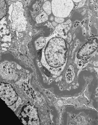
The diagnosis is often suggested by initial inspection of the specimen at low power on the microscope. Some tubules are abnormal, and have striking dilatation, with casts. Other tubules have marked granularity of the cytoplasm. Glomeruli show collapse of capillary loops to different extents, with prominent, vacuolated, granular, visceral epithelial cells on the outside of the collapsed tufts, and material resembling the tubular casts in Bowman’s space (Figs 6.31 and 6.32). A characteristic finding in HIV infection on electron microscopy is aggregation of intracytoplasmic tubular structures within the endoplasmic reticulum, called tubulo-reticular bodies, in glomerular endothelial cells (Fig. 6.33). These structures are induced by interferon alpha, and can also be found in conditions other than HIV infection, most notably in lupus nephritis.
80 |
6 Indication for Biopsy: Nephrotic Syndrome |
Fig. 6.28 Cortex in a renal biopsy specimen from a man of 69 with the nephrotic syndrome. Glomeruli have segmental lesions of various sizes and at various sites. The diagnosis is late classic segmental sclerosing glomerulonephritis, which can also be called late classic focal segmental glomerulosclerosis
In the Columbia classification, this abnormality is called the collapsing variant of focal segmental glomerulosclerosis. Another variant, called the cellular variant, seems similar or identical to the collapsing variant.
Usually, collapsing glomerulopathy progresses rapidly to renal failure.
Other Findings in the Kidney in Human Immunodeficiency
Viral Infection
Collapsing glomerulopathy in people with HIV is often called HIV-associated nephropathy. Many renal disorders have been described in these people, who are at risk of other infections and the renal complications of them, but who could also have any renal disorder found in people without HIV.
Other Findings in the Kidney in Human Immunodeficiency Viral Infection |
81 |
Fig. 6.29 Outer cortex in a renal biopsy specimen from a girl of 3 with the nephrotic syndrome. Glomeruli are possibly enlarged, but show no other abnormality. In particular, there are no segmental lesions. The inner cortex is illustrated in Fig. 6.30
The conditions particularly seen include membranous nephropathy, acute postinfective glomerulonephritis, IgA nephropathy, subendothelial membranoproliferative glomerulonephritis, fibrillary-immunotactoid glomerulopathy, and hemolytic uremic syndrome. These resemble the conditions in people without HIV, although there may be additional features, such as tubulo-reticular bodies, and there can be combinations of the conditions. HIV-infected people may have a condition that resembles lupus nephritis, with heavy deposition of all immunoproteins in several sites in glomeruli, but without serologic and clinical features of systemic lupus erythematosus. Another disorder that has been described overlaps with the lupuslike nephritis, and is apparently intermediate between acute postinfective glomerulonephritis and membranous nephropathy, with mesangial and subepithelial immune deposits.
Drugs used to treat HIV can have effects on the kidney, such as damage to proximal tubular cells.
82 |
6 Indication for Biopsy: Nephrotic Syndrome |
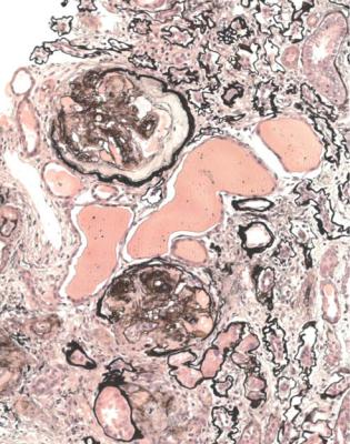
Fig. 6.30 Inner cortex and arcuate vessels next to the medulla in the renal biopsy specimen from the girl of 3 whose outer cortex is seen in Fig. 6.29. Both figures are at the same magnification. A couple of glomeruli have areas of segmental sclerosis, arrowed. This is the typical appearance of the childhood form of late classic segmental sclerosing glomerulonephritis, which can also be called late classic focal segmental glomerulosclerosis
Is the Pathologist Told that the Person Biopsied Has Diabetes Mellitus? Is There Evidence of Diabetic Glomerulopathy?
The pathologist may not be informed that the person biopsied has diabetes mellitus, either by oversight or because the nephrologists are not aware of this, but almost always the pathologist is told.
Diabetic renal disease is a useful term that covers several effects of diabetes mellitus on the kidney, including changes in blood vessels, tubules, and the medulla. Diabetic glomerulopathy is a more specific term for the characteristic changes in glomeruli.
A rule is that although people with diabetes mellitus and clinical renal abnormalities usually have diabetic glomerulopathy, they may have any other renal disease, either alone or in combination with diabetic glomerulopathy.
Value of Renal Biopsy in Diabetes Mellitus |
83 |
Fig. 6.31 Cortex in a renal biopsy specimen of a man of 49 with the nephrotic syndrome, acute renal failure, and HIV infection. Many tubules are dilated and contain casts. Glomeruli are shrunken to different extents, and some appear solid. The diagnosis is collapsing glomerulopathy
Value of Renal Biopsy in Diabetes Mellitus
Renal biopsy establishes whether there is diabetic glomerulopathy or another disorder, the finding of which may affect clinical management. The prevalence of diseases other than diabetic glomerulopathy in biopsy specimens in diabetes mellitus depends upon the policy of nephrologists. If many or most people with diabetes mellitus and a clinical renal disorder have a biopsy, other findings are uncommon. Selective biopsy of people with diabetes mellitus and an atypical clinical renal disorder will show other findings more commonly, but may miss a few in the diabetic population without a biopsy. People with diabetic retinopathy are highly likely to have diabetic glomerulopathy, and many nephrologists consider a renal biopsy unnecessary in these people.
Renal biopsy allows assessment of the extent of chronic damage, and indicates the prognosis for the kidney.
84 |
6 Indication for Biopsy: Nephrotic Syndrome |
Fig. 6.32 Glomerulus in a renal biopsy specimen from a woman of 23 with the nephrotic syndrome, but without HIV infection. The glomerulus shows collapse of the tuft as the mesangium has disappeared. Epithelial cells are enlarged and vacuolated. The diagnosis is collapsing glomerulopathy
Diagnosis of Diabetic Glomerulopathy
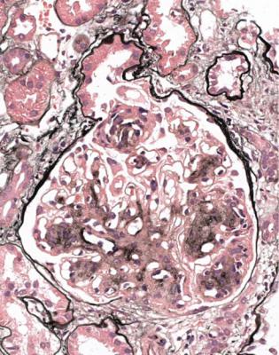
Well-established diabetic glomerulopathy is easy to diagnose. There is usually extensive chronic damage in the cortex. Surviving glomeruli appear large, with thickened basement membranes, although not so thick as those in well-established membranous nephropathy. There is strikingly variable increase in mesangial matrix, in which round areas have a peripheral ring of nuclei and are called Kimmelstiel– Wilson nodules (Fig. 6.34). These nodules are sites of healing of microaneurysms (Fig. 6.35). The nodules persist a long time and can be seen in shrunken glomeruli, and even in globally sclerosed glomeruli (Figs 6.36 and 6.37).
Other features are seen in well-established diabetic glomerulopathy, both in glomeruli and in other parts of the kidney. Glomeruli can have areas of hyalinosis stained by eosin, called hyaline caps or lipohyaline caps when they are on the tuft, and called capsular drops when they are on the inner surface of Bowman’s capsule (Fig. 6.38). Arterioles often have marked hyalinosis (Fig. 6.39). This change
Diagnosis of Diabetic Glomerulopathy |
85 |
Fig. 6.33 Electron micrograph showing a tubulo-reticular body in a glomerular endothelial cell. This is frequently found in HIV infection, but is also seen in other conditions, such as lupus nephritis, which is the diagnosis in this renal biopsy specimen, from a woman of 37 with proteinuria
affecting both afferent and efferent arterioles at the hilum of glomeruli is probably only seen in diabetes mellitus (Fig. 6.40). Especially in the old days, when diabetes mellitus was untreated, parts of tubules had accumulation of glycogen, that made the tubular cells appear clear, although this is hardly ever seen now. This is called the Armanni–Ebstein lesion (Fig. 6.41).
Less marked diabetic glomerulopathy is usually still recognisable on light microscopy, even if there are no Kimmelstiel–Wilson nodules. The main feature is increase in mesangial matrix, without evidence of mesangial deposition of immunoproteins, particularly IgA. If there is deposition of IgA, this indicates either pure IgA nephropathy, or the combination of the two conditions, which is commoner than pure IgA nephropathy in diabetes mellitus, and is shown by evidence of typical diabetic features. Glomerular basement membranes in diabetic glomerulopathy may show linear deposition of IgG, but not of complement (Fig. 6.42).
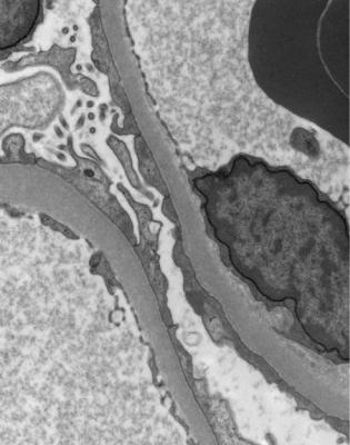
In specimens that appear normal or close to normal, electron microscopy is necessary to show the first sign of diabetic glomerulopathy, which is uniform thickening of glomerular basement membranes (Figs 6.43 and 6.44).
86 |
6 Indication for Biopsy: Nephrotic Syndrome |
Fig. 6.34 Glomerulus in a renal biopsy specimen from a woman of 52 with proteinuria in the nephrotic range, chronic renal failure, and a 4-year history of insulin resistant or type two diabetes mellitus. There is expansion of the mesangium with formation of Kimmelstiel–Wilson nodules, typical of diabetic glomerulopathy.
Kimmelstiel, usually pronounced kimul-steel in English, and Wilson, pronounced in the usual way, described the postmortem kidneys of eight adults, seven of whom were known to have had diabetes mellitus. They found hyaline masses in the glomerular intercapillary tissue, now called the mesangium. They called this intercapillary glomerulosclerosis, but did not use the word nodule (Kimmelstiel P, Wilson C. Intercapillary lesions in the glomeruli of the kidney. American Journal of Pathology 1936; 12: 83–97). Nodule, coming from the Latin for a little knot, was in use by 1948, and the term Kimmelstiel–Wilson lesion was in use by 1951, although who was the first to think of these is difficult to discover. In Latin, the word diabetes meant a passing through or a siphon, and mellitus meant honeyed or sweet. Paul Kimmelstiel (1900–1970) was born in Hamburg, Germany, and became a pathologist there in the department of Theodor Fahr, who with the physician Franz Volhard was important in the development of understanding of the kidney through their clinicopathologic studies, first published in 1914. Kimmelstiel began to study glomerular lesions, but was forced to leave Germany in 1933 by the Nazis, and went to the United States. Clifford Wilson (1906–1997) qualified in medicine at the London Hospital, United Kingdom. In 1934, he went to Harvard University as a Rockefeller Travelling Fellow and met Kimmelstiel, who was an instructor in the Mallory Institute of Pathology. Later, Kimmelstiel worked as a pathologist in several cities in the United States. Wilson became Professor of Medicine at the London Hospital in 1946, succeeding Arthur William Mickle Ellis (1883–1966). Ellis developed a classification of renal disease that was influential for many years. In this, there were only two numbered types of
Diagnosis of Diabetic Glomerulopathy |
87 |
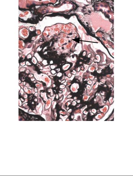
Fig. 6.35 Glomerulus in a renal biopsy specimen from a man of 58 with proteinuria in the nephrotic range, chronic renal failure, and 10 years of insulin resistant or type two diabetes mellitus. There is an increase in mesangium with a microaneurysm, arrowed. This type of lesion heals to form a Kimmelstiel–Wilson nodule
Usually, if the reason for the biopsy is the nephrotic syndrome and the explanation is diabetic glomerulopathy, this diagnosis is immediately evident. If it is not, another diagnosis is likely, such as membranous nephropathy.
Fig. 6.34 nephritis, type one of abrupt onset, short duration, and recovery, and type two of insidious onset, long duration, and no recovery. Ellis did not mean that there were only two kinds of renal disease, because he separated other disorders, such as acute focal nephritis, amyloid, and essential hypertension (Ellis A. Natural history of Bright’s disease: clinical, histological and experimental observations. Lancet 1942; 1: 1–7, 34–36, 72–76). Ellis’s ideas were influenced by Dorothy Stuart Russell (1895–1983), who before she became a famous neuropathologist worked on renal disease, and in 1929 published a classification of kidney disorders that were then called Bright’s disease. Pathologists used this to mean chronic, nonsuppurative inflammation of the kidney. Russell predicted the existence of entities not identified in her series, but necessary to complete the neatness of the classification
88 |
6 Indication for Biopsy: Nephrotic Syndrome |
Fig. 6.36 Cortex in a renal biopsy specimen from a woman of 48 with the nephrotic syndrome, chronic renal failure, and many years of insulin resistant or type two diabetes mellitus. There is extensive chronic renal damage. Glomeruli are globally sclerosed, but advanced changes of diabetic glomerulopathy, including Kimmelstiel–Wilson nodules, are still evident
Diabetic glomerulopathy seems a consequence of persistently high concentration of glucose in the blood, which stimulates increased production and decreased degradation of mesangial matrix and glomerular basement membrane components.
Diabetic glomerulopathy indicates a high risk of progression to renal failure, and also of death from cardiovascular causes, particularly myocardial infarction.
Differential Diagnosis of Diabetic Glomerulopathy
Nodules in glomeruli can be seen in a few conditions other than diabetic glomerulopathy.
The commonest disorder that has glomerular nodules resembling diabetic glomerulopathy is light chain glomerulopathy, or nodular light chain glomerulopathy (Fig. 6.45). Clues to this diagnosis are that the person biopsied is not known
Differential Diagnosis of Diabetic Glomerulopathy |
89 |
Fig. 6.37 Glomerulus in a renal biopsy specimen from a woman of 66 with proteinuria in the nephrotic range, and 8 years of insulin resistant or type two diabetes mellitus. There is severe ischemic shrinkage of the tuft, but a Kimmelstiel–Wilson nodule is still seen, adherent to Bowman’s capsule just at the origin of the tubule
to have diabetes mellitus, and has a disorder of immunoglobulins, although this may not have been detected before the biopsy. Immunohistologic staining to detect kappa and lambda light chains should show deposition of only one type in glomeruli and in tubular basement membranes (Figs 6.46 and 6.47). Kappa chains are much more often found than lambda.
The nodules in light chain glomerulopathy are usually all approximately the same size within a glomerulus, and most glomeruli look the same, although in diabetic glomerulopathy there is often much variation within a glomerulus and between glomeruli. There is usually heavy deposition of complement in nodules on immunohistologic study in light chain glomerulopathy, but not in diabetic glomerulopathy. Electron microscopy usually shows a line of deposited material in the inner or subendothelial aspect of glomerular basement membranes (Fig. 6.48).
Amyloid may give a similar nodular appearance, but the Congo red stain identifies this (Fig. 6.49).
90 |
6 Indication for Biopsy: Nephrotic Syndrome |
Fig. 6.38 Glomerulus in a renal biopsy specimen from a woman of 58 with proteinuria, chronic renal failure, and insulin resistant or type two diabetes mellitus. Hyaline material is seen in capillary loops, including in a globally sclerosed glomerulus, and there is a large capsular drop on the inside of Bowman’s capsule of the surviving glomerulus
Nodules may occur in advanced stages of subendothelial membranoproliferative or mesangiocapillary glomerulonephritis, also called membranoproliferative or mesangiocapillary glomerulonephritis type one (Fig. 6.50). In this condition, staining with periodic acid-methenamine silver shows doubled glomerular basement membranes, which are not seen in diabetic glomerulopathy.
Is the Pathologist Told that the Person Biopsied Has Systemic Lupus Erythematosus? Is There a Possibility of Lupus Nephritis?
Almost always, systemic lupus erythematosus is diagnosed clinically before renal biopsy. If not, before a specimen is examined, the pathologist should think of lupus nephritis as a possible diagnosis in any young woman with the nephrotic syndrome, or indeed any other indication for a renal biopsy.
Value of Renal Biopsy in Systemic Lupus Erythematosus |
91 |
Fig. 6.39 Cortex in the renal biopsy specimen from the woman of 58 with diabetic glomerulopathy that is illustrated in Fig. 6.38. Arterioles have severe hyalinosis
Lupus nephritis is easy to diagnose, but is made complicated and difficult for pathologists in most texts. This is because there are elaborate systems of classification.
Value of Renal Biopsy in Systemic Lupus Erythematosus
A renal biopsy is hardly ever done in someone with lupus, but without clinical renal disease. The value when there is clinical renal disease is to confirm that there is lupus nephritis, although any other finding is rare, to show the type of renal disease, and to allow an assessment of the severity and proportions of active and late changes, which may be used in decisions about clinical management. A prognostic feature is the amount of chronic damage, assessed by methods such as measurement of the index of chronic damage (Fig. 5.6).
Repeat renal biopsy is perhaps commoner in lupus than in any other condition, if there is reactivation of clinical renal disease, or another change in its clinical
92 |
6 Indication for Biopsy: Nephrotic Syndrome |
Fig. 6.40 Glomerulus in a renal biopsy specimen from a woman of 54 with the nephrotic syndrome and insulin resistant or type two diabetes mellitus. An immunoperoxidase method to detect C9 shows that both arterioles at the glomerular hilum have hyalinosis, a feature of diabetic renal disease
characteristics. Lupus nephritis can change appearances between biopsies, and the new pathological findings influence management.
Diagnosis of Lupus Nephritis
Lupus nephritis can give almost any glomerular abnormality, and almost any combination of abnormalities. A combination of glomerular disorders in a specimen from a person not reported to have systemic lupus erythematosus should make the pathologist think about lupus nephritis as a possible diagnosis. Similar changes may be seen in mixed connective tissue disorders, in which there are various clinical features of systemic lupus erythematosus, scleroderma or systemic sclerosis, and polymyositis.
Diagnosis of Lupus Nephritis |
93 |
Fig. 6.41 Outer medulla in a renal biopsy from a girl of 12 with proteinuria and 8 years of insulin deficient or type one diabetes mellitus that was poorly controlled. Some tubules, probably straight parts of proximal tubules, are lined by clear cells that contain glycogen. This is the Armanni– Ebstein lesion, named after Luciano Armanni (1839–1903), pronounced ar-man-ee, a pathologist in Naples, Italy, who was diabetic himself later in life, and first saw the abnormality in autopsy kidneys in 1872, and Wilhelm Ebstein (1836–1912), pronounced eb-stine, a German physician, who independently saw this in 1882
The pathologist should assess the extent of chronic damage, which may be anywhere between none and widespread (Figs 6.51 and 6.52). Common findings in glomeruli in lupus nephritis are membranous nephropathy, mesangial expansion to various extents, subendothelial membranoproliferative or mesangiocapillary pattern with doubled basement membranes, and segmental lesions of vasculitic type (Figs 6.53–6.57). When the renal disease is active, there is often an interstitial infiltrate of mixed inflammatory cells, with evidence of acute damage to tubules (Fig. 6.57). Blood vessels occasionally show changes of small vessel vasculopathy, also called thrombotic microangiopathy, with thrombosis and fibrinoid necrosis in arterioles, and loose concentric intimal thickening in small arteries (Fig. 6.58).
Immunohistology in lupus nephritis often shows heavy deposition of all immunoproteins in glomeruli, in a distribution that depends upon the pattern of structural
94 |
6 Indication for Biopsy: Nephrotic Syndrome |
Fig. 6.42 Glomerulus in the renal biopsy specimen from the woman of 52 whose diabetic glomerulopathy is illustrated in Fig. 6.34. An immunoperoxidase method to detect IgG shows linear deposition of IgG in glomerular basement membranes (arrowed), sometimes found in diabetic glomerulopathy. Granules of IgG are seen in proximal tubular cells, a finding in glomerular proteinuria
changes (Figs 6.59–6.62). IgG is usually evident. Mesangial deposition of IgG is a clue to the diagnosis of lupus nephritis, because few other conditions give this. The finding of IgA in membranous nephropathy is another clue that there is lupus nephritis. If the disease is late or serologically inactive, there may be little or no deposition of immunoproteins.
Electron microscopy in lupus nephritis confirms the distribution of immune deposits in glomeruli. Tubulo-reticular bodies are often found in glomerular endothelial cells (Fig. 6.33).
There are many immunologic abnormalities in systemic lupus erythematosus, but the cause is not known. One problem may be that fragments of apoptotic cells are not removed in the normal way, but stimulate an immune response to nuclear antigens, particularly nucleosomes, which are positively charged histone proteins complexed to deoxyribonucleic acid. Circulating histones or nucleosomes may attach to the negatively charged glomerular basement membrane, and allow antibodies against these antigens to fix in glomeruli.
Classification of Lupus Nephritis |
95 |
Fig. 6.43 Glomerulus in a renal biopsy specimen from a woman of 18 with proteinuria and 7 years of insulin deficient or type one diabetes mellitus. The glomerulus is close to normal, but has a little mesangial expansion which suggests early diabetic glomerulopathy, confirmed by electron microscopy, as in Fig. 6.44
A pathologist should always give a description of the changes in lupus nephritis, even if a classification system is used. The description should always apply, although the classification will change.
Classification of Lupus Nephritis
There have been a few numerical systems of classification of lupus nephritis, mainly published by the World Health Organization, WHO. Derived from these was a system published in 2003 by the International Society of Nephrology and the Renal Pathology Society ISN/RPS. The intention of these schemes is to ensure uniformity of reporting of lupus nephritis. Any numerical system has difficulties, and the main problems are these.
96 |
6 Indication for Biopsy: Nephrotic Syndrome |
Fig. 6.44 Electron micrograph of part of the glomerular basement membrane in a renal biopsy specimen from a woman of 46 with the nephrotic syndrome, and insulin deficient or type one diabetes mellitus for 7 years. The membrane is from 700 to 900 nm thick, which is more than twice the normal thickness. This is confirmation of early diabetic glomerulopathy. A normal glomerular basement membrane at the same magnification is illustrated in Fig. 6.6
1.Systems come and go. The WHO system changed every few years, and has been superseded. Changes in classification may mean that pathologists and physicians are not using the same system, and that findings of trials of treatment, for example, based on old systems, may not be applicable to new systems.
2.A pathologist cannot work out from first principles what a number in a numerical system means. Without other knowledge, the meaning is obscure of such terms as type one diabetes mellitus or class three lupus nephritis.
3.Glomeruli often show more than one abnormality in lupus nephritis, and there can be marked variation between glomeruli in a specimen. One number may not accurately reflect the combination of changes, and a pathologist may not be able to reconstruct the appearances of a particular specimen just from this number. This is true even when a system allows more than one number to be used.
4.Systems are not reliably used by pathologists, and have imperfect reproducibility. The ISN/RPS system has better reproducibility in some respects than WHO schemes, but is still not used consistently.
Classification of Lupus Nephritis |
97 |
Fig. 6.45 Cortex in a renal biopsy specimen from a man of 60 with the nephrotic syndrome. Appearances resemble diabetic glomerulopathy, but the diagnosis is light chain glomerulopathy. The man was not diabetic, and had myeloma, which was found after the biopsy. There was a Bence Jones protein in the urine, consisting of dimeric monoclonal kappa light chains, with an IgG kappa paraprotein in the serum. Although monoclonal light chains in the urine are called Bence Jones proteins, pronounced benss and jones in the usual way, the name should be Jones proteins, or even MacIntyre proteins. Henry Bence Jones (1813–1873) did not hyphenate his name and was called Jones. He qualified in medicine at St George’s Hospital in London, United Kingdom, where he became a physician, and specialised in what would now be called clinical chemistry. In 1847 and 1848, he reported an unusual protein in the urine of a man aged 45, under the care of Dr Thomas Watson and Dr William MacIntyre. The properties of the urine were discovered by MacIntyre in 1845, but not published by him until 1850. Myeloma was named in 1873, and its relation to Jones’s proteinuria was noted in 1889. About four-fifths of people with myeloma have this type of proteinuria, but few have the glomerulopathy
5.A numerical system has the implication that there is a progression in severity or in time from low numbers to high numbers. This is by analogy with staging systems in cancer. Some pathologists assume that the highest numbers in a classification are the worst conditions.
6.Only glomerular changes are considered when a class number is given to a biopsy specimen with lupus nephritis, although another feature, for example a small vessel vasculopathy, may be much more important.
98 |
6 Indication for Biopsy: Nephrotic Syndrome |
Fig. 6.46 Glomerulus in a renal biopsy specimen from a man of 61 with acute renal failure and myeloma, with monoclonal lambda light chains in serum and urine. There is a nodular appearance. Immunoperoxidase staining to detect kappa light chains shows no significant deposition in the kidney
International Society of Nephrology/Renal Pathology Society 2003 Classification of Lupus Nephritis
No pathologist should know classification systems. If they are used, the pathologist should refer to the guidelines for them, such as those following, and should specify in a report which system is used.
The 2003 RPS/ISN classification is given here, with an explanation of each class:
Class one – Minimal mesangial lupus nephritis: Glomeruli appear normal on light microscopy, but have mesangial immune deposits on immunohistology. If glomeruli have no abnormality at all, there is no lupus nephritis, and no class number is used.
International Society of Nephrology/Renal Pathology Society 2003 Classification |
99 |
Fig. 6.47 Glomerulus in the renal biopsy specimen from the man of 61 shown in Fig. 6.46. Immunoperoxidase staining to detect lambda light chains shows heavy deposition in mesangium, glomerular basement membranes, tubular basement membranes, and in casts. This confirms light chain glomerulopathy
Class two – Mesangial proliferative lupus nephritis: Mesangial hypercellularity or expansion of mesangial matrix is seen with mesangial immune deposits (Figs 6.53 and 6.60).
Class three – Focal lupus nephritis: Focal is defined here to mean that fewer than half of all the glomeruli in a specimen are affected by lesions. These are usually segmental but occasionally global, and are of vasculitic type, active or healed. In the active stage, these show different combinations of endocapillary thrombosis or aggregates of immune complexes, hypercellularity and degeneration of cells in capillary loops, disruption of glomerular basement membranes, and cells in Bowman’s space (Fig. 6.54). These lesions heal to give segmental areas of sclerosis that are sharply outlined, and cross part of the glomerular tuft and Bowman’s space. Class 3(A) means active lesions, 3(C) means healed, chronic, inactive lesions, and 3(A/C) means a mixture. The proportion of glomeruli affected by active or healed lesions is
100 |
6 Indication for Biopsy: Nephrotic Syndrome |
Fig. 6.48 Electron micrograph of a glomerular basement membrane in a renal biopsy specimen from a woman of 44 with proteinuria and renal failure. There is a line of deposited material (arrowed), which indicates a light chain glomerulopathy. After the biopsy, monoclonal kappa chains were found in serum and urine
reported. Class three is often associated with segmental areas of subendothelial immune deposits, and may have mesangial alteration, even affecting all glomeruli.
Class four – Diffuse lupus nephritis: Segmental or global lesions affect at least half of all the glomeruli in a specimen. Segmental in this class is defined as a lesion sparing half of the tuft. Class 4S has more widespread lesions than class three, but is otherwise similar, and is divided into similar subtypes, 4S(A), 4S(A/C), and 4S(C). The global form, class 4G, usually has endocapillary and mesangial hypercellularity, often with a subendothelial
International Society of Nephrology/Renal Pathology Society 2003 Classification |
101 |
Fig. 6.49 Cortex in a renal biopsy specimen from a man of 67 with chronic renal failure and myeloma. Glomeruli contain nodules that are shown by Congo red staining to contain amyloid
membranoproliferative or mesangiocapillary pattern (Figs 6.55–6.57, 6.61, and 6.62). There are usually widespread subendothelial immune deposits, and sometimes these are the main abnormality. Class 4G may show healing to give segmental or global areas of sclerosis, and the subtypes are 4G(A), 4G(A/C), and 4G(C).
Class five – Membranous lupus nephritis: Subepithelial immune deposits are seen, with or without mesangial alteration (Figs 6.51 and 6.59). This can occur with either class three or class four, but class five is only reported as well as the other class if at least half of the surface area of glomerular capillary loops in at least half of glomeruli shows membranous nephropathy (Figs 6.55 and 6.62).
Class six – Advanced, globally sclerosed lupus nephritis: At least nine-tenths of glomeruli are globally sclerosed (Fig. 6.52).
A test of the ISN/RPS 2003 classification showed that class four was by far the commonest class used, and was diagnosed much more often than in a WHO
102 |
6 Indication for Biopsy: Nephrotic Syndrome |
Fig. 6.50 Glomerulus in a renal biopsy specimen from a man of 22 with hematuria and proteinuria. There are nodules. Periodic acid-methenamine silver staining shows double basement membranes, with other features of subendothelial membranoproliferative glomerulonephritis. Subendothelial membranoproliferative glomerulonephritis was found to be the diagnosis in two of the three kidneys in the Gordon Museum at Guy’s Hospital in London, United Kingdom, that had been described by Bright, who for many years was honoured by the term Bright’s disease that was applied to clinical renal disorders (Fig. 6.34). Richard Bright (1789–1858), whose surname is pronounced in the usual way, was born in Bristol, United Kingdom, qualified in medicine in Edinburgh, and became a physician at Guy’s Hospital at the same time as Addison and Hodgkin, who also had diseases named after them. In 1827, Bright published a book called “Reports of medical cases selected with a view of illustrating the symptoms and cure of diseases by a reference to morbid anatomy”. He showed the association of edema, then called anasarca or dropsy or dropsical affection, with proteinuria and disorders of the kidney, and separated edema due to renal disorders from edema due to diseases of the heart and liver. The third kidney in the Gordon Museum showed amyloid secondary to pulmonary tuberculosis
classification. There was poor reproducibility between pathologists of assessment of activity and chronicity. Active features in glomeruli are endocapillary hypercellularity, infiltration of leukocytes, disruption of nuclei called karyorrhexis, thrombosis, rupture of basement membranes, cells in Bowman’s space, subendothelial immune deposits, and intraluminal immune aggregates. Chronic features in glomeruli are
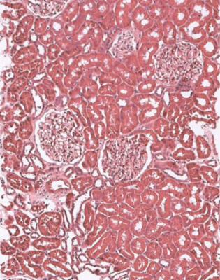
International Society of Nephrology/Renal Pathology Society 2003 Classification |
103 |
Fig. 6.51 Cortex in a renal biopsy specimen from a woman of 21 with the nephrotic syndrome, and clinical reactivation of systemic lupus erythematosus that had been treated for 2 years. There is no acute or chronic tubular damage. Glomeruli have pure membranous nephropathy as a sign of lupus nephritis. In Latin, the word lupus meant a wolf. In English, the word was used for many centuries to mean a skin ulcer. This was well defined by Barrough in 1590: “Lupus is a malignant ulcer quickly consuming the neather parts; and it is very hungry like unto a woolfe”. Different types of ulcer became recognised, such as lupus vulgaris caused by tuberculosis. The name systemic lupus erythematosus had this basis rather than other suggested origins, such as the supposed resemblance of the facial rash to a wolf’s face. Systemic lupus erythematosus is thought to have caused renal failure and death in the American writers Jack London (1876–1916) and Flannery O’Connor (1925–1964), and the ruler of the Philippines, Ferdinand Marcos (1917–1989), who died after two renal allografts
segmental or global sclerosis, fibrosed adhesions between tuft and Bowman’s capsule, and fibrosis in Bowman’s space.
Active lupus nephritis usually responds to immunosuppression, although some types are more difficult to treat, particularly segmental vasculitic lesions, in classes three and four, and global membranoproliferative changes, in class four. Late changes in any class, as in any renal disorder, are not likely to improve.
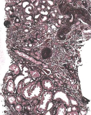
104 |
6 Indication for Biopsy: Nephrotic Syndrome |
Fig. 6.52 Cortex in a renal biopsy specimen from a woman of 52 with hematuria, proteinuria, chronic renal failure, and a 12-year history of systemic lupus erythematosus. There is severe late damage, with global sclerosis of nearly every glomerulus
Are Glomeruli Abnormal? Is There Amyloid?
Amyloid is often an unexpected finding in renal biopsy specimens, and may be missed if the diagnosis is not considered, and no specific stain is requested. Because of this, and because amyloid is one of the few things that a simple stain can identify, there is justification to use Congo red as a routine stain on every renal biopsy specimen (Fig. 6.63).
Diagnosis of Amyloid
Amyloid is easy to diagnose. The diagnosis can be made on one glomerulus, or even if there are no glomeruli in the specimen (Fig. 4.5).
There are various appearances on light microscopy, but usually there is expansion of mesangium and glomerular basement membranes by acellular material that stains
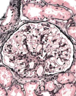
Diagnosis of Amyloid |
105 |
Fig. 6.53 Glomerulus in a renal biopsy specimen from a woman of 39 with hematuria, proteinuria, and a short history of systemic lupus erythematosus. There is lupus nephritis, seen as mild mesangial expansion
palely with eosin (Fig. 6.64). This may stain black with periodic acid-methenamine silver. Often there are spikes or bristles projecting from the outside of glomerular capillary loops on sections stained with periodic acid-methenamine silver. These do not look like the spikes in membranous nephropathy, because in amyloid the projections are longer and coarser, and are found in groups rather than uniformly throughout glomeruli (Fig. 6.65). Material similar to that in glomeruli is often seen in walls of small blood vessels, tubular basement membranes, and interstitial tissues.
The diagnosis is given by Congo red staining. A control section known to contain amyloid should always be included when sections are stained by Congo red.
Amyloid appears various shades of red on orthodox light microscopy, depending on the amount of amyloid, thickness of sections, and technique of Congo red staining (Fig. 6.63). Sections thicker than the usual 3 μm are helpful in the study of amyloid, especially if there is not much in a specimen (Figs 6.66 and 6.67). Small amounts of amyloid can be missed, and the diagnosis in the nephrotic syndrome may be thought to be minimal change nephropathy.
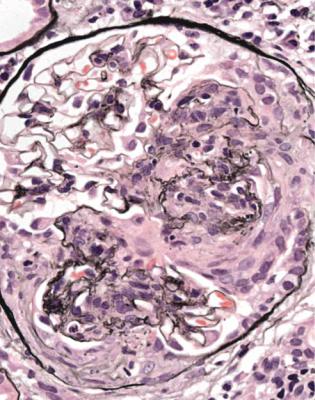
106 |
6 Indication for Biopsy: Nephrotic Syndrome |
Fig. 6.54 Glomerulus in a renal biopsy specimen from a woman of 40 with the nephrotic syndrome, hematuria, hypertension, acute renal failure, and systemic lupus erythematosus for over 10 years, but previously without any renal disorder. There is lupus nephritis, seen as a sharply outlined segmental lesion of vasculitic type, with disruption of basement membranes in capillary loops, and cells in Bowman’s space
Not all material that stains with Congo red is amyloid. For instance, the internal elastic lamina of arteries stains in normal arteries. Polarising microscopy is necessary to confirm amyloid in sections stained by Congo red. Amyloid can be diagnosed satisfactorily by light microscopy, and electron microscopy is not essential for the diagnosis.
Principles of Polarising Microscopy
To confirm amyloid, microscopic examination is needed with polarising filters below and above the section, called polariser and analyser. When one filter is rotated, the background changes from light to dark, with the maximum darkness when the planes of polarisation of the filters are at right angles to each other, and the filters
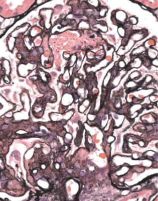
Principles of Polarising Microscopy |
107 |
Fig. 6.55 Glomerulus in a renal biopsy specimen from a woman of 23 with hematuria, proteinuria, and clinical reactivation of systemic lupus erythematosus that had been treated for 4 years. There is lupus nephritis, seen as a combination of membranoproliferative pattern with doubled basement membranes, membranous pattern, and early segmental vasculitic pattern, with thrombosis and disruption of a capillary loop
are said to be crossed. At this point and near it, birefringent structures lying at about 45◦ to the planes of polarisation appear bright against the dark background.
Birefringent means that a structure has two refractive indices for transmitted light. The refractive index is the ratio of velocity of light in a vacuum, or in air, to velocity in the material. A birefringent structure has asymmetry of its component parts. Light polarised in one plane interacts with components orientated in that plane, and its velocity is reduced or retarded more than light polarised at right angles. At 45◦ to the plane of polarisation, a birefringent structure resolves the linearly polarised light beam into two components, or vectors, at right angles to each other. As these vectors pass through the material, one is retarded more than the other. When these components emerge from the structure, their velocity returns to that in air, and they merge to produce elliptically polarised light. This means that the light waves, if viewed end on along their direction of travel, are no longer vibrating in
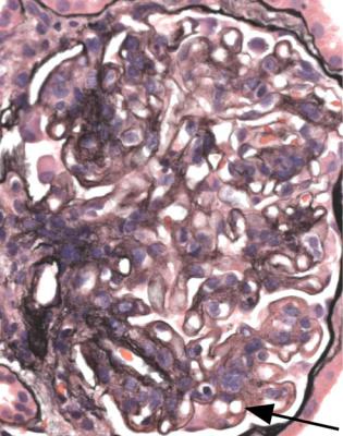
108 |
6 Indication for Biopsy: Nephrotic Syndrome |
Fig. 6.56 Glomerulus in a renal biopsy specimen from a woman of 45 with the nephrotic syndrome, acute renal failure, and clinical reactivation of systemic lupus erythematosus that had been treated for 9 years. There is lupus nephritis, with the appearance of subendothelial membranoproliferative glomerulonephritis, including subendothelial immune deposits (arrowed) that are called wire loops, because they resemble the tools used in microbiology to spread organisms on culture dishes
just one plane, but the tip of the combined vector rotates to trace an ellipse. Part of the light can now pass through a crossed analyser, and the structure appears bright against a dark background.
This explains why materials, such as starch particles or some other crystalline structures, have an appearance of Maltese crosses when examined between crossed polariser and analyser. There are four quadrants, in each of which there is maximal effect of the birefringence on polarised light (Fig. 6.68).
Characteristic of amyloid stained by Congo red is the appearance of various colours under these conditions (Figs 6.69–6.72). These may be called anomalous colours, because they include colours other than the red seen under unpolarised illumination. The only colour usually reported is green, often called “apple green birefringence”, although yellow and blue, or shades between these and green, are commonly seen. These colours alternate when the slide is rotated.
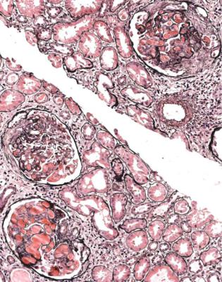
Principles of Polarising Microscopy |
109 |
Fig. 6.57 Cortex in a renal biopsy specimen from a woman of 22 with the nephrotic syndrome, renal failure that appeared acute, and clinical reactivation of systemic lupus erythematosus that had been treated briefly 1 year before. There is lupus nephritis with acute interstitial nephritis, various amounts of mesangial expansion, active and healing glomerular vasculitic lesions, and large intracapillary masses in glomeruli that are often called hyaline thrombi, although they are immune aggregates rather than thromboses
The explanation of anomalous colours is that molecules of Congo red are orientated on fibrils of amyloid, so that they absorb and retard light polarised in one plane, but not light polarised at right angles. This means that amyloid stained by Congo red is birefringent. The birefringence is not the same for every wavelength of light. Birefringence is maximal around the absorption peak of Congo red, which is in the blue–green part of the spectrum. Wavelengths shorter than the peak are transmitted with one direction of rotation of elliptically polarised light, giving blue, and longer wavelengths have an opposite direction of rotation, giving yellow.
When conditions are perfect for polarising microscopy, blue and yellow are both transmitted, and the net appearance is green. When conditions are not perfect, additional birefringence in the optical system may remove one of the colours, partly or completely, by the process called compensation, and shades of blue or yellow
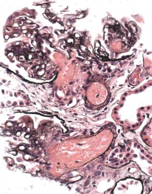
110 |
6 Indication for Biopsy: Nephrotic Syndrome |
Fig. 6.58 Cortex in a renal biopsy specimen from a woman of 19 with acute renal failure, hematuria, proteinuria, and relapse of systemic lupus erythematosus that had been treated for 5 years. There is lupus nephritis with a small vessel vasculopathy, also called thrombotic microangiopathy, seen as thrombosis and fibrinoid necrosis in arterioles
appear. Which one is removed depends on the relative orientation of the amyloid and the compensating agent. The extraneous birefringence could be in microscope lenses, the glass slide, or the coverslip.
Birefringence alone does not completely account for the anomalous colours, because as light passes through Congo red, there is absorption, particularly in the blue–green. The net effect of birefringence and absorption accounts for the colours seen in Congo red at 45◦ to the polariser, between crossed polariser and analyser.
As the polariser or analyser is rotated from this position, other colours appear. This test is useful in practice, to confirm amyloid. Increasing rotation in one direction gives yellow, orange, bright red, and dull red, in some areas of orientated amyloid. The other direction gives light green, white, and colourless in these areas. Specimens show both changes at the same time, because amyloid has many different orientations in a section.
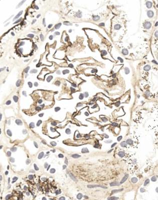
Principles of Polarising Microscopy |
111 |
Fig. 6.59 Glomerulus in a renal biopsy specimen from a woman of 44 with proteinuria in the nephrotic range, and reactivation of systemic lupus erythematosus that had been quiescent for many years. An immunoperoxidase stain to detect C9 shows the appearances of membranous nephropathy, with uniform, tiny granules on the outside of every capillary loop
These colours are produced by the combination of progressive decline in birefringence, and progressively strengthened effects of absorption on directly transmitted light. More light is transmitted directly as the polariser and analyser are uncrossed. Light passing directly through orientated molecules of Congo red in one plane is absorbed, and the appearance is of white lacking blue–green, which is red, against a white background. Light at right angles to this plane is not absorbed, and there is white, or no colour.
This change in appearance, related to a difference in the plane of illumination with polarised light, is called dichroism. Dichroism is detected in a material with use of a polariser only, without an analyser, or vice versa. Rotation of the slide, or the polariser or analyser, will show areas that change intensity of colour. Amyloid stained by Congo red is dichroic, but this is usually difficult to detect, because there are only small areas in a section where the amyloid has a uniform orientation.
112 |
6 Indication for Biopsy: Nephrotic Syndrome |
Fig. 6.60 Glomerulus in the renal biopsy specimen from the woman of 39 with lupus nephritis, which is illustrated in Fig. 6.53. An immunoperoxidase stain to detect IgG shows only mesangial deposition
Determination of the Type of Amyloid
Amyloid is not one substance, but a group of misfolded proteins with the similar property of formation of extracellular insoluble fibrils, which bind serum amyloid P glycoprotein. The commonest type found in the kidney is AL amyloid, derived from a monoclonal immunoglobulin light chain, and this is much more commonly lambda than kappa. The finding of amyloid consistent with AL type is often the first indication that there may be paraproteinemia, although this may be difficult to detect.
The second commonest type is AA amyloid. This is derived from serum amyloid A protein, which is an apolipoprotein of high-density lipoproteins, and is an acute phase reactant produced by the liver in many inflammatory conditions. AA amyloid is often associated with rheumatoid arthritis and other rheumatologic disorders, but also occurs in chronic infections, such as tuberculosis, bronchiectasis, osteomyelitis, and persistent abscesses, such as those found in the skin of some
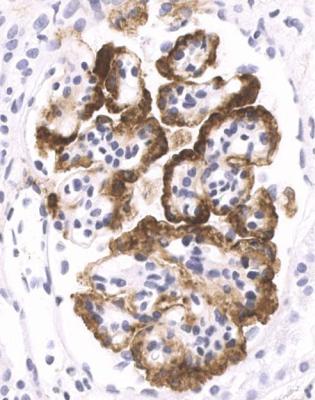
Determination of the Type of Amyloid |
113 |
Fig. 6.61 Glomerulus in a renal biopsy specimen from a man of 32 with the nephrotic syndrome, renal failure, and reactivation of systemic lupus erythematosus that had been quiescent for 2 years. An immunoperoxidase stain to detect C9 shows heavy subendothelial deposition.
intravenous drug users. This type also occurs in people with leprosy and schistosomiasis in tropical countries. Two inflammatory disorders which do not usually have a rise in concentration of serum amyloid A protein, and are not associated with amyloidosis, are systemic lupus erythematosus and ulcerative colitis. Persistent infections outside the kidney explain the common finding of AA amyloid in cattle and dogs.
AA amyloid is also found in familial mediterranean fever, and other inherited periodic fever disorders, in which it may respond to treatment. Other familial types of amyloid are associated with genetic abnormalities of fibrinogen Aalpha chain, lysozyme, transthyretin, apolipoprotein A1, gelsolin, and other proteins, but these are all rare. The commonest type found in the kidney is fibrinogen. Investigation of these types is not usually possible in most laboratories. Some other types of amyloid do not affect the kidney, or are not investigated by renal biopsy, such as the amyloid derived from beta two microglobulin found in people on hemodialysis,
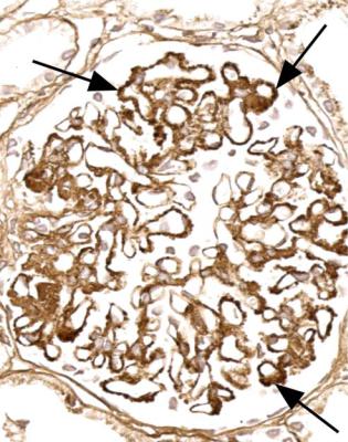
114 |
6 Indication for Biopsy: Nephrotic Syndrome |
Fig. 6.62 Glomerulus in the renal biopsy specimen of the woman of 23 with lupus nephritis that is illustrated in Fig. 6.55. An immunoperoxidase stain to detect IgG shows deposition in mesangium, on the outside of many capillary loops in a membranous pattern, and inside a few capillary loops, arrowed
or the cerebral amyloid derived from amyloid precursor protein in Alzheimer’s disease.
When amyloid is discovered, the pathologist should try to define the type. This will help clinical investigation and management. If AL amyloid is found, appropriate immunologic investigations should be arranged by nephrologists. The prognosis of renal amyloidosis depends upon first, the extent of chronic renal damage, which is an indicator of the length of survival until renal replacement therapy is needed, and second, the underlying disorder, which has more relation to the death rate. AL amyloid has a higher mortality rate than other types, related to the underlying neoplastic disease and the damage to other organs, particularly the heart.
Immunohistology using an antibody to amyloid A protein usually confirms or excludes AA amyloid unequivocally (Figs 6.73 and 6.74). Antibodies to kappa and lambda light chains are not so straightforward, because they do not always react with the abnormal light chains in AL amyloid, but if available they should be used
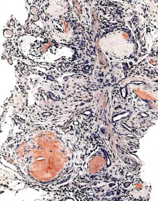
Determination of the Type of Amyloid |
115 |
Fig. 6.63 Cortex in a biopsy specimen of a renal allograft in a woman of 38, 12 years after transplantation for chronic renal failure that developed in childhood due to a segmental sclerosing glomerular disorder. Examination of a section stained with Congo red shows deposits of amyloid. This was unexpected. The amyloid was shown to be AL type, and further clinical investigation showed that the woman had myeloma
(Figs 6.75 and 6.76). Amyloid that does not react with an antibody to amyloid A protein, nor with antibodies to light chains, should be reported as non-AA amyloid, most likely AL, or consistent with AL, unless the pathologist is able to show that there is one of the rarer types.
Occasionally, especially when immunohistology is unavailable, another method of differentiation between AA amyloid and other types may be used. This is examination of thick sections by Congo red staining, with and without pretreatment of sections with potassium permanganate. Potassium permanganate always reduces the extent of Congo red staining, but if all staining is abolished, the amyloid is probably AA type. If any staining remains, the type is probably AL, but could be one of the rare types. This method is cruder than immunohistology, and may be unreliable when there are small amounts of amyloid.
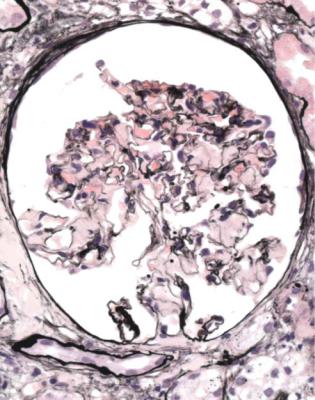
116 |
6 Indication for Biopsy: Nephrotic Syndrome |
Fig. 6.64 Glomerulus in a renal biopsy specimen from a man of 71 with the nephrotic syndrome. The mesangium contains material that is not stained by periodic acid-methenamine silver. The diagnosis was shown to be amyloid of AL type. After the biopsy, a paraprotein was found in serum and urine
Differential Diagnosis of Amyloid
Diagnosis of amyloid is usually straightforward. Provided the staining with Congo red is reliable, there should be little difficulty.
An uncommon group of disorders that can resemble amyloid is differentiated by a lack of reactivity with Congo red. These disorders include fibrillary glomerulopathy and immunotactoid glomerulopathy, although these names are used inconsistently, and one name or the other, or fibrillary-immunotactoid glomerulopathy, may be sometimes applied to conditions with deposition of extracellular microfibrils or microtubules, without reactivity with Congo red, and in the absence of cryoglobulins, systemic lupus erythematosus, or diabetes mellitus.
These disorders can have various appearances on orthodox light microscopy, but usually there is mesangial expansion which resembles amyloid, but does not
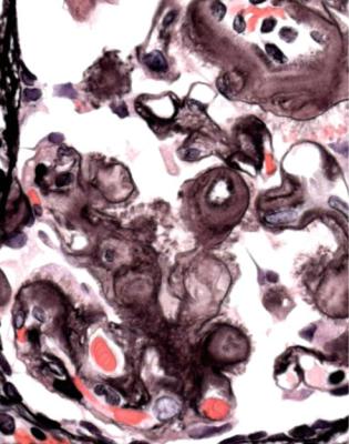
Differential Diagnosis of Amyloid |
117 |
Fig. 6.65 Glomerulus in a renal biopsy specimen from a man of 66 with the nephrotic syndrome, and a paraprotein in serum and urine. The mesangium contains material stained by periodic acidmethenamine silver, with groups of coarse bristles projecting from basement membranes in places. A further investigation is shown in Fig. 6.74
stain with Congo red (Fig. 6.77). Immunohistologic study shows IgG and complement in mesangium, and sometimes elsewhere. The deposits are usually polyclonal, containing both kappa and lambda light chains, but monoclonal deposits may be found, particularly in the immunotactoid type. Electron microscopy shows filamentous or microtubular deposits in mesangium and glomerular basement membranes. The appearance and size of the deposits is used to differentiate fibrillary glomerulopathy from immunotactoid glomerulopathy, in which there is the appearance of microtubular structures (Fig. 6.78). Fibrils in fibrillary glomerulopathy are about 10–30 nm thick, and microtubules in immunotactoid glomerulopathy are about 20–50 nm thick. In comparison, fibrils in amyloid are about 8–12 nm thick.
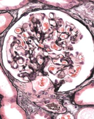
118 |
6 Indication for Biopsy: Nephrotic Syndrome |
Fig. 6.66 Glomerulus in a renal biopsy specimen from a man of 65 with the nephrotic syndrome and acute renal failure. This is close to normal on periodic acid-methenamine silver staining. Congo red staining is shown in Fig. 6.67
Have Minimal Change Nephropathy, Membranous Nephropathy, Segmental Sclerosing Disorders, Diabetic Glomerulopathy, Lupus Nephritis, Amyloid, and Conditions that Could be Confused with Them Been Excluded?
Particularly in developing countries there are two other glomerular disorders that can commonly produce the nephrotic syndrome.
Both give the appearance on low power microscopy of glomerular hypercellularity, which affects the whole of each glomerulus uniformly. These global and diffuse disorders are acute postinfective glomerulonephritis, and subendothelial membranoproliferative or mesangiocapillary glomerulonephritis, which is also sometimes called membranoproliferative or mesangiocapillary glomerulonephritis type one.
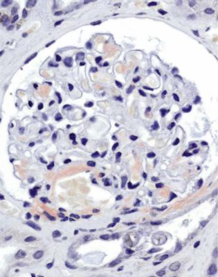
Differential Diagnosis of Amyloid |
119 |
Fig. 6.67 Glomerulus in the renal biopsy specimen from the man of 65 illustrated in Fig. 6.66. Congo red staining shows deposits of amyloid in mesangium, and also in an arteriolar wall and adjacent Bowman’s capsule. Further investigations are shown in Figs 6.75 and 6.76
Acute postinfective glomerulonephritis typically presents several days after an infection as acute nephritis, with edema, proteinuria, hematuria, hypertension, and acute renal failure. Serum complement component C3 has a reduced concentration. The commonest cause is infection of the pharynx or skin by Streptococcus pyogenes. This is why the term acute poststreptococcal glomerulonephritis is sometimes used, although infection with other microorganisms can precede the disorder. These include Staphylococcus aureus, Gram-negative bacilli, and, less commonly, virtually every other pathogenic organism. The presentation may be atypical, and the nephrotic syndrome may predominate.
Acute postinfective glomerulonephritis is probably not due to deposition of circulating immune complexes, but to an antibody response to antigens deposited in glomeruli early in the course of an infection. Most people with this disorder recover to normal.
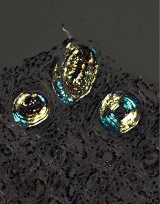
120 |
6 Indication for Biopsy: Nephrotic Syndrome |
Fig. 6.68 Tubules in a renal biopsy specimen from a woman of 65 with myeloma kidney, or light chain cast nephropathy, stained by Congo red, and examined between crossed polariser and analyser. There is a Maltese cross appearance produced by birefringence, although the cast material is not amyloid. There are anomalous colours, blue–green and yellow
Subendothelial membranoproliferative glomerulonephritis may also follow an infection, or more usually occurs during the course of a persistent infection, such as infection with hepatitis B or C viruses, malaria, schistosomiasis, syphilis, infective endocarditis, longstanding abscesses, or infected shunts draining cerebral ventricles. This can also be seen associated with sickle cell disease, various neoplasms such as chronic lymphocytic leukemia, and several other conditions. There is usually a mixture of clinical renal abnormalities.
Subendothelial membranoproliferative glomerulonephritis is probably due to deposition of immune complexes in glomeruli, either because there is prolonged heavy generation of immune complexes, or because immune complexes cannot be cleared when there are deficiencies of complement components. Various complement components, particularly C3, may have a reduced concentration in serum. Remission is unlikely. Generally, there is gradual progression to renal failure.
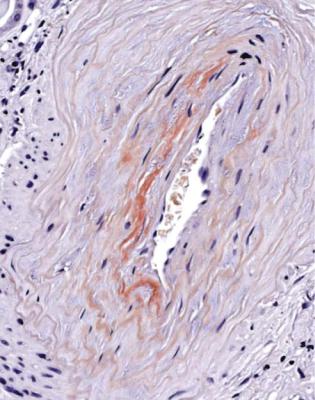
Acute Postinfective Glomerulonephritis |
121 |
Fig. 6.69 Artery in a renal biopsy specimen from a man of 85 with proteinuria, hematuria, and acute renal failure, stained by Congo red, which shows amyloid. Fig. 6.70 shows its appearance between crossed polariser and analyser
Differentiation Between Acute Postinfective Glomerulonephritis
and Subendothelial Membranoproliferative Glomerulonephritis
Both disorders give global and diffuse glomerular hypercellularity, and both may have an infiltrate of neutrophils in the tuft at early stages. One of the features that helps the pathologist to distinguish between them is the appearance of glomerular basement membranes on sections stained by periodic acid-methenamine silver. In acute postinfective glomerulonephritis, these appear single, but in subendothelial membranoproliferative glomerulonephritis, these appear double, with interposition of mesangial cells between the two membranes (Figs 6.20, 6.79, and 6.80). The glomerular tuft in subendothelial membranoproliferative glomerulonephritis may appear in distinct lobules, and there may be mesangial nodules (Fig. 6.50).
On immunohistologic study, acute postinfective glomerulonephritis has irregular coarse granules, particularly of complement and sometimes of IgG, mainly on the outside of glomerular capillary loops, unlike the uniform fine granules
122 |
6 Indication for Biopsy: Nephrotic Syndrome |
Fig. 6.70 The microscopic field in Fig. 6.69, stained by Congo red, between crossed polariser and analyser, showing an anomalous green colour. Pure green is not always seen under these conditions, despite a widespread belief that it is
seen in membranous nephropathy (Fig. 6.81). Subendothelial membranoproliferative glomerulonephritis has large deposits, particularly of complement but often of IgG and IgM as well, in various proportions on the inner aspect of capillary loops, and sometimes in mesangium (Fig. 6.82). Electron microscopy confirms the distribution of immune deposits, and shows whether glomerular basement membranes are single or double, in which case, there is interposition of mesangial cells (Figs 6.83 and 6.84).
Other Findings in the Nephrotic Syndrome
Other glomerular disorders may present with the nephrotic syndrome, but these are unusual. In particular, IgA nephropathy, which is often the commonest single finding in any series of renal biopsy specimens, hardly ever presents in this way. The finding of mesangial IgA deposits, but no other abnormality on light microscopy, in a child with the nephrotic syndrome, suggests a diagnosis of coincidental minimal change nephropathy and IgA nephropathy. Henoch–Schönlein nephritis, which
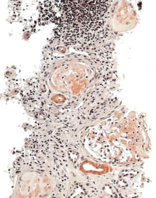
Other Findings in the Nephrotic Syndrome |
123 |
Fig. 6.71 Cortex in a renal biopsy specimen from a man of 68 with the nephrotic syndrome, chronic renal failure, a paraprotein in serum, and paraplegia, after a road traffic accident 12 years previously. Congo red staining shows amyloid in glomeruli and walls of blood vessels. Fig. 6.72 shows the appearance between crossed polariser and analyser
is the combination of IgA nephropathy and vasculitic glomerulonephritis, usually presents with acute renal failure, but may be associated with proteinuria in the nephrotic range at the time of presentation.
The nephrotic syndrome in young children may have findings not seen at other ages. The term congenital and infantile nephrotic syndrome covers several conditions associated with the nephrotic syndrome in young children. Congenital strictly means present at birth, but congenital nephrotic syndrome is used for onset of the syndrome between birth and 3 months. Onset after 3 months and up to 1 year is called infantile nephrotic syndrome. The commoner of these is congenital. By far, the commonest finding is the Finnish type of congenital nephrotic syndrome, an autosomal recessive condition in which the protein nephrin is abnormal in glomerular epithelial cells. Diffuse mesangial sclerosis is the next most frequent, and may be associated with mutation of the Wilms’ tumor gene, WT1. Specimens are mostly seen in specialised centres, and are often difficult to interpret, particularly if the biopsy is within a few months of birth. The diagnosis is reached by a combination of investigations, which may include genetic studies.
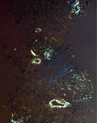
124 |
6 Indication for Biopsy: Nephrotic Syndrome |
Fig. 6.72 The microscopic field in Fig. 6.71, stained by Congo red, between crossed polariser and analyser, showing anomalous colours, blue–green and yellow. A further investigation is shown in
Fig. 6.73
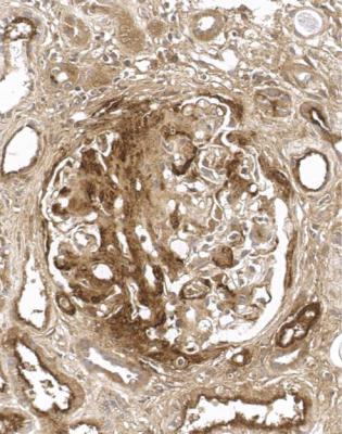
Other Findings in the Nephrotic Syndrome |
125 |
Fig. 6.73 Glomerulus in the renal biopsy specimen from the man of 68 that is illustrated in Figs 6.71 and 6.72. Immunoperoxidase study to detect amyloid A protein shows heavy deposition in the glomerulus. The staining in tubular epithelium is seen in every kidney, and is ignored. Despite the paraprotein, the diagnosis was amyloid of AA type. This was attributed to a persistent inflammatory response to bed sores of long duration
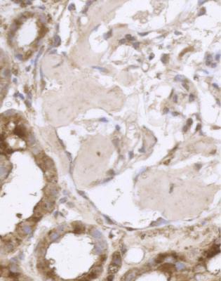
126 |
6 Indication for Biopsy: Nephrotic Syndrome |
Fig. 6.74 Glomerulus in the renal biopsy specimen from the man of 66 with amyloid, illustrated in Fig. 6.65. Immunoperoxidase study to detect amyloid A protein shows no deposition in the glomerulus. Although there was no definite deposition of kappa or lambda light chains in glomeruli, the diagnosis was non-AA amyloid, consistent with AL type
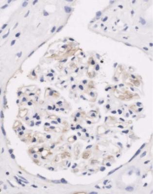
Other Findings in the Nephrotic Syndrome |
127 |
Fig. 6.75 Glomerulus in the renal biopsy specimen from the man of 65 with amyloid, which is illustrated in Figs 6.66 and 6.67, stained by an immunoperoxidase method using an antibody to kappa light chains. This shows no significant deposition. Fig. 6.76 shows the comparable immunostaining for lambda light chains
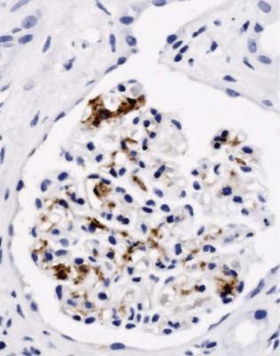
128 |
6 Indication for Biopsy: Nephrotic Syndrome |
Fig. 6.76 Another section of the same glomerulus as Fig. 6.75, immunostained for lambda light chains. There is deposition in the mesangium, which indicates AL amyloid. After the biopsy, an IgG lambda paraprotein and monoclonal lambda light chains were found in the man’s serum
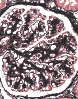
Other Findings in the Nephrotic Syndrome |
129 |
Fig. 6.77 Glomerulus in a renal biopsy specimen from a man of 36 with heavy proteinuria and episodes of hematuria. There is mesangial expansion, but no reactivity with Congo red. Electron microscopy shows fibrillary deposits in mesangium, with the appearance of fibrillary glomerulopathy
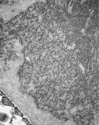
130 |
6 Indication for Biopsy: Nephrotic Syndrome |
Fig. 6.78 Electron micrograph of part of a glomerulus in a renal biopsy specimen from a man of 37, with HIV infection and the nephrotic syndrome. There are microtubular structures in the mesangium, indicating immunotactoid glomerulopathy
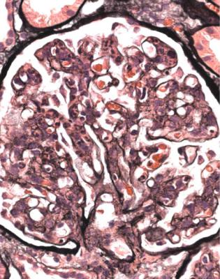
Other Findings in the Nephrotic Syndrome |
131 |
Fig. 6.79 Glomerulus in a renal biopsy specimen from a man of 31 with the nephrotic syndrome and acute renal failure, after an illness with fever. The glomerulus appears hypercellular, with swollen endocapillary cells and a heavy infiltrate of neutrophils. Basement membranes are normal. The diagnosis is acute postinfective glomerulonephritis. The man recovered to normal
132 |
6 Indication for Biopsy: Nephrotic Syndrome |
Fig. 6.80 Glomerulus in a renal biopsy specimen from a man of 45 with the nephrotic syndrome, hematuria, and chronic renal failure. There is mesangial expansion, and most basement membranes appear double on the section stained by periodic acid-methenamine silver. The diagnosis is subendothelial membranoproliferative glomerulonephritis
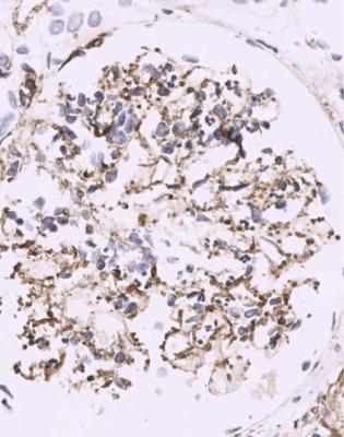
Other Findings in the Nephrotic Syndrome |
133 |
Fig. 6.81 Glomerulus in a section stained by an immunoperoxidase method to detect C9, in a renal biopsy specimen from a man of 67 with acute postinfective glomerulonephritis. There are coarse granules of complement scattered irregularly on the outside of capillary loops, with finer deposits in mesangium
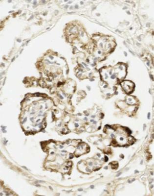
134 |
6 Indication for Biopsy: Nephrotic Syndrome |
Fig. 6.82 Glomerulus in a renal biopsy specimen from a woman of 64 with the nephrotic syndrome and chronic lymphocytic leukemia, stained by an immunoperoxidase method to detect IgG. There are large areas of subendothelial deposition, which confirms subendothelial membranoproliferative glomerulonephritis
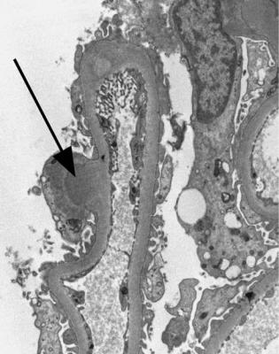
Other Findings in the Nephrotic Syndrome |
135 |
Fig. 6.83 Electron micrograph of a glomerulus in a renal biopsy specimen from a man of 38 with acute postinfective glomerulonephritis, showing a large subepithelial deposit (arrowed)
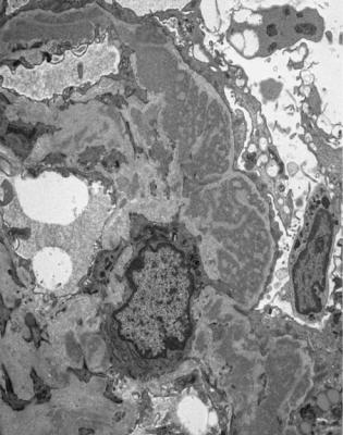
136 |
6 Indication for Biopsy: Nephrotic Syndrome |
Fig. 6.84 Electron micrograph of a glomerulus in a renal biopsy specimen from a man of 44 with subendothelial membranoproliferative glomerulonephritis, showing many large subendothelial deposits
Summary: Nephrotic Syndrome
The nephrotic syndrome always indicates a glomerular disorder.
Extent of tubular damage should be assessed in the nephrotic syndrome. Severe acute tubular damage may be a sign of renal vein thrombosis.
In adults with the nephrotic syndrome, membranous nephropathy, segmental sclerosing disorders, and minimal change nephropathy are usually the commonest findings.
Diabetic glomerulopathy, lupus nephritis, and amyloid often account for nearly all other biopsy specimens in adults with the nephrotic syndrome. Acute postinfective glomerulonephritis and subendothelial membranoproliferative glomerulonephritis may be found, especially in developing countries.
In those children with the nephrotic syndrome who have a biopsy, most have either minimal change nephropathy or a segmental sclerosing disorder.
Further Reading: Nephrotic Syndrome |
137 |
Further Reading: Nephrotic Syndrome
D’Agati VD, Fogo AB, Bruijn JA, Jennette JC. Pathologic classification of focal segmental glomerulosclerosis: a working proposal. American Journal of Kidney Diseases 2004; 43: 368– 382. This gives the Columbia classification of segmental sclerosing glomerular disorders.
D’Agati VD, Jennette JC, Silva FG. Non-Neoplastic Kidney Diseases. Atlas of Nontumor Pathology, first series, fascicle 4. Washington, DC:American Registry of Pathology and Armed Forces Institute of Pathology, 2005. Chapters 3, 5–7, 9–11, 13, 17.
Howie AJ, Pankhurst T, Sarioglu S, Turhan N, Adu D. Evolution of nephrotic-associated focal segmental glomerulosclerosis and relation to the glomerular tip lesion. Kidney International 2005; 67: 987–1001.
Jennette JC, Olson JL, Schwartz MM, Silva FG. Heptinstall’s Pathology of the Kidney. Sixth ed. Philadelphia:Lippincott Williams and Wilkins, 2007. Chapters 4–9, 12, 18, 19.