

Chapter 1
Introduction
Aims of the Book
This book is meant to be a practical guide to interpretation of renal biopsy specimens. Two things follow.
First, the book has much less text than most others on renal pathology. Only the most essential points about pathogenesis are included. There is little on other matters not directly relevant to interpretation. Diseases are not mentioned that are not investigated by biopsy, such as many developmental and urologic disorders of the kidney. Rare conditions are generally mentioned briefly, if at all. Large and comprehensive texts can be consulted where necessary.
The second thing is that this book has a different approach from standard texts in pathology. These consider each disease separately as identified, known, defined entities. This is excellent if a pathologist knows what to call the disease in a particular specimen, and wishes to confirm the diagnosis or find out more about it, but is little use if the pathologist has no idea about the diagnosis. Most pathologists have experience of the search through many descriptions of diseases, to try to find the name that seems the most appropriate for the abnormalities in a specimen. This book should be seen as a guide into the standard texts, which may be used as references for further information, once a diagnosis has been made.
This book tries to help the pathologist by the use of two rules, common things are common, and people with diseases present in characteristic clinical ways. The book is arranged by clinical presentation rather than by diseases, and concentrates on common conditions. Although there are hundreds of disorders that may affect the kidney, few are common, and these usually have typical clinical features. There is deliberate repetition, so that a pathologist should be able to give a diagnosis even if the clinical presentation is unusual or, as is more common, the clinical information is misleading.
The importance is stressed of how much the pathologist relies on the information supplied on request forms, and how much the interpretation depends upon the clinical circumstances. An apparently normal kidney in a biopsy specimen should be given a different diagnosis if there is microscopic hematuria than if there is the nephrotic syndrome. The pathologist can give most help to nephrologists and renal transplant surgeons when the clinical information provided is full and accurate.
A. J. Howie, Handbook of Renal Biopsy Pathology. |
1 |
C Springer 2008 |
|
2 |
1 Introduction |
Introduction to Renal Disease from a Pathologist’s Point of View
The kidney is mysterious and frightening to many medical students and junior physicians. The organ cannot normally be felt or seen, the anatomy and physiology seem difficult and complicated, clinical features of renal disorders are indirect and not easily investigated, and there seems a secret language used by renal pathologists. Evidence that renal diagnoses are thought unnecessarily elaborate is that one core curriculum for medical students recommended that the pathologic classification of glomerular disorders should not be included. This book ignores classification as far as possible, and concentrates on correlation between clinical features and pathologic findings.
The kidney is essential for life, although not so immediately essential as the heart, lungs, or brain. Some types of interference with the structure or function of those organs can cause death within seconds or minutes, such as a dysrhythmia, strangulation, or an intracerebral hemorrhage. Removal of two normal kidneys would lead to death, not immediately but in a few days, from effects of accumulation of sodium and water, causing pulmonary edema and interference with gas exchange in the lungs, and effects of abnormal concentrations of solutes in body fluids, particularly potassium and hydrogen ions, causing dysrhythmias.
This shows that the main function of the kidney is to control the amount and electrolyte composition of body fluids.
Blood is filtered by glomeruli. The filtrate is altered in tubules by appropriate reabsorption and secretion of substances. Various chemical tests can be made to see how effective the kidney is at this function, which is called renal excretory function, or simply renal function. These tests are direct or indirect measures of the glomerular filtration rate, which is the volume of fluid filtered through glomeruli in a unit of time. If the rate is reduced, there is said to be renal impairment or renal failure.
If the kidney fails, this function can be partly replaced by either an artificial semipermeable membrane in hemodialysis, or the peritoneal membrane in peritoneal dialysis, with enough function to support life. Transplantation of a kidney is the best way that this function and other functions of the kidney can be restored.
Part of the normal function of the kidney is to filter the blood, but to prevent leakage into the filtrate of plasma proteins, especially albumin, the plasma protein in the highest concentration, and of blood cells, especially red cells, the most numerous blood cell. Disorders of the kidney, particularly of glomeruli, can cause appearance of proteins or red cells in the urine. These are called proteinuria and hematuria. These can occur together or independently, may be caused by disorders outside the kidney, such as elsewhere in the urinary tract, and even if caused by a kidney disorder, may not be associated with renal failure.
At the simplest level, pathologists will see renal biopsy specimens taken as part of the investigation of at least one of these abnormalities, renal failure, proteinuria, and hematuria. Biopsy of a renal allograft is usually because it is not working properly, which in equivalent to renal failure.
Further Reading: Introduction |
3 |
Among other functions of the kidney are the control of blood pressure, and production of hormones such as erythropoietin, but renal biopsy is hardly ever done to investigate disorders of these functions in the absence of renal failure or proteinuria or hematuria.
The Value of a Renal Biopsy
A renal biopsy is sometimes seen as a dangerous and unnecessary investigation that gives no help in clinical management. An aim of this book is to emphasise the usefulness of information that a pathologist can give on a specimen. There are two particularly important pieces of information.
1.The diagnosis. The disease in a kidney, more than anything else, determines the likely clinical course. The diagnosis, which means the name put on the disease, is the most important guide to further investigations, treatment, and management. For some diseases, the diagnosis includes an assessment of how severe and active are the changes in the kidney.
2.The amount of chronic damage in the kidney. This gives a strong indication of the length of time that a kidney is likely to survive.
Summary: Introduction
This book is meant to help pathologists to give a clinically useful interpretation of renal biopsy specimens, and to help others to understand pathologists’ reports on these specimens.
Clinically, the term renal function refers to the excretory function of the kidney. Abnormalities of the kidney may give impairment of renal function, or leak of blood or proteins into the urine.
The two main factors that determine the clinical outcome are the disease in a kidney, and the amount of chronic damage at diagnosis.
Further Reading: Introduction
Adu D, Tse WY, Howie AJ. Clinical investigation. In: Shena FP, editor-in-chief. Clinical Medicine Series: Nephrology. London: McGraw-Hill, 2001: 17–32.
Lote CJ. Principles of renal physiology. Fourth edition. Dordrecht: Kluwer Academic, 2000.
Chapter 2
General Points About Renal Biopsy Specimens
Introduction to General Points
A renal biopsy specimen is the best example of a specimen that a pathologist cannot interpret without clinical information.
From information about age and sex of the person biopsied, reason for biopsy, and other clinical features, the pathologist should have an idea of likely appearances in the specimen, before any sections are seen on a microscope.
This is because there are two general rules.
1.Common things are common.
2.Diseases present in characteristic ways.
Strictly speaking, the second rule should state that people with diseases present in characteristic ways, because diseases do not exist on their own, but in medicine, conventionally, diseases are considered to be independent entities. For simplicity, this convention will be followed.
Common Things Are Common and Diseases Present
in Characteristic Ways
This work approaches interpretation of renal biopsy specimens with these rules. They are more helpful in real life than the standard textbook approach, which is to describe diseases, rather than to suggest how a pathologist can give a name to something that is unknown before the specimen is examined. Lists of diseases often give each one equal prominence whether they are rare or common, which is little use in practice.
Things to Remember About the General Rules
Diseases come and go. The findings given here will become out of date. New diseases will appear.
A. J. Howie, Handbook of Renal Biopsy Pathology. |
5 |
C Springer 2008 |
|
6 |
2 General Points About Renal Biopsy Specimens |
Diseases have different prevalences between the sexes, at different ages, and in different parts of the world.
There may be more than one disease in a kidney. In children there is usually only one, but in adults there is often more than one, if only because many have ischemic damage associated with age or hypertension, as well as something else.
There are rare conditions, but if a specimen appears out of the ordinary, the pathologist should think of an unusual form of a common disorder, or a combination of common disorders, before something rare.
A renal biopsy specimen is a tiny proportion of a kidney, and may not be representative of it. A needle biopsy specimen weighs under 100 mg, a kidney in an average adult weighs about 150 g, and so a biopsy specimen is much less than one thousandth part of a kidney by weight. A specimen may contain about twenty glomeruli, out of the several hundreds of thousands in a typical kidney. Even so, renal biopsy is helpful in most conditions, and indeed there are several in which only one glomerulus is needed to make the diagnosis.
Importance of the Request Form to the Pathologist
The pathologist relies on information written on the request form, which may be wrong, or incomplete, or give a misleading emphasis, especially if written by an inexperienced physician. Ideally, the pathologist should be able to discuss clinical features of every person biopsied with a physician who understands what is going on, but this is not always possible.
Indications for Renal Biopsy
There are only a few indications for renal biopsy, meaning the reasons why a biopsy is taken.
One indication is presence of protein in the urine, called proteinuria, which may be asymptomatic, and detected by chance or a deliberate search, or may be associated with edema and other features that are collectively called the nephrotic syndrome. Another is presence of blood in the urine, called hematuria. Others are states of reduced renal excretory function, called acute renal failure or acute renal impairment, and chronic renal failure or chronic renal impairment. Most biopsies of renal allografts are to investigate acute or chronic impairment of graft excretory function.
There are often combinations of these features. The terms nephritic syndrome and acute nephritis are sometimes applied to an acute illness with edema, hematuria, proteinuria, hypertension, and acute renal failure. Because the features of the nephritic syndrome may be incomplete or atypical and one feature may dominate, and the term is sometimes used in a loose and ambiguous way, there is not a separate
Indications for Renal Biopsy |
7 |
section on it, and the disorders that can present in this way are covered in the sections on the nephrotic syndrome, acute renal failure, and hematuria.
In someone who has had a renal biopsy, the procedure may be repeated to check on the progress of the disease, or to look for effects of treatment, or to see if another condition has developed. Repeat specimens can be considered in the same ways as initial specimens, with the advantage that there is previous material for comparison.
To simplify the approach to interpretation of renal biopsy specimens, the indications are considered in a way that allows for all possible combinations. If there is the nephrotic syndrome, irrespective of other features, that is regarded as the main indication. If there is acute renal failure, irrespective of features apart from the nephrotic syndrome, acute renal failure is regarded as the main indication. If there is chronic renal failure, without the nephrotic syndrome or acute renal failure, and irrespective of proteinuria or hematuria, chronic renal failure is regarded as the main indication. If there is hematuria with or without proteinuria, but with normal renal function, hematuria is regarded as the main indication. The last indication is asymptomatic proteinuria on its own.
To summarise, the indications for biopsy are considered in this order, and the first one that applies to a specimen is regarded as the main one:
first, nephrotic syndrome second, acute renal failure third, chronic renal failure fourth, hematuria
fifth, asymptomatic proteinuria
The scheme followed in this work is not rigid, and should allow a diagnosis to be made on specimens with misleading indications written on the request form, such as one with hematuria and proteinuria in which there is chronic renal failure, although it is not noted on the request form, or one with renal failure that is considered acute clinically but actually is chronic, or a specimen on which the pathologist is not clear which is the dominant indication.
These indications may be called medical reasons for a renal biopsy. Biopsy of a mass in or near the kidney is uncommon, and may be called a surgical or urologic reason for a renal biopsy.
There are other clinical features that are not by themselves indications for a renal biopsy, although they may be present with features that are indications. These include hypertension, loin pain or other abdominal pain, and anemia. Suspected or definite urinary tract infection is usually considered a contraindication to biopsy.
A pathologist may see renal biopsy specimens taken for unusual or accidental reasons. Kidney may be seen in attempted biopsy of other structures, particularly of liver and masses in the region of the kidney. The specimen should be examined as if it were an intentional biopsy of the kidney, because abnormalities may be found (Fig. 2.1). Occasionally, there are none of the common indications, but a biopsy is taken to investigate the possibility of subclinical disease in the kidney, such as vasculitis, or there is a familial renal problem, or a kidney is assessed as a possible
8 |
2 General Points About Renal Biopsy Specimens |
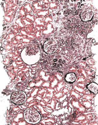
Fig. 2.1 Attempted biopsy of a liver allograft in a woman of 45, at annual review. This is a specimen not of liver, but of renal cortex. There is chronic damage, particularly seen as tubular atrophy. One potential explanation is effects of a calcineurin inhibitor, used as an immunosuppressant. IgA nephropathy is another common finding when attempted liver biopsy specimens include kidney. Renal comes from the Latin for kidney, nephrocomes from Greek for kidney, and the word kidney is of obscure origin. Biopsy comes from Greek words meaning life and vision. In Latin, cortex meant bark of a tree. Tubule is derived from the Latin for a little pipe or tube
graft, or there is a known or suspected metabolic or tubular disorder, especially in children. During surgical exploration of a graft for a vascular or ureteric problem, a biopsy may be done simply because the surgeon is there.
Different nephrologists and renal transplant surgeons have different indications for biopsy. Some may not biopsy people with hematuria alone, or asymptomatic proteinuria, or chronic renal failure. Not all children with the nephrotic syndrome have a renal biopsy, and in some places this is also true of adults. In some transplant units, allografts are biopsied at set times irrespective of function. Different renal biopsy practices will give different frequencies of findings, but the findings given in this work are likely to be comparable with those in many centres. Findings in some parts of the world, such as in developing or low income countries, may be markedly different.
Summary: General Points About Renal Biopsy Specimens |
9 |
When allografts are excluded, the frequencies of indications for biopsy are different at different ages, and in different parts of the world. In some countries in adults, chronic renal failure, acute renal failure, and nephrotic syndrome each account for nearly a third of specimens, in descending order of commonness, with fewer taken for hematuria, and even fewer for asymptomatic proteinuria. In some countries in children, nearly half are taken for hematuria, and the order of indications for the rest is nephrotic syndrome, acute renal failure, asymptomatic proteinuria, and chronic renal failure. In some parts of the world, almost the only indication for renal biopsy is the nephrotic syndrome.
A satisfactory specimen for a pathologist is one with definite features, on which a clear cut diagnosis can be made. When allografts are excluded, the most satisfying indications from the pathologist’s point of view are nephrotic syndrome and acute renal failure, because an adequate diagnosis should almost always be possible. The next are hematuria and chronic renal failure. Proteinuria is the indication most likely to frustrate the pathologist’s intention to give an unequivocal diagnosis.
Complications of a Renal Biopsy
The commonest serious complication of a biopsy is macroscopic hematuria, reported to follow 5–10% of procedures in most series. Hemorrhage into retroperitoneal tissues around the kidney is common, but is clinically evident after about 1% of biopsies. Either form of hemorrhage may require blood transfusion, or less commonly radiologic embolisation of material into renal arteries to block them, or even less commonly nephrectomy if there is otherwise uncontrollable bleeding. A puzzling observation is that the pathologist is hardly ever able to predict when there will be clinically significant bleeding after a renal biopsy. Large blood vessels may be seen in specimens without clinically obvious bleeding afterwards, while there may be no large vessels in specimens complicated by hemorrhage. Other complications include development of arteriovenous fistulas and consequences of puncture of intra-abdominal organs.
Summary: General Points About Renal Biopsy Specimens
The two general rules are that common things are common, and diseases present in characteristic ways.
The main indications for a renal biopsy are the nephrotic syndrome, acute renal failure, chronic renal failure, hematuria, and asymptomatic proteinuria. Combinations of these occur.
Biopsy of a renal allograft is usually to investigate acute or chronic graft failure. Renal biopsy may also be done to investigate a mass in the kidney.