

Chapter 7
Indication for Biopsy: Acute Renal Failure
Introduction to Acute Renal Failure
This means that so far as can be determined clinically, renal excretory function was normal until the last few days or weeks, when renal function was noted to be abnormal. This acute reduction of glomerular filtration rate may also be called acute renal impairment, or acute kidney injury.
In most people with acute renal failure, the symptoms and signs are those of the underlying condition that explains the renal dysfunction, rather than specifically of the dysfunction, which may just give the symptom of not feeling completely well. Renal failure is assessed by investigations such as serum creatinine concentration, which is an indirect measure of the glomerular filtration rate. This means that renal failure, although it may be suspected clinically, is a condition that is diagnosed by clinical chemistry.
Renal function is a continuous variable, and there are arbitrary divisions between normal function and abnormal function, and between mild renal impairment and severe renal impairment that few would disagree should be called frank acute renal failure. The pathologist should not be concerned with these problems, but should approach any renal biopsy specimen in the way suggested here, if the request form gives the information of acute renal impairment, or acute renal failure, or similar terms, such as acute rise in serum creatinine concentration.
Few people are known to have had definitely normal renal function before they are found to have abnormal function. Nephrologists rely on clues from the clinical history, and other features such as size of the kidneys, which should be normal in acute renal failure, to decide whether this dysfunction is acute or chronic, but occasionally they may be in doubt. A sudden deterioration of renal function can occur in people with pre-existing, stable, longstanding renal impairment, as well as in those with previously normal renal function. This shows that there is no simple definition of acute renal failure.
If the request form mentions renal failure but not whether this is acute or chronic, or hints that the failure could be acute or chronic, the pathologist should approach interpretation with the assumption that the failure is acute. Assessment of the amount of chronic, irreversible damage in the biopsy specimen should immediately
A. J. Howie, Handbook of Renal Biopsy Pathology. |
139 |
C Springer 2008 |
|
140 |
7 Indication for Biopsy: Acute Renal Failure |
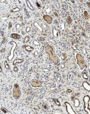
clarify whether there is longstanding renal failure, rather than acute dysfunction, or whether there is a mixture of both (Figs 4.4 and 7.1).
In acute renal failure, the person biopsied may have small urine output, called oliguria, or no urine output, called anuria, and may be on dialysis. There may be hematuria, proteinuria that may be heavy enough to give the nephrotic syndrome, and hypertension that clinically may be in the accelerated phase. If the nephrotic syndrome is present, the biopsy specimen should be assessed in the way suggested in Chapter 6.
Renal biopsy specimens are always abnormal in acute renal failure, and the pathologist should almost always be able to give a satisfactory diagnosis.
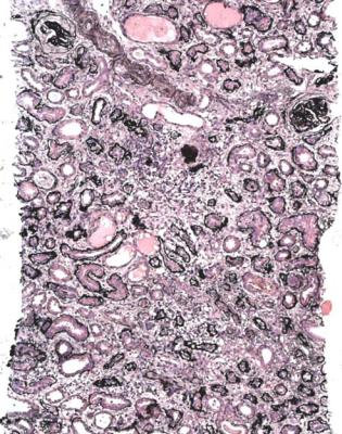
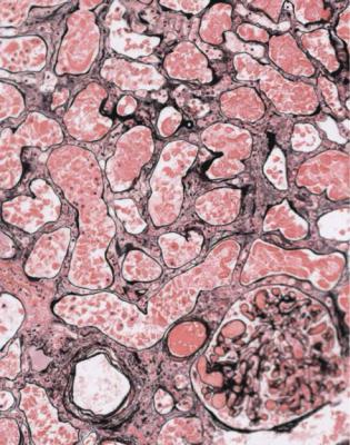
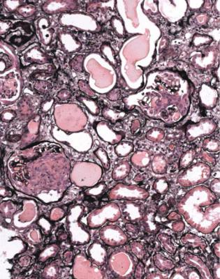
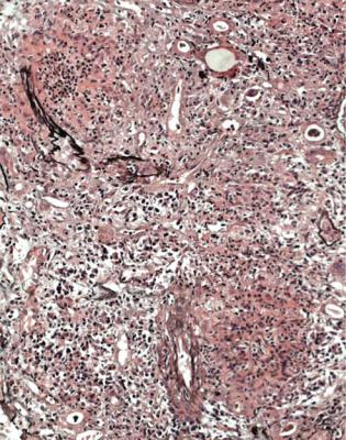
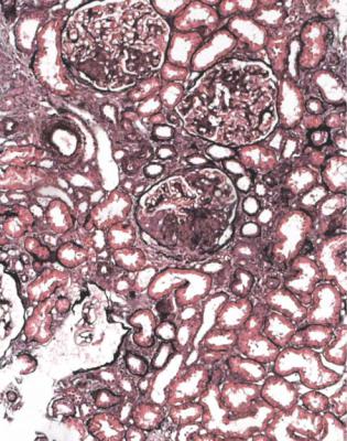
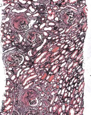
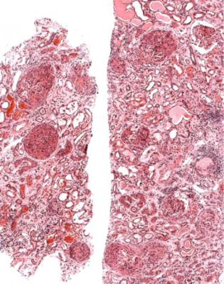
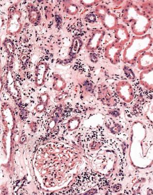
Fig. 7.1 Cortex in a renal biopsy specimen from a man of 30 with severe hypertension, two kidneys of normal size, and renal failure that clinically appeared acute. There is advanced early tubular atrophy, and much of the damage appears chronic and irreversible, rather than acute and potentially reversible. Glomeruli have evidence of IgA nephropathy, and there is a small vessel vasculopathy, also called thrombotic microangiopathy, consistent with effects of accelerated hypertension. Renal function did not recover
Value of a Renal Biopsy in Acute Renal Failure |
141 |
Most people with acute renal failure do not have a renal biopsy. This is usually because the cause seems adequately explained by findings in the clinical history, clinical examination, and investigations.
Causes of Acute Renal Failure
Traditionally, causes of acute renal failure are divided into prerenal, renal or intrarenal, and postrenal. Prerenal means reduced blood flow to the kidney, renal means direct damage to the kidney, and postrenal means obstruction of the urinary tract. Reality is more complicated than this. For instance, reduced blood flow and obstruction of the urinary tract will lead to damage to the kidney.
Causes are different between the sexes, at different ages, and in different parts of the world. Neonates may have acute renal failure from effects of dehydration, hypotension from congenital heart disease, and other conditions. Developmental disorders of the urinary tract may give renal failure, but this is more likely chronic than acute, and in severe disorders the child is stillborn. Children are most likely to have acute renal failure from diarrhea-associated hemolytic uremic syndrome, but there are many other causes, including many of those that are found in adults. Adults can have acute renal failure from many causes, including hypotension from blood loss, dehydration, septic shock or left ventricular dysfunction, toxic damage for instance from gentamicin, and urinary tract obstruction, for example from prostatic enlargement in old men, although there is likely to be chronic renal damage in people who present with urinary tract obstruction. Contrast media injected intravascularly for radiologic investigations can induce acute renal failure, but there is usually pre-existing chronic renal damage.
In developing countries, acute renal failure may be a complication of infective diarrheal illnesses, obstetric problems, intravascular hemolysis from glucose-6- phosphate dehydrogenase deficiency, hemolytic and other complications of malaria, and many other disorders.
Value of a Renal Biopsy in Acute Renal Failure
Renal biopsy is used to investigate acute renal failure when the explanation is not considered adequately explained by clinical findings. The biopsy specimen gives information that will help nephrologists in clinical management. A diagnosis may be given that requires further clinical investigation, such as myeloma. The most important help is in the decision whether to begin aggressive treatment of an underlying disease, such as vasculitis. Acute renal failure may recover, provided the changes in the kidney are reversible, and the underlying cause is treatable and treated.
Renal biopsy in acute renal failure should be considered an urgent matter. This is one of the few circumstances when the pathologist can have an immediate
142 |
7 Indication for Biopsy: Acute Renal Failure |
influence on clinical management. For instance, if renal vasculitis is unrecognised and untreated, survival rates are worse than for almost all cancers.
A General Rule: Renal function is more closely correlated with structural changes in tubules than with any other changes. Acute renal failure is associated with acute abnormalities in tubules.
One reason for this is a consequence of the fact that the kidney, particularly the cortex, has a large blood flow that is necessary for its function. The kidney mass is about half a hundredth of the body mass, about 300 g in a person weighing 60 kg, and yet the renal blood flow is almost one-quarter of the cardiac output.
Renal blood flow can be reduced severely in the short term to maintain the blood supply to the heart, lungs, and brain, in severe hemorrhage, for example. This necessarily reduces the glomerular filtration rate, and also has an effect on tubules. These have a high metabolic rate, and are sensitive to a reduction in blood flow, which is ischemia. If the blood volume and renal blood supply are restored quickly, there may be no tubular damage, and renal function can return to normal. If there has been ischemic tubular damage, there are mechanisms that prevent effective glomerular filtration until the tubules recover. These mechanisms also explain the acute renal failure that follows an initial toxic effect on tubules, for instance from gentamicin and other aminoglycoside antibiotics, some antineoplastic drugs such as cisplatin, some types of immunoglobulin light chains, and accidental or deliberate poisoning with many chemical agents, such as carbon tetrachloride. There can be a mixture of ischemic and toxic initiating events, for example, in acute renal failure associated with crush injury to skeletal muscle.
Acute Abnormalities in Tubules
Acute abnormalities in tubules can have various appearances.
At the worst, there is infarction of some of the kidney or all of it, and all structures are affected. This means that tubules, glomeruli, interstitial tissues, and blood vessels are necrotic and appear poorly stained, sometimes with surviving areas next to dead areas (Fig. 7.2). The mildest acute abnormalities can be impossible to detect on sections stained by hematoxylin and eosin. There may be patchy loss of the brush border of proximal tubules, which can be shown by periodic acid Schiff staining or immunohistologic methods.
Between these limits, there can be changes in tubules such as vacuolation that may be fine or coarse, irregularity of epithelial thickness, loss of cells to leave bare basement membranes, and accumulation of material in the lumen that may be cells, cellular debris, or acellular casts (Figs 6.1, 6.3, 7.3, and 7.4). Not all tubules necessarily show the same features, and some can appear normal while others appear abnormal. Most of these changes can be seen in acute tubular damage of any cause, and they rarely by themselves indicate a precise cause. Single, large vacuoles in many tubular cells are consistent with prolonged hypokalemia, but may
Acute Abnormalities in Tubules |
143 |
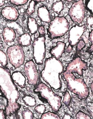
Fig. 7.2 Two pieces of cortex in a renal biopsy specimen from a woman of 75 with acute renal failure and atrial fibrillation. One piece is mostly necrotic, while the other has acute tubular damage. There was assumed to be embolisation of left atrial thrombus into branches of the renal artery, but there was no direct evidence of this in the specimen. With appropriate treatment, there was partial recovery of renal function
occur in other circumstances, and probably reflect local conditions in the affected tubules.
Interstitial tissues often appear increased in acute renal failure, and a common explanation is edema (Fig. 7.3). This may still be seen after recovery from acute renal failure (Fig. 7.5).
The range of possible abnormalities reflects the range of renal function, which does not have just two states, definitely normal and definitely abnormal, but can vary along a continuous scale. Renal failure is not all or none, but has a gradation of severity.
Provided acutely abnormal tubules are not in an infarcted area of cortex, they may recover to normal. Infarcted areas and atrophic tubules will never recover.
144 |
7 Indication for Biopsy: Acute Renal Failure |
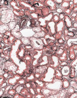
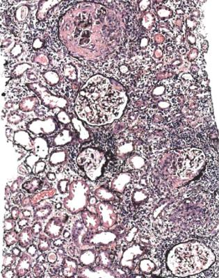
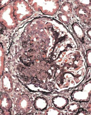
Fig. 7.3 Cortex in a right renal biopsy specimen from a man of 67 with acute renal failure, after surgery to relieve obstruction of the left renal pelvis. Most tubules are acutely damaged, with irregular flattening of the epithelium. There is interstitial edema. The cause of the acute tubular damage is not apparent in the specimen, but was later thought to be septicemia
The term often used for tubular changes in acute renal failure is acute tubular necrosis. This is not strictly accurate, and not particularly useful for pathologists, because identifiable necrosis of tubular cells is hardly ever seen. Tubular cells may be lost from tubules by apoptosis, or by being shed into the tubular fluid, to be passed in urine. Lost cells are replaced by division of remaining cells, and there may be nuclear abnormalities during this regenerative stage (Fig. 7.6). A better term than acute tubular necrosis is acute tubular damage, which covers the range of abnormalities short of infarction of the cortex. Acute tubular injury is also used.
Because nearly every biopsy specimen taken to investigate acute renal failure shows acute tubular damage, this should not be given as a diagnosis, unless it is the only abnormality. This means that in many biopsy specimens, the explanation of acute tubular damage is evident, and should be given as the diagnosis.
Approach to the Diagnosis in Acute Renal Failure |
145 |
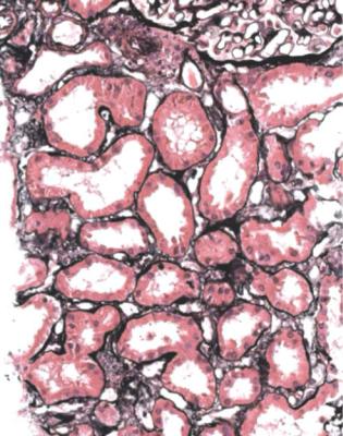
Fig. 7.4 Cortex in a renal biopsy specimen from a man of 42 with acute renal failure, after heart transplantation with many complications. Some tubules appear normal, some have fine vacuolation of the epithelium, and a few are atrophic. The fine vacuolation may be an effect of a calcineurin inhibitor used as an immunosuppressant, but similar changes can be seen in people with acute tubular damage, not treated with this type of drug (Fig. 5.9)
Approach to the Diagnosis in Acute Renal Failure
Is There Necrosis in the Kidney?
In a biopsy specimen taken to investigate acute renal failure, the first thing for the pathologist to assess is whether there is necrosis of the kidney, particularly of the cortex. This means death of all structures, meaning tubules, glomeruli, interstitial tissues, and blood vessels, not necessarily uniformly throughout the specimen (Fig. 7.2). This is a rare finding, but has serious implications for prognosis, because if there is necrosis of most or all of both kidneys, renal function will never recover.
The most likely causes of necrosis are these:
1.A thrombosing condition, such as disseminated intravascular coagulation, with thrombi in glomerular capillaries or arterioles or larger vessels, or a small vessel
146 |
7 Indication for Biopsy: Acute Renal Failure |
Fig. 7.5 Cortex in a renal biopsy specimen from a woman of 57 who had recovered from acute renal failure by the time of biopsy. Most tubules appear normal, but there is interstitial edema
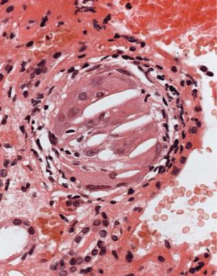
vasculopathy, also called thrombotic microangiopathy, in which there are acute abnormalities in vessels, particularly loose, concentric, intimal thickening in small arteries, and fibrinoid necrosis of arterioles (Figs 6.58 and 7.7–7.9). Various disorders, such as the group called thrombotic thrombocytopenic purpura and hemolytic uremic syndrome, can give these appearances, and usually the pathologist cannot differentiate between the disorders.
2.Infarction caused by blockage of a large artery, which is not often included in the specimen. Embolic material in arteries is a clue to this diagnosis, but is much more commonly seen in specimens of chronically ischemic kidneys without necrosis (Fig. 7.10).
3.The condition renal cortical necrosis, caused by acute, severe underperfusion of the kidney, without blockage of arteries. Renal cortical necrosis may be incomplete, and there may be surviving tubules and other structures among necrotic tissues. If there are no clues such as this feature or hints of a small vessel
Approach to the Diagnosis in Acute Renal Failure |
147 |
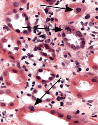
Fig. 7.6 Medulla in a biopsy specimen of a renal allograft present for 9 days in a man of 48. Tubules are recovering from acute damage, and three mitotic figures are arrowed
vasculopathy, also called thrombotic microangiopathy, or embolic material in arteries, the pathologist may not be able to determine the cause of necrosis in a specimen.
Is There the Expected Number of Tubules in the Kidney?
In the usual specimen in which there is no necrosis, the pathologist should decide whether there is the expected number of tubules, when allowance is made for the age of the person biopsied. Assessment of the amount of tubules and atrophy is done by inspection at low power on the microscope. In an old person, there may be patches of atrophic tubules that should not be seen in a young person. Occasionally, there may be such extensive atrophy that despite the clinical presentation with acute renal failure, the pathologist can say that there is a relatively longstanding renal disorder that is unlikely to recover, rather than an acute one that may recover (Fig. 7.1). A combination of acute damage and chronic changes can also occur (Fig. 6.27).
148 |
7 Indication for Biopsy: Acute Renal Failure |
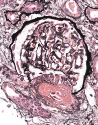
Fig. 7.7 Cortex in a renal biopsy specimen from a man of 72 with acute renal failure. There is necrosis, with thrombosis of glomeruli and arterioles. Clinically, the eventual diagnosis was thrombotic thrombocytopenic purpura
Are There Clues to the Cause of Acute Tubular Damage?
In acute renal failure that appears genuinely acute to the pathologist, there are tubules in normal numbers or nearly normal numbers, even though they may be more widely separated then normal, and they are expected to be structurally abnormal. The next problem for the pathologist is to decide whether there are changes in the specimen that give clues to the cause of the tubular damage. These clues can be in the tubules themselves, or in glomeruli, interstitial tissues, or blood vessels.
Common Findings in Renal Biopsy Specimens in Acute
Renal Failure
The pathologist should examine the specimen to see if there is evidence of one of the common conditions expected in renal biopsy specimens in acute renal failure.
Common Findings in Renal Biopsy Specimens in Acute Renal Failure |
149 |
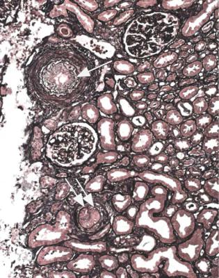
Fig. 7.8 Cortex in a renal biopsy specimen from a woman of 44 with acute renal failure. There is a small vessel vasculopathy, also called thrombotic microangiopathy, with loose concentric intimal thickening in a small artery (single arrow), and thrombus in another (double arrow). Clinically, the diagnosis was a mixed connective tissue disorder, with features of scleroderma and systemic lupus erythematosus
These conditions are not the same as those that are common in all people with acute renal failure, because most do not have a biopsy.
In adults, the commonest finding, generally in more than a quarter of specimens, is vasculitis. Other findings, in descending order of commonness, are pure acute tubular damage not explained by anything in the specimen, acute interstitial nephritis, small vessel vasculopathy, also called thrombotic microangiopathy, myeloma kidney, and acute postinfective glomerulonephritis. Other disorders together generally account for less than a tenth of specimens.
In children, over half the specimens have one of three conditions, Henoch– Schönlein nephritis, hemolytic uremic syndrome, and acute interstitial nephritis. Other likely findings are lupus nephritis, pure acute tubular damage not explained by anything in the specimen, and acute postinfective glomerulonephritis, with a
150 |
7 Indication for Biopsy: Acute Renal Failure |
Fig. 7.9 Cortex in a renal biopsy specimen from a woman of 29 with acute renal failure, 2 years after a liver transplant. An arteriole has fibrinoid necrosis of its wall. The explanation was hemolytic uremic syndrome, possibly due to cyclosporine treatment
range of disorders in the few remaining specimens. Myeloma kidney is not seen in children. Vasculitic conditions other than Henoch–Schönlein nephritis occur, but are rare.
Exceptionally, as well as frank necrosis of the cortex, a few other uncommon disorders can be detected on low power microscopy used by the pathologist to assess the relative amounts of acute and chronic tubular damage. These include infiltration by a lymphoma or other neoplasm (Fig. 7.11).
Is There Evidence of Renal Vasculitis?
Because the most likely finding in adults who are biopsied in acute renal failure is vasculitis, this should always be considered as a possibility in specimens taken for this indication.
Vasculitis is often a multisystem illness, and there may be clinical clues such as fever, arthralgia, myalgia, weight loss, upper respiratory tract symptoms, neurologic
Diagnosis of Vasculitic Glomerulonephritis |
151 |
Fig. 7.10 A small cortical artery in a renal biopsy specimen from a man of 72, showing blockage of the artery by material including cells and clefts, from which cholesterol has been dissolved during processing of the material. These are the appearances of an atherosclerotic or thromboatherosclerotic embolus
signs, and a rash. Hematuria and proteinuria are common, but not the nephrotic syndrome.
Vasculitis in the kidney almost always takes the form of a glomerular abnormality that can be called vasculitic glomerulonephritis. Other names for this are used, but are less satisfactory. They include focal segmental thrombosing and necrotising glomerulonephritis, glomerulonephritis associated with antineutrophil cytoplasmic antibodies or ANCA-associated glomerulonephritis, glomerulonephritis with crescents or crescentic glomerulonephritis, pauci immune glomerulonephritis, pauci immune crescentic glomerulonephritis, and rapidly progressive glomerulonephritis.
Diagnosis of Vasculitic Glomerulonephritis
Vasculitic glomerulonephritis can range in severity from a small segmental abnormality in one glomerulus to complete destruction of all glomeruli, with all stages in between. This means it can be focal and segmental, as some names for this disorder
152 |
7 Indication for Biopsy: Acute Renal Failure |
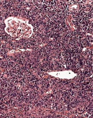
Fig. 7.11 Cortex in a renal biopsy specimen from a woman of 77 with insulin resistant or type two diabetes mellitus, and acute on chronic renal failure. There is a dense infiltrate of lymphoid cells. This is a non-Hodgkin’s lymphoma
describe it, but it can also be focal and global, or diffuse and segmental, or diffuse and global.
This is one of the conditions of the kidney in which one abnormal glomerulus is enough to give the diagnosis (Fig. 7.12).
Vasculitic glomerulonephritis is not a static condition, but changes with time. The earliest stage recognisable by a pathologist is segmental thrombosis in a
glomerulus, with disruption of capillary loops, seen on sections stained with periodic acid-methenamine silver (Fig. 7.13). The next stage is accumulation of fibrin and cells in Bowman’s space over the affected capillary loops (Figs 7.14 and 7.15). The cells are a mixture of macrophages and epithelial cells, sometimes with neutrophils and giant cells. Sometimes Bowman’s capsule is disrupted. Then there is gradual deposition of fibrous tissue and disappearance of cells (Fig. 7.16). The final stage is a scar which is sharply defined, includes part of the glomerular tuft and part of the original Bowman’s space, and is adherent to Bowman’s capsule (Fig. 7.17).
Diagnosis of Vasculitic Glomerulonephritis |
153 |
Fig. 7.12 Cortex in a renal biopsy specimen from a man of 47 with a multisystem illness, a weakly positive antineutrophil cytoplasmic antibody, hematuria, proteinuria, and normal renal function. One glomerulus has a small, acute, segmental vasculitic lesion (arrowed). This is enough to give the diagnosis of renal vasculitis
The glomerular abnormality in vasculitic glomerulonephritis can be of any size from one capillary loop to the whole of the glomerulus (Fig. 7.16). Segmental abnormalities are usually well outlined, with edges that are easily defined (Figs 7.12– 7.15, and 7.17). This feature is a help in identification of vasculitic changes, especially late ones, because other segmental abnormalities have indistinct outlines, and do not cross Bowman’s space, although the tuft may adhere to Bowman’s capsule.
The word crescent is often used in vasculitic disorders, as in the term glomerulonephritis with crescents, but this has difficulties. Generally, this is taken to mean a group of cells in Bowman’s space. A problem for the pathologist is to know how many cells are needed before a group can be called a crescent. Some definitions do not say, and some specify a size of the group, such as half of Bowman’s space. The World Health Organization’s definition is two layers or more of cells that partially or completely fill Bowman’s space. On any definition, crescents
154 |
7 Indication for Biopsy: Acute Renal Failure |
Fig. 7.13 Glomerulus in a renal biopsy specimen from a woman of 73 with a purpuric rash, hematuria, proteinuria, and acute renal failure. There is a small segmental area of thrombosis and disruption of capillary loops, shown by periodic acid-methenamine silver staining. This is the earliest recognisable stage of vasculitic glomerulonephritis
are not a constant feature of vasculitic glomerulonephritis. Crescents can also be found in acute postinfective glomerulonephritis, subendothelial membranoproliferative glomerulonephritis, and other conditions. There is a similar problem with precision of terminology about crescentic glomerulonephritis, which means different things to different people. Some pathologists apply this when at least half of all glomeruli contain what can be considered to be crescents.
Vasculitic abnormalities not only change with time. Different stages can be present at the same time in a biopsy specimen (Figs 7.15–7.17), and even in the same glomerulus (Fig. 7.18). In some people, renal vasculitis seems to be due to a single event, and in others to a prolonged process. There may be relapses at various intervals after the original acute changes have healed.
The pathologist can often see glomerular changes of vasculitis at low power on microscopy. The glomerular count should include the total number and the number of globally sclerosed glomeruli, as usual, and the number of glomeruli with
Diagnosis of Vasculitic Glomerulonephritis |
155 |
Fig. 7.14 Another glomerulus in the renal biopsy specimen from the woman of 73 illustrated in Fig. 7.13. This shows a larger lesion, with thrombosis, disruption of capillary loops, and cells in Bowman’s space
vasculitic abnormalities, which needs high power microscopy. These abnormalities can be divided into stages, such as acute or active if thrombosis or tuft disruption or cells in Bowman’s space are seen, healing if fibres appear amongst the cells, and healed if there are fibrosed areas in glomeruli.
The glomerular tuft away from segmental changes is normal in typical vasculitic glomerulonephritis, with no deposition of immunoproteins away from vasculitic abnormalities on immunohistologic study. This explains the term pauci immune glomerulonephritis, in which the first syllable in pauci, which comes from the Latin word for few, is pronounced either as in lawn or as in harm.
Glomeruli are not the only abnormal structures in renal vasculitis.
1.Tubules: In acute renal failure, there is acute tubular damage. If the disease has been present for a few weeks or more, there may be areas of tubular atrophy, more extensive than expected for the age of the person biopsied. Occasionally, a
156 |
7 Indication for Biopsy: Acute Renal Failure |
Fig. 7.15 Cortex in a renal biopsy specimen from a woman of 67 with nose bleeds, mouth ulcers, infiltrates in the lungs on radiography, arthralgia, a strongly positive antineutrophil cytoplasmic antibody of cANCA type, hematuria, proteinuria, and acute renal failure. Two glomeruli have segmental areas of tuft disruption, with cells in Bowman’s space. This is an active stage of vasculitic glomerulonephritis. The clinical diagnosis was Wegener’s granulomatosis. A common pronunciation of the eponym is vayg-a-ner. At a meeting in 1936, reported in 1937, Wegener described autopsy findings in three people with what he thought was an infective condition. They had granulomatous inflammation in the nose, generalised arteritis, and glomerulonephritis (Wegener F. On generalised septic vessel diseases. Translated in Thorax 1987; 42: 918–919). Friedrich Wegener (1907–1990) qualified in medicine in Germany, and became a pathologist in Kiel, and later elsewhere. His first autopsy was said to be on one of the people he described in 1936. The name Wegener’s granulomatosis seems to have been used first in 1947
renal biopsy is taken in a suspected vasculitic condition in which renal function is normal. In these circumstances, the tubules may be normal (Fig. 7.12).
Most specimens with active vasculitic glomerulonephritis have blood in tubules, with a distinctive appearance. Blood in tubules may be seen in any condition, but has a different appearance. This is due to the trauma of the biopsy procedure, and the blood appears fresh, with identifiable and separate red cells, usually in tubules at the edges of the specimen (Fig. 7.19). Tubules in vasculitic glomerulonephritis
Diagnosis of Vasculitic Glomerulonephritis |
157 |
Fig. 7.16 Glomerulus in the renal biopsy specimen from the woman of 67 that is illustrated in Fig. 7.15. Bowman’s space is filled with cells. Fibres are beginning to appear among the cells, shown by periodic acid-methenamine silver staining. This is early healing of vasculitic glomerulonephritis
often contain blood that seems solid or discolored, with disruption of cells and release of hemoglobin, which is called laking. Tubules in the centre of the specimen are just as likely to be affected as those at the edges (Fig. 7.20). If there is this type of blood on initial sections, but glomeruli appear normal, the pathologist should request extra sections, because there is a chance that at least one glomerulus has vasculitic changes.
2.Interstitial tissues: Interstitial tissues often have an infiltrate of mixed inflammatory cells in vasculitis, sometimes with striking numbers of eosinophils. This is part of the vasculitic disorder, and should not be given an additional diagnosis of acute interstitial nephritis. There may also be interstitial hemorrhage (Fig. 7.21).
3.Blood vessels: Arteritis or arteriolitis may be seen in a renal biopsy specimen, almost always associated with vasculitic glomerulonephritis. The affected vessels may not even be in the cortex or medulla (Figs 4.6 and 7.22). These changes are often patchy. The muscular layer of the vessel may be replaced

158 |
7 Indication for Biopsy: Acute Renal Failure |
Fig. 7.17 Glomerulus in the renal biopsy specimen from the woman of 67 that is illustrated in Figs 7.15 and 7.16. On a section stained by periodic acid-methenamine silver, there is a sharply defined, fibrous, segmental lesion, which includes a disrupted area in the tuft and adjacent Bowman’s space, and is adherent to Bowman’s capsule. This is healed vasculitic glomerulonephritis
by eosinophilic material, a change called fibrinoid necrosis. There may be a thrombus in the lumen, with an infiltrate of various types of inflammatory cells in the wall. This is sometimes called leukocytoclastic vasculitis, if disrupted neutrophils are a prominent feature.
The combined effect of these changes in glomeruli, tubules, interstitial tissues, and vessels makes a specimen with active vasculitis other than the mildest look strikingly abnormal at low power on microscopy (Fig. 7.23).
Clinical Conditions with Renal Vasculitis
Classification of vasculitic disorders is artificial, because there are overlaps between them. Nevertheless, many people with vasculitis can be given a satisfactory clinical diagnosis, even though the findings in a renal biopsy specimen may be the same in several different disorders.
Clinical Conditions with Renal Vasculitis |
159 |
Fig. 7.18 Glomerulus in a renal biopsy specimen from a man of 42 with asthma, neuropathy, arthropathy, hematuria, proteinuria, and normal renal function. On a section stained by periodic acid-methenamine silver, part of the glomerulus has a sharply defined, fibrous segmental lesion, which includes an old disrupted area in the tuft and adjacent Bowman’s space with adhesion to Bowman’s capsule. The rest of the glomerulus has active disruption of capillary loops with cells on the surface. This is active and healed vasculitic glomerulonephritis in the same glomerulus. The clinical diagnosis was Churg–Strauss syndrome, in which the names are commonly pronounced tchurg and strowss. This was described by Jacob Churg, known as Jack (born 1910), and Lotte Strauss (1913–1985), at the Mount Sinai Hospital in New York, where both were pathologists (Churg J, Strauss L. Allergic granulomatosis, allergic angiitis and periarteritis nodosa. American Journal of Pathology 1951; 27: 277–294). The term Churg–Strauss syndrome was in use by 1961
Renal vasculitis, with otherwise normal glomeruli and no significant deposition of immunoproteins, can be seen in several clinical conditions. The pathologist is not able to differentiate these on renal biopsy appearances. Common vasculitic disorders that affect the kidney are Wegener’s granulomatosis and microscopic polyangiitis, also called microscopic polyarteritis. Others are Churg– Strauss syndrome and the vasculitis associated with infective endocarditis. On current definitions, polyarteritis nodosa cannot be diagnosed if there is vasculitic
160 |
7 Indication for Biopsy: Acute Renal Failure |
Fig. 7.19 Cortex in a renal biopsy specimen from a man of 37 with proteinuria, but no hematuria. There are red blood cells in the lumen of several tubules, which is a consequence of trauma from the biopsy procedure. This appearance is unlike that of bleeding into tubules before the biopsy, illustrated in Fig. 7.20
glomerulonephritis. The antithyroid drug, propylthiouracil, and the antihypertensive drug, hydralazine, are rare causes of renal vasculitis.
Despite the name of the condition, granulomas are not seen in the kidney in Wegener’s granulomatosis (Figs 7.15–7.17). The strict pathologic definition of a granuloma is a localised microscopic collection of macrophages, with or without other cells, as seen in tuberculosis or sarcoid. What Wegener called granulomas were glomeruli with global destruction by vasculitis.
Wegener’s granulomatosis is a condition in which there is active chronic inflammation, sometimes with true granulomas and arteritis, affecting the nose, nasal sinuses, middle ear, and other parts of the respiratory tract, sometimes with renal vasculitis, which may include arteritis of vessels up to the size of the main renal artery. There are usually antibodies in the serum against the cytoplasm of neutrophils. These antibodies are detected throughout the cytoplasm in some tests, and are called cytoplasmic antineutrophil cytoplasmic antibodies or cANCA. The
Clinical Conditions with Renal Vasculitis |
161 |
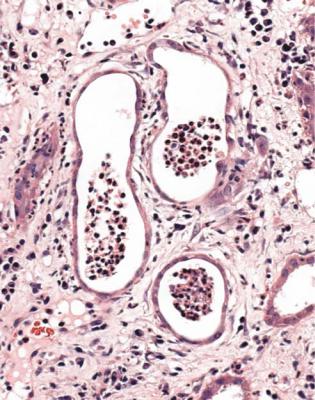
Fig. 7.20 Medulla in a renal biopsy specimen from a man of 52 with a multisystem illness, a weakly positive antineutrophil cytoplasmic antibody, hematuria, proteinuria, and acute renal failure. Many tubules contain blood, which is more solid than in the tubules illustrated in Fig. 7.19. This finding suggests that there is likely to be vasculitic glomerulonephritis. Cortex in this man’s specimen is illustrated in Figs 7.21 and 7.23
antigen recognised by most cANCA is proteinase three. The pathologist should not be influenced by reported presence or absence of these antibodies in determination of whether a biopsy specimen shows vasculitis, because cANCA are not specific for Wegener’s granulomatosis, and the condition may occur without them. This illustrates a problem with the term ANCA-associated glomerulonephritis.
Microscopic polyangiitis is the condition in which there is vasculitis of small vessels in at least one organ, usually the kidney, sometimes with vasculitis of arteries including those the size of the main renal artery, but without the features of Wegener’s granulomatosis in the respiratory tract. If the disorder is only in the kidney, this may be called renal-limited microscopic polyangiitis, or renal-limited vasculitis. There are often antibodies in serum against the cytoplasm of neutrophils, with a characteristic perinuclear distribution in some tests, called perinuclear antineutrophil cytoplasmic antibodies or pANCA. The antigen recognised by
162 |
7 Indication for Biopsy: Acute Renal Failure |
Fig. 7.21 Cortex in the renal biopsy specimen from the man of 52, the medulla in which is illustrated in Fig. 7.20. There is a heavy, patchy infiltrate of inflammatory cells in interstitial tissues, which also contain hemorrhage. These are features associated with vasculitic glomerulonephritis, which is illustrated in this man’s specimen in Fig. 7.23
pANCA is myeloperoxidase. These antibodies are not specific for microscopic polyangiitis.
Although infiltrates of eosinophils in various organs may be seen in Churg– Strauss syndrome, an acute eosinophilic interstitial infiltrate in the kidney can occur in all vasculitic disorders, and is not by itself evidence of Churg–Strauss syndrome. Asthma and eosinophilia in the blood are clinical features of this condition, and often there are antineutrophil cytoplasmic antibodies, either cANCA or pANCA (Fig. 7.18).
Polyarteritis nodosa is now uncommon. Arteries are affected by vasculitis in this condition. If there is vasculitic glomerulonephritis, the clinical diagnosis may have to be amended to one of the other vasculitic disorders. Hepatitis B virus infection is occasionally found in polyarteritis nodosa. There may be giant cell arteritis in any of the vasculitic disorders, but this is uncommon (Fig. 7.24).
Clinical Conditions with Renal Vasculitis |
163 |
Fig. 7.22 Cortex in a renal biopsy specimen from a woman of 58 with nasal ulceration, mononeuritis multiplex, a positive antineutrophil cytoplasmic antibody of cANCA type, and hematuria. An interlobular artery has fibrinoid necrosis of part of its wall, and an infiltrate of inflammatory cells
Infective endocarditis can affect the kidney in several ways. One of the commonest is by production of vasculitis. The vasculitic glomerulonephritis in infective endocarditis used to be called focal embolic nephritis and embolic nonsuppurative focal nephritis. Infective endocarditis can also affect the kidney by causing acute postinfective glomerulonephritis, subendothelial membranoproliferative glomerulonephritis, infarcts due to embolism, renal cortical necrosis due to severe hypotension, and pure acute tubular damage due to hypotension or sepsis. Antibiotics used to treat infective endocarditis may cause acute tubular damage or acute interstitial nephritis. Combinations of any of these conditions may occur. Usually, the pathologist is told on the request form that there is infective endocarditis, and this helps interpretation of the specimen.
Vasculitic glomerulonephritis is probably a consequence of glomerular endothelial damage. This follows interaction with neutrophils activated by antineutrophil cytoplasmic antibodies bound to proteinase three or myeloperoxidase, expressed on the cell membrane of neutrophils primed by inflammatory mediators.
164 |
7 Indication for Biopsy: Acute Renal Failure |
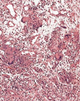
Fig. 7.23 Cortex in the renal biopsy specimen from the man of 52, the medulla in which is illustrated in Fig. 7.20, and cortex in Fig. 7.21. On microscopy at low magnification, there are several abnormalities, which include acute tubular damage, blood in tubules, an interstitial inflammatory infiltrate, acute segmental glomerular lesions, and an acute global glomerular lesion. Some glomeruli have disruption of Bowman’s capsule. These are all features of renal vasculitis
Vasculitic glomerulonephritis may respond well to aggressive treatment with immunosuppression, provided that most of the renal damage is acute rather than chronic. The response also depends upon the severity of vasculitis outside the kidney, and the general state of the person affected.
If There is Renal Vasculitis, Are There Clues to Recognisable Conditions?
Four other disorders can give vasculitic changes in the kidney, with features that allow the pathologist to recognise them, mainly because they have deposits of immunoproteins in glomeruli. The diagnosis of vasculitic glomerulonephritis or pauci immune vasculitic glomerulonephritis in a report implies that there is not
Conditions That Can be Differentiated from Pauci Immune Vasculitic Glomerulonephritis |
165 |
Fig. 7.24 Cortex in a renal biopsy specimen from a man of 69 with a long history of dermatomyositis and pulmonary fibrosis, and recent development of acute renal failure, hematuria, and proteinuria. There is a severe acute granulomatous arteritis. This responded to immunosuppressive treatment
one of these four conditions, which should be named in the diagnosis. These are Henoch–Schönlein nephritis, lupus nephritis, Goodpasture’s disease, and cryoglobulinemic glomerulonephritis.
Conditions That Can be Differentiated from Pauci Immune Vasculitic Glomerulonephritis: 1 Henoch–Schönlein Nephritis
This is a condition in which vasculitic changes occur in glomeruli, but the glomerular tuft away from these changes appears abnormal, with mesangial expansion, sometimes even with a few doubled basement membranes next to the mesangium. On immunohistologic study, there is mesangial deposition of IgA, which indicates Henoch–Schönlein nephritis. This can also be called vasculitic IgA nephropathy, or similar terms (Figs 7.25–7.27). The condition can be regarded as part of IgA
166 |
7 Indication for Biopsy: Acute Renal Failure |
Fig. 7.25 Cortex in a renal biopsy specimen from a woman of 33 with mild acute renal impairment, hematuria, proteinuria, and a history of a rash and arthralgia 2 years before the biopsy. Tubules have a little atrophy and a little acute damage. There are segmental lesions of vasculitic type in glomeruli. Immunohistologic study showed mesangial deposition of IgA, and gave the diagnosis of Henoch–Schönlein nephritis. Glomerular changes are illustrated at higher magnification in Fig. 7.26. The names are often pronounced hee-nock and shern-line. What is now usually called Henoch–Schönlein purpura was first given the name Schönlein–Henoch syndrome in 1948. Sometimes, in English, Schönlein is spelled Schoenlein to avoid use of the umlaut symbol. He preceded Henoch, but now follows him in the name of this disease. Eduard Heinrich Henoch (1820–1910) was born in Berlin, and was a pupil of Schönlein, as was Virchow (Fig. 4.5). In 1868, Henoch, who was what would now be called a pediatrician, described children with attacks of purpura, joint pains, intestinal colic, and intestinal hemorrhage. He later added the association with renal disease. Johann Lukas Schönlein (1793–1864) was Professor of Pathology and Therapy in Würzburg, and then Professor of Medicine in Zurich and Berlin. His accounts of an illness with joint pains and a rash were given in various editions of a textbook written by students based on his lectures. In the 1834 edition, Schönlein mentioned oliguria as a finding in the condition. The first report of the finding of IgA in glomeruli in Henoch–Schönlein nephritis seems to have been in 1968, although the significance of this was overlooked by the authors.There is a strong chance that the composer W A Mozart (1756–1791) had Henoch–Schönlein purpura as a boy, and died of renal failure when he was 35
Conditions That Can be Differentiated from Pauci Immune Vasculitic Glomerulonephritis |
167 |
Fig. 7.26 Glomerulus in the renal biopsy specimen from the woman of 33 with Henoch–Schönlein nephritis, the cortex in which is illustrated in Fig. 7.25. There is an acute segmental lesion with tuft disruption and cells in Bowman’s space, but this is not so sharply defined as the lesions in other types of vasculitis, such as in Figs 7.12, 7.15, and 7.17. The rest of the tuft is not normal, and has mesangial increase, with IgA deposition on immunohistologic study
nephropathy in its widest sense, rather than as a separate condition. In Henoch– Schönlein nephritis, the intensity of immunostaining for IgA may appear less than in most examples of IgA nephropathy without vasculitic lesions, possibly because the deposits are overshadowed by the acute glomerular changes.
Often, Henoch–Schönlein nephritis appears to have characteristic vasculitic changes in glomeruli, but sometimes these seem atypically vasculitic or not fully characteristic, particularly in that they are not so well outlined as usual vasculitic changes (Fig. 7.26). On the initial sections stained by hematoxylin and eosin, the combination of glomerular changes that are vasculitic, either definitely or nearly, and abnormal mesangium suggests a strong likelihood of Henoch–Schönlein nephritis. The diagnosis is made certain by the finding of IgA deposition.
Generally, Henoch–Schönlein nephritis has vasculitic lesions in only a few glomeruli, and these are often of different ages, some active and some healed. If
168 |
7 Indication for Biopsy: Acute Renal Failure |
Fig. 7.27 Cortex in a renal biopsy specimen stained by an immunoperoxidase method to detect IgA from a woman of 34 with acute renal failure, hematuria, and proteinuria. A glomerulus resembles the one in Fig. 7.26, and shows IgA deposition in mesangium away from the vasculitic lesion, with light mesangial deposition of IgA in the other glomerulus. This indicates Henoch–Schönlein nephritis
there is a severe vasculitic glomerulonephritis with lesions all at the same stage, the finding of IgA in glomeruli is more likely to indicate the coincidence of IgA nephropathy and vasculitic glomerulonephritis, in a condition such as Wegener’s granulomatosis, rather than Henoch–Schönlein nephritis (Fig. 7.28). This is an example of the rule that there may be more than one condition in a renal biopsy specimen. Clinical and serologic findings, such as pulmonary vasculitis and a high titre of cANCA, help to differentiate coincidental IgA nephropathy and Wegener’s granulomatosis, for example, from Henoch–Schönlein nephritis.
Henoch–Schönlein nephritis is not the same thing as Henoch–Schönlein purpura, also called anaphylactoid purpura, which is a systemic disorder with various combinations of arthralgia, rash, abdominal pain, melena, hematuria, proteinuria, and acute renal failure. In children, in whom the disorder is recognised most often, renal biopsy is not usually done, at least at the height of the clinical illness, because the disorder generally resolves. If there is a biopsy, the specimen usually shows
Conditions That Can be Differentiated from Pauci Immune Vasculitic Glomerulonephritis |
169 |
Fig. 7.28 Cortex in a renal biopsy specimen from a man of 52 with a multisystem illness, which included upper respiratory tract problems, a strongly positive antineutrophil cytoplasmic antibody of cANCA type, acute renal failure, hematuria, and proteinuria. There are acute vasculitic lesions in glomeruli. On immunohistologic study, there is mesangial deposition of IgA. Even so, the diagnosis was considered to be vasculitic glomerulonephritis, consistent with Wegener’s granulomatosis, with coincidental IgA nephropathy, rather than Henoch–Schönlein nephritis. This was because the vasculitic lesions were more widespread and more sharply defined than usually seen in Henoch– Schönlein nephritis, the overall effect on the kidney was more severe, and the lesions were all at the same stage. Clinical and serologic findings supported this interpretation
Henoch–Schönlein nephritis, but may show IgA nephropathy without vasculitic changes.
A diagnosis of Henoch–Schönlein purpura is less common in adults, and if there is a biopsy, about half have Henoch–Schönlein nephritis, and the other half have IgA nephropathy without vasculitic changes. Henoch–Schönlein nephritis is just as often seen in adults who do not have obvious clinical features of Henoch–Schönlein purpura outside the kidney, as in those with a vasculitic rash or other systemic features at the time of biopsy (Figs 7.25–7.27). About one in ten of people with IgA nephropathy, in its widest sense, in a renal biopsy specimen, but no systemic features of Henoch–Schönlein purpura, have Henoch–Schönlein nephritis.
170 |
7 Indication for Biopsy: Acute Renal Failure |
Conditions That Can be Differentiated from Pauci Immune Vasculitic Glomerulonephritis: 2 Lupus Nephritis
The next condition that can have vasculitic changes is lupus nephritis. Almost always, the clinical diagnosis of systemic lupus erythematosus has been made before the biopsy, usually in a young woman. There are often combinations of changes in glomeruli in lupus nephritis, and the finding of such combinations is a clue to the diagnosis (Figs 6.54–6.57). Deposition of all immunoproteins on immunohistologic study confirms the diagnosis (Fig. 6.62).
Conditions That Can be Differentiated from Pauci Immune Vasculitic Glomerulonephritis: 3 Goodpasture’s Disease
Changes of severe acute vasculitic glomerulonephritis in initial sections of a biopsy specimen, with diffuse global abnormalities all at the same stage, should make the pathologist suggest the possibility of Goodpasture’s disease (Fig. 7.29). This is the disorder associated with antibodies to glomerular basement membranes. The diagnosis is given by the finding of linear deposition of IgG and complement in glomerular basement membranes on immunohistologic study (Fig. 7.30).
Goodpasture’s disease can be a difficult diagnosis to confirm by immunohistology when virtually all the glomerular basement membranes are destroyed. The disease is rare, can occur at any age, and presents with acute renal failure, with hematuria and proteinuria. There may be hemoptysis, which indicates pulmonary hemorrhage. Not every example of renal vasculitis that either has diffuse and global glomerulonephritis, or is associated with hemoptysis, is due to Goodpasture’s disease. The terms Goodpasture’s syndrome, or pulmonary/renal vasculitic syndrome, can be used if there is renal vasculitis with pulmonary hemorrhage, but no antibodies against glomerular basement membranes.
Occasionally, both antibodies to glomerular basement membranes and antibodies to neutrophil cytoplasmic antigens, or ANCA, are found. Arteriolitis and arteritis are more likely in the kidney and other organs if both antibodies occur, than if there are only antibodies to basement membranes.
Goodpasture’s disease has strong associations with some DR alleles of HLA, which is the abbreviation for human leukocyte antigens, or human lymphocyte antigens, or histocompatibility linked antigens. These alleles determine the ability of antigen presenting cells to bind peptides derived from either the noncollagenous domain of the alpha three chain of collagen type four, or similar microbial antigens. This leads to the generation of antibodies to collagen type four, which is found in glomerular basement membranes.
Goodpasture’s disease is more difficult to treat than other types of vasculitis, and is more likely to progress rapidly to irreversible renal failure.
Conditions That Can be Differentiated from Pauci Immune Vasculitic Glomerulonephritis |
171 |
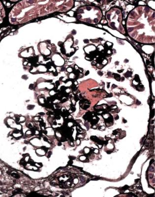
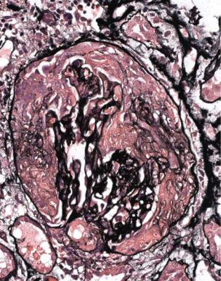
Fig. 7.29 Cortex in a renal biopsy specimen from a man of 69 with acute renal failure, hematuria, proteinuria, and no other systemic features. Nearly every glomerulus has global vasculitic changes at the same early stage. Immunoperoxidase study, shown in Fig. 7.30, confirmed antiglomerular basement membrane antibody disease or Goodpasture’s disease. Good and pasture in the eponym are pronounced in the usual way. Ernest William Goodpasture (1886–1960) was a pathologist in the United States Naval Hospital in Chelsea, Massachusetts, when he did autopsies on two young men who caught influenza at the height of the pandemic in September 1918, but he was in Harvard Medical School when he described them (Goodpasture EW. The significance of certain pulmonary lesions in relation to the etiology of influenza. American Journal of the Medical Sciences 1919; 158: 863–870). Only one of the two men had disease outside the lungs. He was 18 and had pulmonary hemorrhage, vasculitis in the spleen and small intestine, and a glomerular disorder with a fibrinous exudate in Bowman’s space and proliferation in tufts. In 1958, M C Stanton and J D Tange suggested the name Goodpasture’s syndrome for the combination of pulmonary hemorrhage and glomerulonephritis, and they took no notice of the vasculitis in the spleen and intestine in the man reported by Goodpasture. Later, the term Goodpasture’s disease began to be applied when Goodpasture’s syndrome was associated with antiglomerular basement membrane antibodies, after linear deposition of IgG in glomerular basement membranes was reported in 1964. The man with renal disease reported by Goodpasture had Goodpasture’s syndrome, but may not have had antibodies to glomerular basement membranes, now considered essential for the diagnosis of Goodpasture’s disease
172 |
7 Indication for Biopsy: Acute Renal Failure |
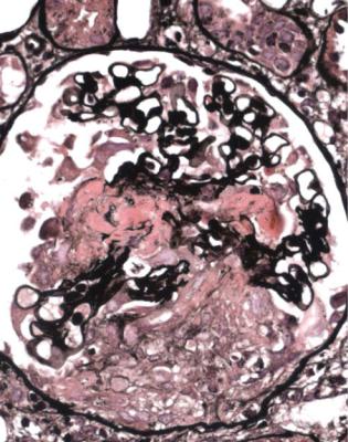
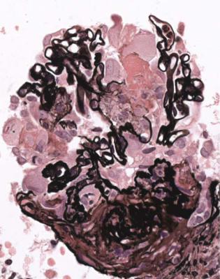
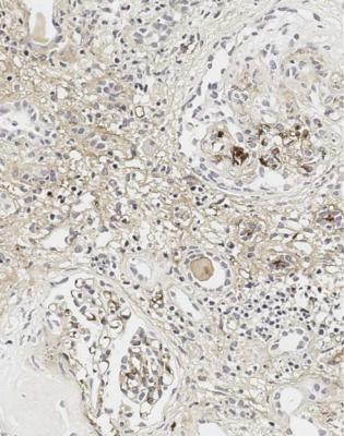
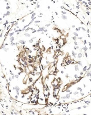
Fig. 7.30 Glomerulus in the renal biopsy specimen of the man of 69 shown in Fig. 7.29. An immunoperoxidase method to detect C9 shows linear deposition in the remains of the glomerular basement membrane, which is disrupted in places, with cells throughout Bowman’s space. The distribution of IgG is similar. These features indicate antiglomerular basement membrane antibody disease or Goodpasture’s disease
Conditions That Can be Differentiated from Pauci Immune Vasculitic Glomerulonephritis: 4 Cryoglobulinemic Glomerulonephritis
In cryoglobulinemic glomerulonephritis, there may rarely be vasculitic changes in glomeruli that have other abnormalities, particularly expansion of mesangial areas, and doubled basement membranes with the appearance of subendothelial membranoproliferative glomerulonephritis. There is also deposition of acellular material in capillary loops, which suggests the diagnosis (Figs 7.31 and 7.32). This material consists of aggregated immunoglobulins, and on immunohistologic study contains IgM and often IgG (Fig. 7.33). Arteritis and arteriolitis are more commonly seen than vasculitic glomerulonephritis.
Conditions That Can be Differentiated from Pauci Immune Vasculitic Glomerulonephritis |
173 |
Fig. 7.31 Cortex in a renal biopsy specimen from a man of 53 with acute renal failure, hematuria, and proteinuria. Glomeruli appear solid and hypercellular. Further investigations, illustrated in Figs 7.32 and 7.33, gave the diagnosis of cryoglobulinemic glomerulonephritis. This was due to precipitation of a monoclonal IgM paraprotein with polyclonal IgG
Cryoglobulins are immunoglobulins that clinical immunologists detect by cooling serum to see if there is a precipitate. Cryo comes from the Greek word for frost. The pathologist should not be concerned if immunologists cannot find cryoglobulins in blood, although the pathologist’s diagnosis is more readily believed if they do. The abnormal immunoglobulins that precipitate in glomeruli when plasma proteins are suddenly concentrated, as water is filtered, may not precipitate when serum is cooled. Complement components C4 and C3 are usually in low concentration in serum in cryoglobulinemia.
Cryoglobulins are divided into a few types. The pathologist cannot differentiate these easily, but clinical immunologists are usually able to determine the type from findings in the serum. The first type is a monoclonal IgM paraprotein, which precipitates by itself, and there is often Waldenström’s macroglobulinemia, due to a nonHodgkin’s lymphoma. Sometimes, a renal biopsy specimen in this condition just shows the glomerular deposits, with little else. In the second type of cryoglobulins,
174 |
7 Indication for Biopsy: Acute Renal Failure |
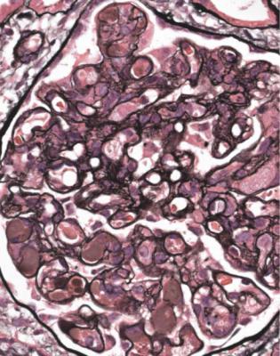
Fig. 7.32 Glomerulus in the renal biopsy specimen from the man of 53 with cryoglobulinemic glomerulonephritis, the cortex in which is illustrated in Fig. 7.31. Staining with periodic acidmethenamine silver shows deposits of material in several capillary loops, with double basement membranes in several loops, to give the appearance of subendothelial membranoproliferative glomerulonephritis. Immunohistologic findings are illustrated in Fig. 7.33
the precipitates are mixtures of a monoclonal IgM paraprotein and polyclonal IgG, which means that the paraprotein has rheumatoid factor activity, acting as an antibody to IgG. Many of the people with this type have chronic infection with hepatitis C virus, and a few have Waldenström’s macroglobulinemia. In the third type of cryoglobulins, which are rarely associated with the glomerular disorder, the precipitates are mixtures of polyclonal IgM with rheumatoid factor activity, and polyclonal IgG. Rheumatoid arthritis and related diseases are the usual underlying explanations of this type.
In the past, the term essential mixed cryoglobulinemia was sometimes used when there were IgM and IgG cryoglobulins, usually of the second type, without a lymphoma, although now most are recognised to be due to hepatitis C virus infection. The prognosis of cryoglobulinemic glomerulonephritis is that of the underlying disease.
Diagnosis of Acute Postinfective Glomerulonephritis |
175 |
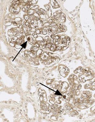
Fig. 7.33 Glomeruli in the renal biopsy specimen from the man of 53 with cryoglobulinemic glomerulonephritis that is illustrated in Figs 7.31 and 7.32. An immunoperoxidase method to detect IgM shows solid aggregates of IgM in a few capillary loops (arrowed)
Are Glomeruli Abnormal but There Is No Evidence of Vasculitis?
In acute renal failure, if glomeruli are abnormal, but do not have features of vasculitic disorders, the most likely condition is acute postinfective glomerulonephritis.
Diagnosis of Acute Postinfective Glomerulonephritis
Clues to this diagnosis are that every glomerulus is affected equally, and the whole of each glomerulus is affected. Glomeruli appear large, solid, and filled with cells, a mixture of neutrophils, monocytes, and swollen cells inside basement membranes (Fig. 6.79). These endocapillary cells are mesangial and endothelial, which are not easily differentiated. Glomerular tufts may be so large that they prolapse into the origin of the proximal tubule (Fig. 6.20). Unusually, there may be cells in Bowman’s
176 |
7 Indication for Biopsy: Acute Renal Failure |
space forming structures that can be called crescents, but if the features are otherwise typical of acute postinfective glomerulonephritis, the prognosis should still be good.
Confirmation of acute postinfective glomerulonephritis requires sections stained by periodic acid-methenamine silver and by an immunohistologic method. Silver staining shows that glomerular basement membranes are single. If membranes appear double, the diagnosis is subendothelial membranoproliferative glomerulonephritis (Fig. 6.80). Immunohistologic staining in acute postinfective glomerulonephritis shows granular deposits, of various sizes, of IgG and complement in mesangium, and on the outside of glomerular basement membranes (Fig. 6.81). These differ from the large deposits seen in subendothelial membranoproliferative glomerulonephritis (Fig. 6.82). Complement persists longer than IgG and may be the only type of immunoprotein detected in acute postinfective glomerulonephritis. Electron microscopy will also help to differentiate these two conditions (Figs 6.83 and 6.84).
IgA is not found in pure acute postinfective glomerulonephritis, and its presence suggests either the coincidence of IgA nephropathy and acute postinfective glomerulonephritis, or lupus nephritis, most easily differentiated by serologic studies.
Diagnosis of Subendothelial Membranoproliferative
Glomerulonephritis
Subendothelial membranoproliferative glomerulonephritis is sometimes called type one membranoproliferative or mesangiocapillary glomerulonephritis, to distinguish it from a condition that in practice has little resemblance to it, better called dense deposit disease than type two membranoproliferative or mesangiocapillary glomerulonephritis. The use of these type numbers is unfortunate and misleading, suggesting a connection between unrelated conditions.
Subendothelial membranoproliferative glomerulonephritis resembles acute postinfective glomerulonephritis in several ways. Every glomerulus is affected, and the whole of each is affected. Glomeruli appear larger, more solid, and more cellular than normal (Fig. 6.80). There may be cells in Bowman’s space forming structures which can be called crescents.
The differences from acute postinfective glomerulonephritis are that many or all glomerular basement membranes appear double on silver staining, and there is heavy deposition of immunoproteins in glomeruli, in a different distribution from that in acute postinfective glomerulonephritis. There are coarse deposits of IgG, IgM, and complement, mainly on the inside of glomerular capillary loops, and sometimes in mesangium (Fig. 6.82). Lupus nephritis can give the appearance of subendothelial membranoproliferative glomerulonephritis, but immunohistologic and serologic findings should indicate lupus (Figs 6.55 and 6.56).
Impression of Glomerulonephritis Given by Ischemic Glomeruli |
177 |
Dense deposit disease has distinctive appearances on orthodox microscopy, immunohistology, and electron microscopy (Figs 6.16–6.18).
Membranoproliferative glomerulonephritis is an overused diagnosis. Three conditions that may be given this name are acute postinfective glomerulonephritis, membranous nephropathy, and nodular light chain glomerulopathy, but there are others. Satisfactory silver staining and immunohistology should settle the diagnosis, plus electron microscopy, if necessary.
Membranoproliferative glomerulonephritis has been the subject of classifications based on subtle and often subjective distinctions. The disorder sometimes called membranoproliferative or mesangiocapillary glomerulonephritis type three is the combination of subendothelial membranoproliferative glomerulonephritis and membranous nephropathy. Type three is also sometimes applied to rare examples of subendothelial membranoproliferative glomerulonephritis with prominent intramembranous deposits that are not so dense, uniform, and well defined as those in dense deposit disease. These are examples of the difficulties associated with classifications that use numbers rather than descriptions.
Impression of Glomerulonephritis Given by Ischemic Glomeruli
If there is no glomerulonephritis, glomeruli in acute renal failure usually appear shrunken and smaller than normal in proportion to Bowman’s capsule. This is because they are perfused at less than their usual pressure. Such ischemic shrinkage makes glomeruli appear more solid than normal, and basement membranes appear thick, because they are wrinkled (Fig. 5.9). These features can give an impression of glomerulonephritis, but there are no segmental changes of vasculitis, there is not the tuft enlargement of acute postinfective glomerulonephritis, and silver staining shows the wrinkling of basement membranes.
Has a Glomerular Disorder Been Excluded in Acute Renal
Failure?
If there is no glomerulonephritis, the explanation of acute renal failure may be seen in interstitial tissues, tubules, or blood vessels.
Has a Glomerular Disorder Been Excluded? Is There a Clue in Interstitial Tissues to the Cause of Acute Renal Failure?
Interstitial tissues are usually abnormal even in pure acute tubular damage, and show edema, with separation of tubules by loose tissue which contains few cells (Fig. 7.3).
178 |
7 Indication for Biopsy: Acute Renal Failure |
An interstitial infiltrate of inflammatory cells is common in acute renal failure associated with glomerular disorders such as vasculitic glomerulonephritis, lupus nephritis, and acute postinfective glomerulonephritis (Figs 6.57, 7.21, and 7.23). The term acute interstitial nephritis should not be used in these conditions, but only in the absence of glomerulonephritis.
Diagnosis of Acute Interstitial Nephritis
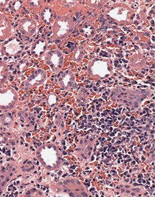
In acute interstitial nephritis, also called acute tubulo-interstitial nephritis, there is acute tubular damage with interstitial edema and an interstitial infiltrate of inflammatory cells. There are lymphocytes, plasma cells, macrophages, and eosinophils, in various proportions (Fig. 7.34). Often there are inflammatory cells inside tubular epithelial cells, and cells and cellular debris in the lumen of tubules, although these features are not essential for the diagnosis (Fig. 7.35).
Fig. 7.34 Cortex in a renal biopsy specimen from a man of 60 with acute renal failure. There is an interstitial infiltrate of inflammatory cells, mostly lymphocytes. A few cells enter the epithelium of tubules. Fig. 7.35 illustrates other tubules in this specimen. The diagnosis was acute interstitial nephritis, attributed to a response to antibiotics used to treat salmonella enteritis
Diagnosis of Acute Interstitial Nephritis |
179 |
Fig. 7.35 Cortex in the renal biopsy specimen from the man of 60 with acute interstitial nephritis, which is also illustrated in Fig. 7.34. A few tubules contain cells, at least some apparently polymorphs. Differentiation may be impossible between cellular debris produced by infiltration of tubules as part of acute interstitial nephritis, illustrated in Fig. 7.34, and pus as a sign of coincidental ascending infection, illustrated in Fig. 7.36
Neutrophil polymorphs may be present, but if they are the dominant cell line, or if there are groups of neutrophils forming pus inside tubules, the pathologist should consider whether the changes are likely to be those of ascending infection of the kidney, giving acute pyelonephritis (Fig. 7.36). This is usually in radial streaks through the cortex, compared with a more even distribution of acute interstitial nephritis, and may have detectable bacteria or fungi in tubules. Abscesses from infection carried to the kidney in the blood are also filled with neutrophils, and may contain detectable microorganisms, but are randomly scattered in the kidney. Both acute interstitial nephritis and ascending or blood-borne infection can occur in the same renal biopsy specimen, and features of purulent infection can be seen in any specimen. This is an example of the general rule that there may be more than one condition in a renal biopsy specimen. The pathologist should tell nephrologists
180 |
7 Indication for Biopsy: Acute Renal Failure |
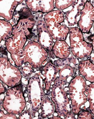
Fig. 7.36 Cortex in a biopsy specimen of kidney, taken 6 months after transplantation into a man of 44. There are groups of tubules distended by pus. A Gram stain showed Gram-positive cocci in these tubules. Staphylococcus aureus was found in the urine. This is acute pyelonephritis
whenever ascending or other infection seems a possibility. Urinary infection may not be easy to investigate in people without urine output, and untreated infection could have severe effects if immunosuppression were to be used.
Eosinophils are not always present, but are helpful to the pathologist, because they usually indicate significant acute interstitial nephritis. If eosinophils are easily seen or are the main cell type, the term acute eosinophilic interstitial nephritis can be used.
Occasionally, in acute interstitial nephritis, there are groups of macrophages, sometimes with a few giant cells, generally forming small, poorly defined collections, when the condition can be called acute granulomatous interstitial nephritis
(Fig. 7.37). Granulomas that are large, or well defined, or contain many giant cells are unlikely in an acute disorder, and are more likely in a biopsy specimen taken in investigation of chronic renal failure (Fig. 7.38).
Causes and Associations of Acute Interstitial Nephritis |
181 |
Fig. 7.37 Cortex in a renal biopsy specimen from a man of 65 with acute renal failure. There is severe acute interstitial nephritis with several granulomas, which include giant cells. The diagnosis is acute granulomatous interstitial nephritis, attributed to a response to antibiotics used to treat a liver abscess. When this appearance is seen, exclusion of tuberculosis may be impossible, but is approached by a combination of the clinical features, lack of acid-fast bacilli on Ziehl–Neelsen staining, and knowledge that there is almost always chronic renal damage in granulomatous conditions such as tuberculosis and sarcoid, illustrated in Fig. 7.38
Causes and Associations of Acute Interstitial Nephritis
Acute interstitial nephritis, including eosinophilic and granulomatous variants, can have several causes or associations. The pathologist is not usually able to determine the underlying explanation.
Common causes and associations are these.
1.Allergic responses to drugs, such as nonsteroidal anti-inflammatory drugs and antibiotics, particularly penicillin derivatives, although almost every drug that has ever been used has been said to cause acute interstitial nephritis, usually on inadequate evidence.
182 |
7 Indication for Biopsy: Acute Renal Failure |
Fig. 7.38 Cortex in a renal biopsy specimen from a woman of 37 with chronic renal failure and systemic features suggestive of sarcoid. There is chronic damage with a small granuloma, which contains giant cells. Appearances are consistent with sarcoid in the kidney
2.Infections, such as leptospirosis and hantavirus diseases.
3.The condition called tubulo-interstitial nephritis with uveitis, abbreviated to TINU. This condition is almost always in young women, and the renal problem may be detected at the same time as the eye problem, or may be before or after it.
Sjögren’s syndrome, which is the combination of dry eyes and mucous membranes with arthritis, can be associated with acute interstitial nephritis, but there is often late damage at presentation. This is almost always with chronic renal failure, rather than acute renal failure, although there may also be disordered function of some segments of the nephron (Fig. 7.39). Acute granulomatous interstitial nephritis may occur in immunodeficiency disorders, such as inherited or acquired hypogammaglobulinemia.
Causes and Associations of Acute Interstitial Nephritis |
183 |
Fig. 7.39 Cortex in a renal biopsy specimen from a woman of 31, with Sjögren’s syndrome, proteinuria, hypokalemia, and renal tubular acidosis. There is a patchy lymphocytic infiltrate, mainly around collecting ducts. The disorder is named after Henrik Samuel Conrad Sjögren (1899–1989), a Swedish ophthalmologist. The name is often pronounced shyo-gren
Has a Glomerular Disorder Been Excluded? Is There a Clue in Tubules to the Cause of Acute Renal Failure?
In acute renal failure, if the renal biopsy specimen shows no signs of glomerulonephritis, nor of acute interstitial nephritis, the pathologist should examine tubules for evidence of a disorder other than pure acute damage, or of a clue to the cause of acute damage.
Only if there is no indication of a cause, either in the tubules themselves or in other parts of the kidney, should the diagnosis be given as acute tubular damage, or acute tubular necrosis, because such damage is expected to be found in every specimen in acute renal failure. For example, there is no need to give the diagnosis as acute interstitial nephritis with acute tubular damage, because acute interstitial nephritis implies that there is acute tubular damage.
184 |
7 Indication for Biopsy: Acute Renal Failure |
If acute tubular damage is the only finding, the appearances are similar in acute renal failure caused by tubular toxins, which include drugs such as gentamicin, and ischemia, as in hypovolemia, hypotension, and sepsis, which is one cause of the systemic inflammatory response syndrome, in which there are many mechanisms leading to underperfusion of the kidney. These include increased vascular permeability in many organs, with reduced blood volume, vasoconstriction in the kidney, and myocardial damage, causing hypotension.
The clinical term hepatorenal syndrome is sometimes used when there is acute renal failure in liver failure. Renal biopsy is rarely done in liver failure because there is a risk of bleeding, but may be necessary to exclude conditions such as vasculitis. If there is no recognisable condition, a specimen will show acute tubular damage, with casts stained by bile. The renal failure is due to ischemia from severe vasoconstriction in the kidney.
Findings in Tubules: Myeloma Kidney
In particular, the pathologist should think about the possibility of effects of myeloma on tubules, especially in old people. A renal biopsy specimen sometimes gives the diagnosis of myeloma before other investigations can be done. The shorthand term myeloma kidney means the condition in which an abnormal immunoglobulin is filtered by glomeruli and damages tubules, although myeloma can affect the kidney in other ways. Other names for this include immunoglobulin light chain cast nephropathy and myeloma cast nephropathy.
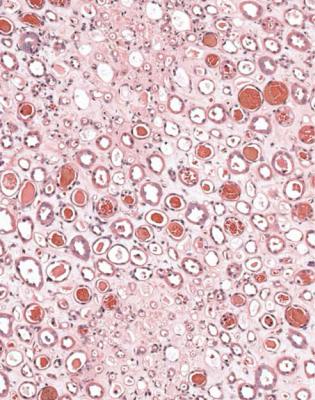
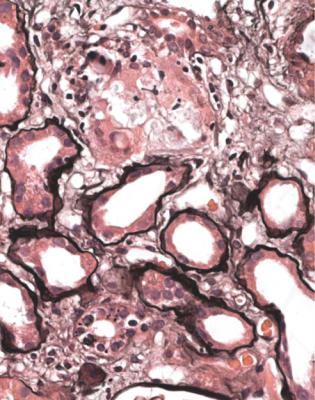
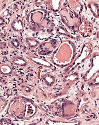
The clue to the diagnosis of myeloma kidney is the presence in tubules of solid material that differs from the cast material commonly seen. In myeloma, the casts appear dry, palely stained, brittle, and cracked, unlike the more usual casts that appear wet, shiny, and deeply stained. In myeloma, the casts are surrounded by giant cells within tubules, and sometimes by polymorphs (Fig. 7.40). The giant cells are fused macrophages. Occasionally, there are needle-shaped casts. There may be an interstitial infiltrate of various types of inflammatory cells. On periodic acid Schiff staining, myeloma casts are virtually unstained, while the usual or ordinary casts found in almost all conditions, including in some tubules in myeloma kidney, are strongly stained (Fig. 7.41).
When these changes are well developed, the diagnosis can be given on sections stained by hematoxylin and eosin, and the pathologist should not be dissuaded by failure of the nephrologists to make the diagnosis clinically before biopsy. Clinical immunologic investigation of serum and urine should confirm the diagnosis.
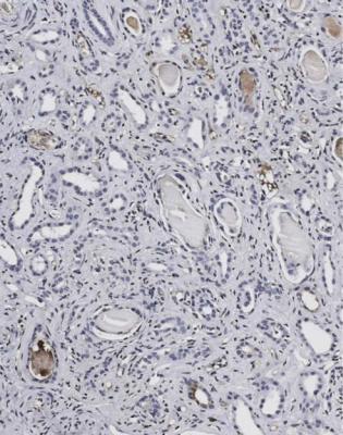
If the changes are slight, immunohistologic study of the specimen is necessary to confirm the diagnosis. This should show disproportionate amounts of either kappa or lambda light chains in casts and in tubular epithelial cells (Figs 7.42 and 7.43). The casts sometimes stain with Congo red, and may give anomalous colours when the section stained with Congo red is examined between crossed polariser and analyser (Fig. 6.68). This does not mean there is amyloid in the kidney.
Findings in Tubules: Myeloma Kidney |
185 |
Fig. 7.40 Cortex in a renal biopsy specimen from a woman of 65 with acute renal failure. Some tubules are atrophic, which suggests that the renal failure is acute on chronic. There are casts in several tubules, several surrounded by giant cells. These gave the diagnosis of myeloma kidney. After the biopsy, a lambda light chain paraprotein was found in the woman’s serum
Renal failure from myeloma kidney rarely improves.
There are several other ways in which myeloma and related paraproteinemias can affect the kidney, although just as people with diabetes mellitus can have renal disease that is not due to diabetic glomerulopathy, people with a paraprotein can have renal disease that is unrelated to their paraprotein. Paraproteinemia is a common finding in old people, and even if the pathologist is told on the request form about a paraprotein, this may be irrelevant to the findings in a renal biopsy specimen.
The main ways, other than myeloma kidney, in which paraproteinemia can affect the kidney include amyloidosis of AL type (Figs 6.65–6.67, and 6.74–6.76), nodular light chain glomerulopathy (Figs 6.45–6.48), and cryoglobulinemic glomerulonephritis (Figs 7.31–7.33). An interesting observation is that these are not often seen in combination, either with each other or with myeloma kidney. Rarely, the neoplastic cells in myeloma or another lymphoma may be seen in or around the kidney (Figs 4.2 and 7.11).
186 |
7 Indication for Biopsy: Acute Renal Failure |
Fig. 7.41 A renal biopsy specimen from a woman of 58 with acute renal failure and myeloma. Periodic acid Schiff staining shows the difference between myeloma casts, which are palely stained and surrounded by giant cells, and the common or ordinary type of cast found in many conditions, including myeloma. The ordinary cast is deeply stained, and has no cellular reaction around it (arrowed)
Findings in Tubules: Myoglobinuria or Crystals
Tubules may contain things other than myeloma casts that give the diagnosis. They may have casts that appear orange or brown on sections stained by hematoxylin and eosin, which suggest myoglobinuria (Fig. 7.44). This is confirmed by immunohistologic staining for myoglobin (Fig. 7.45). The request form may give a history of injury to skeletal muscles, or myositis, for instance in viral infections, or exposure to drugs known to cause rhabdomyolysis, such as statins, which are used to lower lipid concentrations in the blood, or cocaine. Hemoglobin may be seen in tubules, if there is severe hemolysis, for instance, following a mismatched blood transfusion.
The finding of many oxalate crystals in acutely damaged tubules suggests poisoning, particularly by ethylene glycol which is used as antifreeze, although a few crystals may be seen in any specimen with acute or chronic tubular damage. Many
Findings in Tubules: Myoglobinuria or Crystals |
187 |
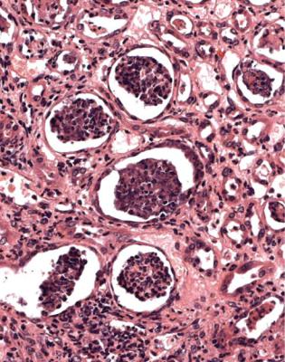
Fig. 7.42 Medulla in a renal biopsy specimen from a man of 71 with acute renal failure and hypercalcemia. Immunoperoxidase staining to detect lambda light chains shows no staining in most casts. An adjacent section stained for kappa light chains is shown in Fig. 7.43
oxalate crystals may also be seen if there is tubular damage, generally chronic rather than acute, associated with either genetic hyperoxaluria, or chronic intestinal or pancreatic disorders with malabsorption of fat. Excess fatty acids in the bowel bind calcium, which cannot combine with oxalic acid, and this allows increased absorption of oxalates. This is called enteric hyperoxalosis.
Oxalate crystals may be seen within tubules on orthodox microscopy, even though they are transparent and do not stain with routine methods. Oxalates are more easily detected when the section is examined between crossed polariser and analyser, because the crystals are birefringent and appear bright (Figs 7.46 and 7.47).
Extensive deposition of calcified material, other than oxalates, within tubular epithelium or basement membranes and elsewhere, may be seen in severe hypercalcemia, which may cause acute renal failure. These deposits are not birefringent but stain with hematoxylin, and can be detected on sections stained with hematoxylin and eosin, and even better on sections stained by Congo red or van
188 |
7 Indication for Biopsy: Acute Renal Failure |
Fig. 7.43 An adjacent section to that in Fig. 7.42. Immunoperoxidase staining to detect kappa light chains shows staining in casts, and in tubular epithelial cells. The renal biopsy specimen showed myeloma kidney and gave the diagnosis of paraproteinemia, which was confirmed later by detection of an IgA kappa serum paraprotein, with a kappa Bence Jones protein, and other evidence of myeloma
Gieson, with hematoxylin counterstain. If necessary, von Kossa stain will confirm calcification, or rather the presence of insoluble phosphate and carbonate, which are assumed to be calcium salts. Calcium oxalate does not react with von Kossa stain.
Has a Glomerular Disorder Been Excluded? Is There a Clue in Blood Vessels to the Cause of Acute Renal Failure?
In acute renal failure, if there is no sign of glomerulonephritis, acute interstitial nephritis, myeloma kidney, or another identifiable tubular disorder, a clue to the cause may be in blood vessels.
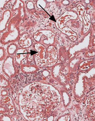
Changes in Blood Vessels: Vasculitis or Embolism |
189 |
Fig. 7.44 Cortex in a renal biopsy specimen from a man of 85 with acute renal failure. There is acute tubular damage, with several casts containing flakes or granules that appear red/orange on hematoxylin and eosin staining (arrowed). This finding suggests myoglobinuria, which is confirmed by immunohistologic study, illustrated in Fig. 7.45. The man was found to have high concentrations of muscle enzymes in the serum, attributed to alcoholic myopathy
Changes in Blood Vessels: Vasculitis or Embolism
Arteritis and arteriolitis are rarely the only evidence of vasculitis in a biopsy specimen, and vasculitic glomerulonephritis is almost always found if they occur (Figs 4.6, 7.22, and 7.24).
Evidence of atherosclerotic or thrombotic embolism may be seen in arteries in acute renal failure, but almost always in old people with more extensive chronic damage than expected for their age, as a reflection of severe chronic ischemia of the kidney (Fig. 7.10). The renal failure is usually an acute worsening of chronic renal failure, rather than acute impairment of previously normal function. Only one artery with embolic material in it has to be found to give the diagnosis, because the implication is that the biopsy is highly unlikely to have hit the only embolus, and that there is widespread embolism in the kidney. Embolism may
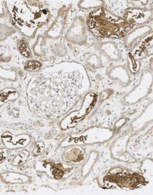
190 |
7 Indication for Biopsy: Acute Renal Failure |
Fig. 7.45 Cortex in the renal biopsy specimen from the man of 85 with myoglobinuria, which is illustrated in Fig. 7.44, stained by an immunoperoxidase method to detect myoglobin. Tubules contain myoglobin, and the diagnosis is confirmed
be found in association with any other renal disorder in old people, an example of the rule that more than one condition may be found in a renal biopsy specimen.
Acute Renal Ischemia
The finding of extensive acute tubular damage or early tubular atrophy, especially if these are uniform and most glomeruli survive, but without clues to a glomerular, tubular or interstitial disorder that would explain the acute changes, suggests that a likely cause is severe ischemia of recent onset (Fig. 7.48).
This could be due to multiple atherosclerotic emboli in the kidney, but the biopsy specimen has not included any to allow the diagnosis to be made. Another possibility is that there is stenosis of the main renal artery, which is usually due to
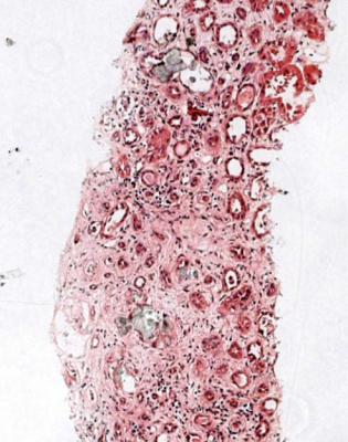
Acute Renal Ischemia |
191 |
Fig. 7.46 Cortex in a renal biopsy specimen from a man of 70 with acute renal failure. There is a mixture of acute and chronic tubular damage, but most appears acute. Examination of this section stained by hematoxylin and eosin between crossed polariser and analyser is illustrated in Fig 7.47, and suggests the diagnosis
atherosclerosis, and the person has been treated with an angiotensin converting enzyme inhibitor. Such treatment may cause a sudden and severe worsening of renal function, by reduction of glomerular filtration. As in atherosclerotic embolism, there is likely to be a background of marked chronic damage. Another possibility is reduced perfusion of the kidney because there is hypotension, perhaps due to a myocardial infarct. Renal biopsy is not often used to investigate acute renal failure in this circumstance.
Renal vein thrombosis causes acute tubular damage from ischemia. This is almost always in the nephrotic syndrome, and thrombus is hardly ever seen in the small veins included in a renal biopsy specimen (Figs 6.1 and 6.2).
The most common explanation of acute renal ischemia is usually suggested by findings in small blood vessels in the specimen. This is a small vessel vasculopathy, also called thrombotic microangiopathy.
192 |
7 Indication for Biopsy: Acute Renal Failure |
Fig. 7.47 The section illustrated in Fig. 7.46, examined between crossed polariser and analyser. There are crystals in several tubules. These are oxalates. The man had malabsorption from chronic pancreatitis. The diagnosis was predominantly acute tubular damage as a complication of enteric hyperoxalosis
Diagnosis of Small Vessel Vasculopathy, also Called Thrombotic Microangiopathy
In a small vessel vasculopathy, also called thrombotic microangiopathy, small arteries in the renal cortex have concentric intimal thickening, which differs from changes seen with age or with nonaccelerated hypertension, sometimes called essential or benign hypertension, although benign is not an appropriate term for a potentially fatal condition. The intimal thickening appears loose, mucoid, and poorly stained by routine methods, and is sometimes called onion skin change (Fig. 7.8).
Arterioles have another abnormality, and show replacement of parts of their wall by material that has few cells, stains deeply with eosin, and is called fibrinoid necrosis. The lumen of affected arterioles may be blocked by thrombus (Fig. 7.9). There may be hemorrhage into the walls of small arteries or arterioles (Fig. 7.49). The fibrinoid necrosis and the thrombosis may extend into glomerular tufts.
Diagnosis of Small Vessel Vasculopathy, also Called Thrombotic Microangiopathy |
193 |
Fig. 7.48 Cortex in a renal biopsy specimen from a man of 58 with acute renal failure and severe ischemic heart disease, for which he was under consideration for heart transplantation. There is severe tubular damage, which mostly appears early atrophy, and affects the cortex uniformly. Glomeruli appear close to normal. These findings suggest severe recent ischemia of the kidney
Glomeruli often show only severe shrinkage from ischemia, but they may have loose material on the inside of capillary loops between the endothelium and the basement membrane. This can be followed by the appearance of double basement membranes, resembling subendothelial membranoproliferative glomerulonephritis, but there are few or no deposits of immunoproteins. Also, the mesangium may disappear, by the process of mesangiolysis. The vascular changes may be so severe that infarction of cortex may be seen. A lack of inflammatory cells in the walls of affected vessels, the concentric changes, and the lack of fibrinoid necrosis in arteries help to differentiate small vessel vasculopathy, also called thrombotic microangiopathy, from vasculitis.
On immunohistologic study, there is deposition of IgM and complement in small arteries and arterioles, in the intima and sometimes in the media (Fig. 7.50).
Sometimes, only the arterial changes or the arteriolar changes occur. With time, the arterial intimal thickening becomes more fibrous, and the arteriolar necrosis
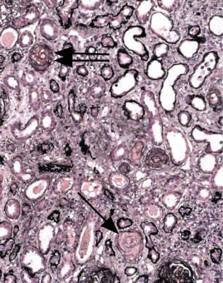
194 |
7 Indication for Biopsy: Acute Renal Failure |
Fig. 7.49 Cortex in the renal biopsy specimen, also illustrated in Fig. 7.1, from the man of 30 with acute renal failure. There is a small vessel vasculopathy, also called thrombotic microangiopathy, with loose intimal thickening in one interlobular artery (single arrow), and intimal hemorrhage in another (double arrow). These changes are consistent with effects of accelerated hypertension
resembles severe hyalinosis (Fig. 7.51). The pathologist can usually still see hints that there is this type of vasculopathy, no longer in its acute or most active stage.
Renal failure from a small vessel vasculopathy, also called thrombotic microangiopathy, may recover, depending on the underlying condition, but often does not.
Disorders Associated with Small Vessel Vasculopathy
This type of vasculopathy can be seen in accelerated hypertension, some examples of hemolytic uremic syndrome and related disorders, and a group of conditions that include systemic lupus erythematosus, the antiphospholipid syndrome, and the one called either scleroderma or systemic sclerosis.
These disorders cannot usually be differentiated by a pathologist, unless there are other features, such as a glomerular disorder, which suggests lupus nephritis. Nephrologists may also be unsure what clinical diagnosis to give to a person with

Disorders Associated with Small Vessel Vasculopathy |
195 |
Fig. 7.50 Cortex in a renal biopsy specimen from a woman of 70 with acute renal failure and clinical features of early scleroderma. An immunoperoxidase method to detect IgM shows heavy deposition in the wall of an arteriole with fibrinoid necrosis, and lighter deposition in the intima of a small artery (arrowed). The diagnosis is small vessel vasculopathy, also called thrombotic microangiopathy
acute renal failure and small vessel vasculopathy, also called thrombotic microangiopathy. For example, scleroderma or systemic sclerosis may have no characteristic clinical features at the time of presentation with acute renal failure (Fig. 7.8).
Accelerated hypertension is sometimes called malignant hypertension. Hypertension with clinical features of the accelerated phase may not be accompanied by pathologic features of small vessel vasculopathy, also called thrombotic microangiopathy, and the vasculopathy may occur without clinically accelerated hypertension.
The vasculopathy due to accelerated hypertension may be the only change in a biopsy specimen, or there may be evidence of a renal disorder, which accounts for the hypertension. The commonest glomerular disorder to be found associated with the vasculopathy is IgA nephropathy, diagnosed on immunohistologic appearances (Fig. 7.1). Two possible explanations of a sudden deterioration of renal function
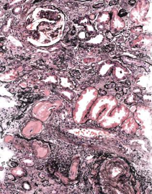
196 |
7 Indication for Biopsy: Acute Renal Failure |
Fig. 7.51 Cortex in a renal biopsy specimen from a man of 67 with chronic renal failure and severe hypertension. Arterioles have marked hyalinosis, suggestive of resolved fibrinoid necrosis, and a small artery has chronic intimal thickening. These findings are consistent with a late small vessel vasculopathy, also called thrombotic microangiopathy, in previously accelerated hypertension. A glomerulus has a segmental area of sclerosis
in someone known to have IgA nephropathy are the development of accelerated hypertension and the development of Henoch–Schönlein nephritis (Figs 7.25–7.27).
A problem for the pathologist, when there is a small vessel vasculopathy, also called thrombotic microangiopathy, is the finding of segmental sclerosing abnormalities in glomeruli, without significant deposition of immunoproteins (Figs 7.51 and 7.52). These segmental glomerular lesions may be a result of accelerated hypertensive effects, by mechanisms such as extension of arteriolar necrosis or thrombosis into the tuft, or hyperfiltration effects following global sclerosis of other glomeruli due to ischemia. Alternatively, the segmental lesions may indicate an underlying, pre-existing glomerular disorder, which explains the accelerated hypertension. Whether the glomerular abnormalities are the result or the cause of accelerated hypertension may be difficult to determine, and the pathologist may only be able to report that there is a small vessel vasculopathy, also called thrombotic microangiopathy, associated with segmental sclerosing glomerular lesions.
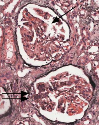
Disorders Associated with Small Vessel Vasculopathy |
197 |
Fig. 7.52 Cortex in a renal biopsy specimen from a woman of 44 with acute renal failure and clinically accelerated hypertension. There is early tubular atrophy, with ischemic shrinkage of one glomerulus (single arrow). The other glomerulus has an area of sclerosis, with adhesion to Bowman’s capsule (double arrow)
Hemolytic uremic syndrome can give the changes of the vasculopathy. These are the usual appearances in adults, although renal biopsy is not often done at the height of the illness, because there is a risk of bleeding. The disorder called thrombotic thrombocytopenic purpura overlaps clinically with hemolytic uremic syndrome, but typically has neurological signs, and is less likely to give acute renal failure.
Both are conditions in which platelets are activated, but coagulation is usually normal. In hemolytic uremic syndrome, endothelial damage from various causes leads to platelet adhesion. In thrombotic thrombocytopenic purpura, there is a deficiency of an enzyme, ADAMTS 13, which stands for a disintegrin-like and metalloproteinase with thrombospondin type one motif. Deficiency, either genetic or due to an acquired antibody, means that multimers of von Willebrand factor circulate in the blood, and cause platelet aggregation. Thromboses in glomeruli and elsewhere in the kidney may be seen (Fig. 7.7). There is evidence of a microangiopathic
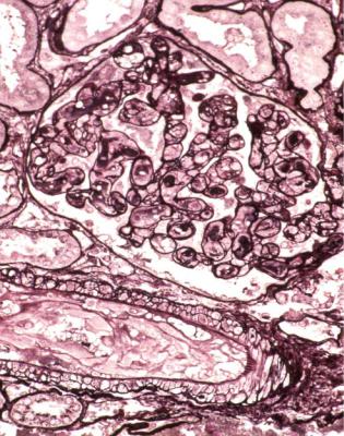
198 |
7 Indication for Biopsy: Acute Renal Failure |
hemolytic anemia, with red cell fragments, reduced haptoglobin concentration, and other features in the blood.
Biopsy is rare in children with hemolytic uremic syndrome associated with diarrhea due to infection with Escherichia coli type O157, because a confident diagnosis can usually be made clinically. The condition is due to a verocytotoxin, or Shiga-like toxin, from the bacteria. There is thrombosis in glomeruli, which sometimes extends into afferent arterioles or arteries. Acute renal failure in this type of hemolytic uremic syndrome generally recovers.
Children and adults with clinically atypical hemolytic uremic syndrome, not associated with diarrhea due to infection with Escherichia coli type O157, have glomerular changes resembling subendothelial membranoproliferative glomerulonephritis, with mesangial increase and doubled basement membranes, but few or no immune deposits. Combinations of glomerular and vascular changes can be seen in a renal biopsy specimen in this type of hemolytic uremic syndrome, in which acute renal failure generally does not recover (Fig. 7.53).
Fig. 7.53 Cortex in a renal biopsy specimen from a boy of 8 with acute renal failure and clinically atypical hemolytic uremic syndrome, not associated with diarrhea. A small artery has loose intimal thickening. A glomerulus has mesangial and endothelial swelling with doubling of basement membranes, which gives the appearance of subendothelial membranoproliferative glomerulonephritis
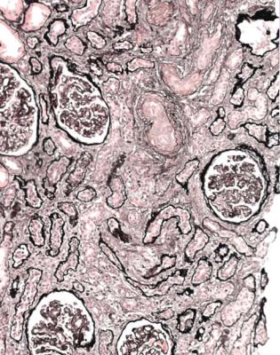
Disorders Associated with Small Vessel Vasculopathy |
199 |
Fig. 7.54 Cortex in a renal biopsy specimen from a woman of 30 with chronic renal failure, systemic lupus erythematosus, a history of repeated spontaneous abortion, and anticardiolipin antibodies in the serum. There is late renal damage with features of a small vessel vasculopathy, also called thrombotic microangiopathy, consistent with the antiphospholipid syndrome
There are several causes of clinically atypical hemolytic uremic syndrome. These include familial disorders, such as hypocomplementemia associated with persistent activation of complement, due to deficiency of regulators of the alternate pathway of complement activation, particularly factor H and factor I, which are both found in normal serum, and membrane cofactor protein or CD46, found on the surface of normal cells. Other causes of the hemolytic uremic syndrome are effects of drugs, such as calcineurin inhibitors and the cytotoxin mitomycin, infections, such as with HIV or the pneumococcus, irradiation, bone marrow transplantation, and some cancers.
In the antiphospholipid syndrome, or antiphospholipid antibody syndrome, there are antibodies in the serum to various phospholipids. The antibodies are also called anticardiolipin antibodies, reagin antibodies, or lupus anticoagulant, depending upon the method of detection. Many people with these antibodies have systemic lupus erythematosus, but some have either incomplete features of lupus or no evidence of it. Women with these antibodies have a high risk of repeated abortion. Other features include venous and arterial thrombosis, which may be seen
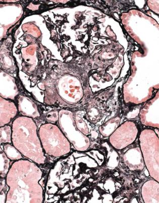
200 |
7 Indication for Biopsy: Acute Renal Failure |
Fig. 7.55 Cortex in a renal biopsy specimen from a woman of 27 with acute renal failure, systemic lupus erythematosus, disseminated intravascular coagulation, and anticardiolipin antibodies. A glomerulus contains thrombosed capillary loops, consistent with the antiphospholipid syndrome
in the kidney. The antiphospholipid syndrome is usually diagnosed when there is late renal damage, and there may be features of a small vessel vasculopathy, also called thrombotic microangiopathy, or late intimal thickening in arteries (Fig. 7.54). Occasionally acute changes are seen, such as thrombosis in glomeruli (Fig. 7.55).
Disseminated intravascular coagulation is due to stimulation of both the coagulation cascade and platelets, often by septicemia, and sometimes by other factors, such as hemorrhagic problems related to pregnancy. Renal biopsy is hardly ever done, but the specimen shows thrombosis in glomeruli or arterioles.
Summary: Acute Renal Failure
Most people with acute renal failure do not have a renal biopsy.
Acute renal failure is associated with acute abnormalities in tubules. Provided acutely abnormal tubules are not in an infarcted area of cortex, they may recover to normal.
Further Reading: Acute Renal Failure |
201 |
The most likely finding in renal biopsy specimens from adults with acute renal failure is vasculitis. This almost always takes the form of vasculitic glomerulonephritis.
Other common findings in acute renal failure are various glomerular disorders, acute interstitial nephritis, tubular disorders such as myeloma kidney, and a small vessel vasculopathy, also called thrombotic microangiopathy.
Only if none of these identifiable conditions is found should the diagnosis be given as acute tubular damage not explained by anything in the biopsy specimen.
Further Reading: Acute Renal Failure
D’Agati VD, Jennette JC, Silva FG. Non-Neoplastic kidney diseases. Atlas of Nontumor Pathology, first series, fascicle 4. Washington, DC: American Registry of Pathology and Armed Forces Institute of Pathology, 2005. Chapters 10–16, 18–22.
Jennette JC, Olson JL, Schwartz MM, Silva FG. Heptinstall’s Pathology of the Kidney. Sixth ed. Philadelphia:Lippincott Williams and Wilkins, 2007. Chapters 7, 8, 10, 12–16, 19, 21, 23, 24.