

Chapter 3
Technical Handling of Renal Biopsy Specimens
Types of Renal Biopsy Specimen
There are two types. One is a needle biopsy specimen, usually taken through the skin, and called a percutaneous specimen. Transjugular specimens are similar specimens, but are taken by an internal approach to the kidney, through a cannula passed from the jugular vein into the renal vein. The other type of specimen is taken directly from a kidney by incision at operation, and often called a wedge biopsy specimen.
Fixation and Processing of Specimens
Different laboratories have different methods of fixation. The simplest is formal saline, which is a solution of one part of 40% formaldehyde in nine parts of 0.9% sodium chloride in water. This is sometimes called 10% formalin, because that is the dilution of the original formaldehyde solution, and sometimes called 4% formalin, because that is the true final dilution of formaldehyde.
Formal saline has several advantages over other fixation methods.
1.All routine histologic staining methods work well after this fixation.
2.Many immunohistologic methods also work well.
3.If necessary before further processing, a part of the specimen can be transferred to another fixative for electron microscopy, with little effect on the quality of the electron microscopic images. This may mean that there is no need for technical staff to attend each time a biopsy is done.
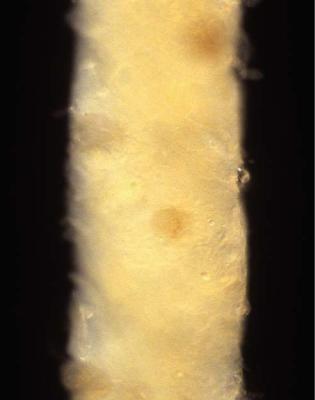
In some centres, immediately after removal, the renal biopsy specimen is examined with a low magnification binocular microscope or other magnifying lens to allow identification of glomeruli, which can be seen as red dots (Fig. 3.1). A small piece, about 1 mm long, containing glomeruli is cut off and put into a solution of glutaraldehyde in buffer suitable for electron microscopy. Another piece is frozen for immunohistologic studies on unfixed sections. The rest of the specimen is then put in a fixative for orthodox light microscopic studies. The term orthodox light
A. J. Howie, Handbook of Renal Biopsy Pathology. |
11 |
C Springer 2008 |
|
12 |
3 Technical Handling of Renal Biopsy Specimens |
Fig. 3.1 Needle biopsy specimen of kidney inspected in reflected light under low magnification on a microscope. Glomeruli are the dark round structures. Small pieces containing a couple of glomeruli can be cut off, and either frozen for immunofluorescence study or put in fixative suitable for electron microscopy. In Latin, glomerulus meant a little ball of thread, and the name was first used in microscopy as a description of what is now called the glomerular tuft. The tuft and the structure that surrounded it, Bowman’s capsule, were originally called a Malpighian corpuscle, in which corpuscle came from the Latin word for a little body. Glomerulus is used now instead of Malpighian corpuscle. Sir William Bowman (1816–1892), whose surname is pronounced with the first syllable as in go, studied medicine in Birmingham, United Kingdom, and became an ophthalmic surgeon in London. His paper on the microscopic structure of the kidney was published in 1842. Marcello Malpighi (1628–1694), pronounced mal-pee-gee with a hard g, was an Italian physician and anatomist, who described the corpuscle in 1666
microscopy in this text means the use of routine pathologic staining methods, without immunohistologic methods.
This collection procedure means that experienced laboratory staff have to be present whenever there is a biopsy, with the risk of delays and inefficiency, or there is a risk that inexperienced medical staff will try to divide the specimen and make mistakes. The advantage is that experienced staff can usually say whether the specimen contains glomeruli, and should be adequate for study by a pathologist. An alternative procedure is that each specimen is put into formal saline by the person
Fixation and Processing of Specimens |
13 |
who takes the biopsy, and then the specimen is taken to the laboratory as soon as possible, with a request form on which there is enough information to allow the pathologist to judge how the specimen should be handled.
There are emergencies whose immediate management can be affected by findings in renal biopsy specimens. Although sections of frozen material can be prepared quickly, they are not usually of satisfactory quality for detailed study of the kidney, and so laboratories should be able to prepare sections of material embedded in paraffin wax within a few hours of reception of specimens. Some departments of pathology offer an emergency service at any time.
The most common emergencies are suspected rejection of a renal allograft, and acute renal failure. Suspected puncture of an organ other than kidney is also an emergency. Nephrotic syndrome, because of its possible complications and implications for urgent management, can be considered an emergency. Specimens taken for these reasons should be processed as rapidly as possible. Most other specimens do not require urgent handling, but if this is available without difficulty, the pathologist should make use of it.
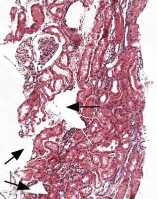
Fig. 3.2 Renal biopsy specimen processed in a cassette packed with a plastic sponge. Artefactual distortion can be seen as indentations at the edge of the section and holes within the section, examples of both of which are arrowed
14 |
3 Technical Handling of Renal Biopsy Specimens |
The sample to be prepared for orthodox light microscopic sections should be processed by the laboratory in its usual way and embedded in paraffin wax. An important technical point is that sponge packing should not be used in cassettes during processing of specimens, because this distorts the tissue, and leaves distinctive holes in sections (Fig. 3.2).
Arrangements for Electron Microscopy of Specimens
If a piece of the specimen has not been fixed specially for electron microscopy at the time of removal, a piece can be removed from the sample fixed in formal saline. This is best done by inspection with a low magnification binocular microscope and transfer of a small piece of cortex into a solution of glutaraldehyde. Electron microscopy may be considered necessary for a diagnosis although all the material has been embedded in paraffin wax, in which case a piece can be removed from the wax, and reprocessed in the appropriate way. Images are not ideal after this, but may still allow the diagnosis to be made.
Some laboratories have a sample of every specimen for electron microscopy and examine them all, some are selective, and some do not have the opportunity to do electron microscopy.
Many diagnoses can be made on renal biopsy specimens without electron microscopy, by orthodox light microscopy, either on its own or with immunohistologic investigation. The most important investigative technique on most renal biopsy specimens is the preparation and study of suitable light microscopic sections, including immunohistology, if necessary.
Electron microscopy is most likely to help in the diagnosis in the following circumstances:
1.When there is hematuria, especially microscopic, with or without proteinuria, when renal excretory function is normal.
2.When there is a family history of renal disease.
3.When there is asymptomatic proteinuria, with normal renal excretory function.
Conditions in which electron microscopic investigation may be helpful, but is not usually essential for the diagnosis, are these:
1.Nephrotic syndrome
2.Acute renal failure
3.Chronic renal failure
4.Renal disease in diabetes mellitus
5.Renal disease in systemic lupus erythematosus
6.Suspected rejection of a renal allograft
7.Repeat specimen when the diagnosis has been made
In children, the safest procedure is to keep a sample of every specimen for possible electron microscopic examination, if this is feasible. In adults, many conditions
Sectioning of Specimens |
15 |
can be investigated adequately without electron microscopy, and this is not usually essential. Features in the clinical history of adults may suggest that there is something unusual, and that electron microscopy may be useful, for instance if the person has diabetes mellitus and hematuria, but normal renal function. If there is any doubt, and if the pathologist has access to an electron microscope, the best course is to keep a sample for possible electron microscopy.
Sectioning of Specimens
Three suggestions about sections of renal biopsy specimens are these.
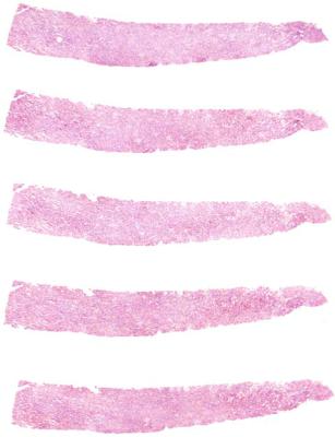
1.Needle biopsy specimens should be embedded as straight as possible, and aligned so that the pieces run across the glass slide, not along its length. This means that the axis of the pieces is at right angle to the long axis of the slide (Fig. 3.3). The reasons for this are that more sections can be mounted on a
Fig. 3.3 Serial sections of a renal biopsy specimen, mounted to run across the slide rather than along its length
16 |
3 Technical Handling of Renal Biopsy Specimens |
slide, and this arrangement of sections makes it easy for a pathologist to follow a structure such as a glomerulus from section to section.
2.Serial sections should be cut, and runs of these should be mounted on consecutively numbered slides. Generally about four to eight sections can be mounted on one slide (Fig. 3.3). If these first two suggestions are followed, glomeruli and other structures can be studied efficiently by the pathologist. Serial sections allow study of glomeruli and other structures in three dimensions, rather than in the two dimensions seen on a single section.
3.Sections should be kept and mounted right from the beginning of appearance of tissue in them. No tissue from a renal biopsy specimen should be discarded. If necessary, extra sections can be cut with little problem, but discarded material is lost for ever.
Staining of Specimens
Most staining methods should be in regular use in pathology laboratories. Stains required on a renal biopsy specimen are hematoxylin and eosin or periodic acid Schiff to give the general appearance, a stain such as periodic acid-methenamine silver to show basement membranes, and a connective tissue stain, such as hematoxylin van Gieson or a trichrome. Because amyloid is sometimes found unexpectedly in renal biopsy specimens, can be shown by specific methods, and may be missed if these methods are not used, a stain such as Congo red may be applied to every specimen. Congo red is also useful because it stains eosinophils, and its counterstain shows calcification. Some pathologists find other stains useful on routine sections, such as Martius scarlet blue. When appropriate, several other stains can be used, such as Gram stain for bacteria, Perls’ Prussian blue for iron deposits, and von Kossa’s method for calcium, or rather, for insoluble phosphate and carbonate, which are usually calcium salts.
Which stains are used and how many sections are cut are usually determined by personal preference of the pathologist. These matters are probably of little importance, compared with the ability of the pathologist to give a diagnosis that corresponds most closely with the disease in the specimen, and to provide other information of clinical significance.
One possible set of slides and staining methods is this. On each of 10 consecutively numbered slides, six to eight serial sections are mounted, most cut at about 3 μm thickness, and those on slides numbered 7 and 8 at 2 μm thickness. Slides numbered 2 and 10 are stained immediately by hematoxylin and eosin. Number 4 is stained by hematoxylin van Gieson, number 6 by Congo red, and the slides with thin sections, numbers 7 and 8, by periodic acid-methenamine silver. Slides numbered 1, 3, 5 and 9 can be stained if necessary in other ways, or further studies can be made with stains already used. All spare slides should be stored with the stained slides, in case they are ever needed. Further sections can be cut from the paraffin block, if necessary.
Immunohistologic Study of Specimens |
17 |
Immunohistologic Study of Specimens
Many renal biopsy specimens can only be analysed properly when studies are made of the distribution of various molecules, particularly immunoglobulins and complement components. This is because many renal diseases are complications of disorders of the immune system. The term immunohistology refers to the method of detection, which is by use of immunoglobulins as antibodies rather than to the substances detected, which are not all immunoproteins.
Laboratories have different practices. Many use immunofluorescence techniques on frozen sections, and routinely stain for antigens such as immunoglobulins G, A, and M, fibrinogen/fibrin, and complement components, particularly C1q and C3. Usually the antibodies to these antigens are directly conjugated to fluorescein isothiocyanate.
Other laboratories use immunoenzyme techniques on paraffin sections, with a variety of ways of detection of antigens, but mostly with a final step including reagents labelled with either peroxidase or alkaline phosphatase. An extra set of sections can be cut routinely from specimens, including those from allografts, if necessary. These sections should be mounted on slides suitable for immunohistologic procedures, meaning that they are coated with reagents such as albumin or poly L lysine, to help adhesion of the sections. The number depends upon the antibodies usually used by the laboratory. One practice is to have four extra slides, each with at least one section. These are stained by an immunoperoxidase method for immunoglobulins G, A, and M, and a complement component C9. Other sections can be cut and stained using antibodies to other substances such as kappa and lambda light chains and amyloid A.
Immunofluorescence methods on frozen sections are technically easier and have been used longer. Immunoperoxidase methods on paraffin sections have the disadvantage that sections have to be treated with proteolytic enzymes before immunostaining, to remove the plasma fixed in blood vessels. Plasma contains immunoproteins which obscure staining of immune deposits, although proteolysis can be controlled, so that the quality of immunostaining can be equivalent to that of immunofluorescence methods on frozen sections. Immunofluorescence methods can also be applied to fixed material after treatment with proteolytic enzymes.
Immunoperoxidase methods on paraffin sections have advantages over immunofluorescence methods on frozen sections. Most of the advantages are also over immunofluorescence methods on paraffin sections of fixed material.
The advantages of immunoperoxidase methods are these. Stained sections are permanent, although fluorescence fades and has to be recorded by photography. Immunoperoxidase sections are examined with an ordinary light microscope rather than a fluorescence microscope, are the same size as the sections stained with routine methods, and can be counterstained so that sites of immunostaining can be identified precisely, unlike on immunofluorescence sections, where the background is dark. Immunoperoxidase methods are generally more economic than immunofluorescence methods, because directly labelled antibodies have to be used at higher concentration than antibodies detected by indirect techniques. Because of technical
18 |
3 Technical Handling of Renal Biopsy Specimens |
arrangements, sometimes immunofluorescence material is examined and reported by people other than the pathologist who examines the orthodox light microscopic sections. Material for immunoperoxidase sections does not need special arrangements for collection and storage. The technique can be applied retrospectively to renal biopsy specimens stored in paraffin wax for many years.
Nearly all illustrations of immunohistologic investigations in this book are of sections stained by immunoperoxidase methods, but findings of immunofluorescence methods are comparable.
Summary: Technical Handling of Renal Biopsy Specimens
Fixation, processing, sectioning, and staining should be designed to give the pathologist the best possible opportunity to study renal biopsy specimens by light microscopy.
Immunohistologic study can be done either by immunofluorescence techniques, usually on frozen sections, or by immunoenzyme techniques on paraffin sections.
Electron microscopy is useful in investigation of some renal biopsy specimens.
Further Reading: Technical Handling of Renal Biopsy Specimens
Furness PN. Renal biopsy specimens. J Clin Pathol 2000; 53: 433–438.
Gregory J, Howie AJ. Protease digestion of renal biopsy specimens for immunohistological study. Journal of Cellular Pathology 1996; 1: 166–169. This and the next reference give practical details of immunoperoxidase methods.
Howie AJ, Gregory J, Thompson RA, Adkins MA, Niblett AJ. Technical improvements in the immunoperoxidase study of renal biopsy specimens. Journal of Clinical Pathology 1990; 43: 257–259.
Jennette JC, Olson JL, Schwartz MM, Silva FG. Heptinstall’s Pathology of the Kidney. Sixth ed. Philadelphia: Lippincott Williams and Wilkins, 2007. Chapter 3.