Добавил:
Опубликованный материал нарушает ваши авторские права? Сообщите нам.
Вуз: Предмет: Файл:

Книги по МРТ КТ на английском языке / Advanced Imaging of the Abdomen - Jovitas Skucas

.pdf
Скачиваний:
2
Добавлен:
20.10.2023
Размер:
21.9 Mб
Скачать

914

ADVANCED IMAGING OF THE ABDOMEN

A CT finding of large, solid peritoneal tumors without an evident primary site in a pediatric patient or a young adult should suggest a small cell tumor.

Neuroendocrine Tumors

One classification of neuroectodermal origin tumors is based on their origin (Table 14.5). Although these tumors can be differentiated pathologically, from an imaging perspective they often have a similar appearance, and many publications lump them together (Fig. 14.25). They are differentiated in this work as an aid to clinical management rather than to imply that each type has a specific imaging appearance, although quite often a specific diagnosis can be suggested from clinical data and tumor location.

Neuroendocrine tumors originate in the extraperitoneum, mesentery, and bowel wall. Except for neuroblastomas and ganglioneuroblastomas these tumors are found mostly in adults. Tumors adjacent to bowel wall are discussed in Chapters 4 and 5.

Ganglioneuroma

Ganglioneuromas are rare neuroendocrine tumors originating from sympathetic ganglia and are found along the paravertebral sympathetic plexus. Most are extraperitoneal in location. These occur mostly in children and young adults. A rare one is hormonally active. Imaging reveals solid tumors that are nonspecific in appearance. Some contain punctate

Table 14.5. Classification of neuroectodermal tumors

Nerve sheath (Schwann cell) origin

Neurilemoma

Neurofibroma

Amputation neuroma

Neurosarcoma

Granular cell tumor

Ganglion cell origin

Ganglioneuroma

Neuroblastoma

Paraganglioma tumors*

Pheochromocytoma

Paraganglioma

* The following are similar tumors. The term pheochromocytoma is used for paraganglioma tumors located in the adrenal medulla.

Figure 14.25. Extensive retroperitoneal poorly differentiated neuroendocrine tumor. A spine metastasis is present (arrow). Primary site of tumor origin could not be determined; both adrenal glands were involved by tumor. (Courtesy of Algidas Basevicius, M.D., Kaunas Medical University, Kaunas, Lithuania.)

calcifications. They vary in contrast enhancement. They are hypointense on T1but of varying intensity on T2-weighted MR images. Hypointense curvilinear bands have been described within these tumors on T2-weighted MR images. Some of these tumors encase blood vessels without narrowing the lumen.

Ganglioneuroblastomas contain both ganglioneuroma and primitive neuroblastoma elements and are considered to be intermediate between benign ganglioneuromas and malignant neuroblastomas. A majority occur in young children. Their imaging appearance varies from a mostly solid to a mostly cystic tumor depending on the degree of cell differentiation.

Neuroblastomas are discussed in Chapter 16.

Paraganglioma (Pheochromocytoma)

Extraadrenal paragangliomas account for about 15% of these tumors. Presumably they arise from embryonic chromaffin cells in parasympathetic tissue and in accessory adrenal glands. A minority are multicentric in origin. Most are detected during the second and third decades of life. An occasional one manifests during pregnancy.

Carney’s syndrome (or triad) consists of a gastrointestinal stromal tumor (often a gastric leiomyosarcoma), an extraadrenal paraganglioma, and a pulmonary chondroma. Whether

915

PERITONEUM, MESENTERY, AND EXTRAPERITONEAL SOFT TISSUES

Carney’s syndrome is an autonomic nervous system abnormality or a multiple endocrine neoplasia syndrome or even a multiple hamartoma syndrome makes for an interesting discussion.

Some paragangliomas are nonfunctional. A paraganglioma can compress and displace an adjacent kidney and induce hypertension (Page kidney). As an example, CT of a 25-year-old woman with suspected appendicitis detected a broad ligament tumor, shown to be a nonfunctioning paraganglioma (67).

Schwannoma (Neurilemoma)

Schwannomas originate from Schwann sheath cells and are most common in the peripheral neural system. Schwann cells are the peripheral neural system’s glial cells. Found mostly in young adults, more often in women, extraperitoneal schwannomas tend to be solid tumors, and their imaging findings are nonspecific. The ones involving the sacral nerves tend to widen the sacral foramina (Fig. 14.26). Preoperative biopsy is often not helpful. A pelvic schwannoma tends to mimic an ovarian neoplasm.

Both MRI and CT are useful in characterizing these tumors (Fig. 14.27). Computed tomography of extraperitoneal schwannomas reveals well-marginated, encapsulated, round or oval tumors. An occasional one undergoes central necrosis, hemorrhage, and central or peripheric calcifications. They show heterogeneous contrast enhancement, with lack of enhancement in cystic regions. Overall, they tend to be somewhat hypovascular. Computed tomography of one pelvic schwannoma revealed only the cyst wall enhancing postcontrast; in some others focal central enhancement is evident.

Neurofibromatous Tumors

Neurofibromatosis type 1 (NF1, von Recklinghausen’s disease) is a relatively common autosomal-dominant hereditary neurocutaneous syndrome consisting of neurofibromas, café-au-lait spots, and other abnormalities involving various body organs. A mutation in the NF1 gene occurs roughly in one of 3500 births; NF1 encodes a tumor suppressor, loss of which leads to neurofibroma formation. Most affected individuals are detected in childhood, but a minority contain a forme fruste type first

Figure 14.26. Schwannoma exiting sacral foramen. Noncontrast CT identifies a soft tissue tumor widening a sacral foramen on the right (arrow). (Courtesy of Patrick Fultz, M.D., University of Rochester.)

manifesting in adulthood. Although a single neurofibroma most often is an isolated event, the presence of multiple neurofibromas or a single plexiform neurofibroma is strong presumptive evidence for von Recklinghausen’s disease. About 50% of affected individuals have spontaneous mutations.

Neurofibromas are complex, noncapsulated tumors originating from Schwann cell lineage in peripheral nerves. Histologically, they differ from neurilemomas. Mast cells appear to play a role in their growth (68). Both somatic and autonomic neural tissue is involved. Because these nerves are ubiquitous and neurofibroma formation sporadic, a wide manifestation of this entity is encountered. Tumors are more common in extraperitoneal tissues, including paraspinal regions, than within the bowel wall. Focal involvement of one organ is unusual but has been reported. Some tumors in the mesentery compress adjacent blood vessels and result in focal ischemia. Extensive extraperitoneal neurofibromas mimic adenopathy.

Neurofibromas tend to be focal, solid, homogeneous, and somewhat hypodense with CT, presumably due to their rich lipid content (Fig. 14.28). Often their appearance mimics that of adenopathy. Cystic degeneration is more common in neurilemomas, but does occur in a minority of neurofibromas. Many are symmetrical bilaterally, with a presacral location also being common. Asymmetry should suggest malignant degeneration. Postcontrast CT reveals a hypovascular soft tissue tumor. Occasional tumors with either peripheral or central contrast-enhancement are described, with the

916

ADVANCED IMAGING OF THE ABDOMEN

A B

C D

Figure 14.27. Neurilemoma. A: Contrast-enhanced CT identifies a hypodense tumor in right adrenal region, suggestive of a cyst. B: T1-weighted MR image reveals a homogeneous hypointense tumor. C: Turbo spin echo T2-weighted image the hyperintense tumor is located posterior to the inferior vena cava. D: Contrast-enhanced T1-weighted image shows inhomogeneous contrast enhancement (arrow). (Source: Rha SE, Byun JY, Jung SE, Chun HJ, Lee HG, Lee JM. Neurogenic tumors in the abdomen: tumor types and imaging characteristics. Radiographics 2003;23:29–43, with permission from the Radiological Society of North America.)

less vascular portion representing lipid-rich Schwann cells and cystic degeneration, while denser segments tend to be more vascular.

Plexiform neurofibromas infiltrate and are poorly marginated. In fact, soft tissue involvement tends to mimic a diffuse infection. Magnetic resonance imaging of some mesenteric plexiform neurofibromas reveals ring-like structures within the tumor on T2-weighted and contrast-enhanced images.

One should keep in mind, however, that mesenteric involvement in von Recklinghausen’s disease needs to be distinguished from hereditary intestinal neurofibromatosis, which is a separate genetic disease.

Carcinoid

A mesenteric origin for a carcinoid is not uncommon. In fact, a distinction between bowel

Figure 14.28. Neurofibroma. Contrast-enhanced CT outlines a tumor with target-like enhancement (arrow). The hyperdense center represents nerve tissue, surrounded by more peripheral myxoid degeneration. (Source: Rha SE, Byun JY, Jung SE, Chun HJ, Lee HG, Lee JM. Neurogenic tumors in the abdomen: tumor types and imaging characteristics. Radiographics 2003;23:29– 43, with permission from the Radiological Society of North America.)

917

PERITONEUM, MESENTERY, AND EXTRAPERITONEAL SOFT TISSUES

and mesenteric origin is often not possible and some of these tumors are simply identified adjacent to a loop of bowel. Because of the oftenassociated dense desmoplastic reaction, even CT often cannot identify the exact site of origin for these tumors.

A typical CT appearance is that of a mesenteric tumor surrounded by radiating stellate folds, and such a finding is presumptive evidence for a carcinoid. Adjacent bowel loops are either infiltrated or simply displaced. Atypical appearances, however, are common. A rare carcinoid presents as a large necrotic mesenteric tumor mimicking a necrotic stromal tumor. Associated enlarged lymph nodes are an inconsistent finding.

A percutaneous biopsy is often nondiagnostic; tumor cells are difficult to obtain because of the exuberant fibrosis.

Other Neuroendocrine Tumors

An extrapancreatic gastrinoma is occasionally located in a lymph node. Primary gastrinomas are rare at this location and a small primary pancreatic tumor should be excluded.

Presacral Tumors

Certain tumors are more common in the presacral soft tissues than in other locations and these are discussed here. Overall, however, presacral tumors are uncommon. They originate from any of the soft tissue elements located in this space and range from solid to cystic. Primary presacral neoplasms can invade the sacrum or rectum. Larger presacral tumors tend to displace the rectum laterally. These tumors tend to be insidious, often are quite large when first detected, and thus are diagnosed late.

A triad of anorectal, sacral, and presacral anomalies was described by Currarino et al. (69) in 1981 and is known as Currarino’s triad. Both a complete triad and an incomplete form exist. A deformed sacrum is common; anorectal anomalies range from stenosis to an imperforate anus and presacral tumors consist of teratomas, meningoceles and various cysts or cystic neoplasms. Conventional radiographs identify an abnormal sacrum, called a scimitar sacrum, but the full extent of abnormalities is better detected with MRI.

Teratoma

The most common presacral tumor in neonates is a teratoma. Most teratomas are benign, although the risk of malignancy increases considerably in older children. Most are large at first presentation. Their imaging appearance is similar to teratomas at other locations and consists of a heterogenous, well-marginated tumor containing both cystic and solid components. The presence of varying amounts of fluid, fat, and calcifications, including teeth, is diagnostic. Malignant ones tend to contain more soft tissue and less of a cystic component than benign ones.

Meningocele

A presacral meningocele is a rare congenital abnormality consisting of herniated meninges and other spinal canal content. An occasional one is familial.Associated abnormalities include sacral abnormalities, spina bifida, and genitourinary defects. Imaging reveals a presacral cystic mass filled with cerebrospinal fluid and containing varying amounts of soft tissue. An associated sacral defect, at times detected even with conventional radiography, should suggest the diagnosis.

Other Presacral Tumors

A chordoma is a low-grade malignant neoplasm resulting in sacral destruction. Extension into the presacral soft tissues is common. A complication after resection of a sacral chordoma is posterior rectal herniation, presumably due to weakness of the posterior pelvic floor.

Widening of a sacral neural foramina should suggest neurofibromatosis.

The rare presacral neuroblastoma has an imaging appearance similar to its counterpart in the adrenal glands.

Ectopic Pregnancy

In general, 1% of all pregnancies are ectopic, and almost all occur in the fallopian tubes; among ectopic pregnancies, about 1% or fewer are intraabdominal. They are thus rare, but their importance lies with the associated high maternal death rate, if undetected. They are believed

918

to represent the sequelae of a missed ruptured ectopic tubal pregnancy.

Some women with an abdominal pregnancy present with an acute abdomen and undergo emergent surgery; others are managed conservatively, at least initially. Placental attachment to the mesentery is incompatible with fetal life, but some fetuses survive if the placenta is attached to the uterus.

If an abdominal pregnancy is clinically suspected, MRI is the imaging modality of choice; US does not detect all abdominal pregnancies. Imaging findings of an abdominal pregnancy include detection of an extrauterine fetus and placenta; fetal body parts are seen outside the uterus, assuming the uterus can be identified. Also, the placenta is located close to maternal bowel and no uterine wall identified between fetus and maternal abdominal wall. The fetus tends to have an unusual position. Peritoneal fluid is not common in a normal pregnancy but is often identified in an abdominal pregnancy.

A rare cesarean scar pregnancy was confirmed by US, MRI, and cystoscopy (70); her pregnancy was terminated by hysterotomy with uterine preservation, followed by methotrexate therapy.

A rare cause of an intraabdominal calcification is a lithopedion (calcified fetus). Most imaging modalities should suggest the diagnosis.

Hernias

Patients with ascites have increased intraperitoneal pressure, and a number of hernias develop in this setting.

A Richter hernia signifies that only a portion of the bowel wall circumference is incarcerated. Strangulation of the involved bowel segment can occur without anatomic lumen obstruction.

Laparoscopic hernia repair has been extensively adopted, although the prevalence of recurrence and complications have raised questions about such an approach.

Diaphragmatic Hernia

Morgagni Hernia

A hernia through the anteromedially located foramen of Morgagni is not common. These defects are lateral to the midline. Occasionally

ADVANCED IMAGING OF THE ABDOMEN

these congenital hernias (called MorgagniLarrey’s hernia in some countries) occur bilaterally. Hernia content ranges from omentum, stomach, and other adjacent structures, and it is surrounded by peritoneum. Some of the smaller ones simply contain fat, at times detectable on chest radiographs and other imaging modalities.

Many of these hernias are discovered incidentally in an otherwise asymptomatic infant or even adult. Symptomatic infants have respiratory distress, recurrent pulmonary infections, or simply failure to thrive. Especially in neonates and infants, other associated anomalies are common. Bowel obstructions in a Morgagni hernia are uncommon.

With gastric herniation, a contrast study with the patient in the lateral position should be diagnostic; a Morgagni hernia is far anterior to the usual esophageal hiatus. Without an associated air–fluid level, conventional chest radiography of a foramen of Morgagni hernia mimics a pericardial cyst or other soft tissue tumor in this location. At times CT is helpful by identifying intestinal content in the chest. Partial diaphragmatic eventration is in the differential diagnosis.

A laparoscopic approach can be used to repair these hernias.

Bochdalek Hernia

A congenital posterolateral diaphragmatic defect, or Bochdalek hernia, is more often detected in neonates on the left side. It varies in size from a barely discernible defect to hemidiaphragmatic agenesis. Almost any intraabdominal organ with sufficient laxity can herniate. Most Bochdalek hernias do not have a hernial sac. If a hernia is large enough, associated pulmonary hypoplasia develops, and it is this hypoplasia that determines the prognosis. Also, these neonates have an increased prevalence of cardiovascular and other anomalies. Pulmonary artery hypertension in this condition is associated with a patent ductus arteriosus.

In neonates a large Bochdalek hernia manifests as respiratory distress. An initial chest radiograph suggests the diagnosis; within several hours air can be identified in the intrathoracic bowel. To prevent further respiratory distress due to bowel distention, once this condition is suspected the stomach should be decompressed with a nasogastric tube. A diag-

919

PERITONEUM, MESENTERY, AND EXTRAPERITONEAL SOFT TISSUES

nosis is not always obvious and a number of developmental lung conditions, such as lobar emphysema and adenomatoid malformation, have a similar appearance. A barium study confirms the intrathoracic location of portions of the intestine.

At times a Bochdalek hernia manifests later in life. Most of these adult hernias are on the right side. Whether they represent a delayed manifestation of a congenital condition or are an acquired focal diaphragmatic defect is a moot point. A chest radiograph reveals a posterior soft tissue tumor adjacent to the diaphragm, a nonspecific finding also seen with lung neoplasms and other lesions. Computed tomography defines the underlying anatomy and also often outlines a diaphragmatic defect. Hernia content ranges from fat, omentum, or a solid organ such as spleen, to bowel.

Other Diaphragmatic Hernias

Hiatal hernia is discussed in Chapter 2. Diaphragmatic trauma and resultant hernias were discussed earlier in this chapter (see Trauma).

A paraesophageal hernia can contain omentum or other adjacent structure. Chest radiography simply detects an abnormal tumor in the posterior mediastinum; CT or MRI confirms the herniation.

An intrapericardial hernia can be congenital or traumatic in origin. Some of these hernias are asymptomatic. With conventional contrast studies they may mimic a hiatal hernia.

Ventral Hernia

Ventral hernias include umbilical hernias, hypogastric hernias (below the umbilicus) and epigastric hernias. The latter are uncommon, found mostly in obese individuals, often contain properitoneal fat and are detected with CT.

An umbilical hernia is common in the pediatric age group where it is usually considered to be congenital. It develops in adults with increased intraabdominal pressure, such as in a setting of ascites or pregnancy. Especially with ascites, the overlying skin can necrose and rupture, a condition associated with a high mortality. An umbilical hernia can also result in bowel incarceration.

Prior to surgical repair of an acquired umbilical hernia, it appears worthwhile to establish its

vascularity with Doppler US. An occasional one contains a herniated paraumbilical collateral vein (71), resulting in major bleeding during repair.

Femoral Hernia

Groin hernias are common in the pediatric age group, but detection of a femoral hernia can be difficult and misdiagnoses are common. Even an incarcerated femoral hernia may be missed clinically.

Computed tomography shows a soft tissue tumor medial to the femoral vein. Problems arise in differentiating a hernia from adenopathy, hematoma, or other tumor.

At times US, including Doppler, aids in differentiating a femoral hernia from other groin masses. Bowel can be identified within a hernia by showing valvulae conniventes, haustrations, or peristalsis.

Inguinal Hernia

Most direct inguinal hernias are acquired, broad-based, and rarely incarcerate. Congenital indirect hernias are due to failure of closure of the processes vaginalis. In adults, an indirect inguinal hernia is acquired and is caused by dilation of the deep inguinal ring. Indirect hernias allow abdominal content to herniate inferiorly. Both direct and indirect hernias can protrude into the scrotum and contain small bowel, large bowel, bladder, omentum, or other contents, including a rare fallopian tube and ovary (72). In general, small bowel is more common in right-sided hernias and colon in left-sided ones, but almost any abdominal organ, including stomach, can herniate (Fig. 14.29). Bowel infarction in a strangulated inguinal hernia is not uncommon; bladder infarction can also develop. Even an acute gangrenous appendicitis has developed in an inguinal hernia. These hernias are accentuated during pregnancy.

Inguinal herniation of the bladder, or inguinal cystocele, can result in urinary obstruction and renal insufficiency. It can result in clinical prostatism without bladder outlet obstruction. Although occasionally identified with CT (Fig. 14.30) the condition is best studied during the filling phase of urodynamic studies with the patient standing.

920

Figure 14.29. Right inguinal hernia containing sigmoid colon, an uncommon finding. Partial colonic obstruction was present at the hernial ring.

Herniography involves contrast injection into the peritoneal cavity and was performed to detect hernias in infants; it is rarely done today. Peritoneal scintigraphy also detects inguinal hernias. More often either CT or US is performed and should be diagnostic. A bowelcontaining hernia is identified if bowel is present distal to the inguinal ring. Fluid adjacent to bowel or bowel wall thickening suggests ischemia, but keep in mind that occasionally a

ADVANCED IMAGING OF THE ABDOMEN

septated hydrocele or hematocele has a similar appearance within a hernia.

Multidetector CT should readily detect these hernias. Indirect hernias occur lateral to the inferior epigastric vessels, in distinction to direct hernias which are located medial to these vessels.

Ultrasonography using a 7.0-MHz transducer should detect whether an inguinal hernia in present in most children; the US criteria used to define these hernias are a widened internal inguinal ring or inguinal canal fluid or organs either at rest or during straining. Ultrasonography cannot readily differentiate between a direct and an indirect hernia.

Magnetic resonance using T1and T2weighted sequences and dynamic sequences readily identifies inguinal rings, including the inferior epigastric vessels.

Most of these hernias are corrected surgically without preprocedure imaging. If the diagnosis is in doubt, US is useful in excluding a hernia containing bladder or other inguinal or testicular tumors. Childhood inguinal herniorrhaphy is one of the causes of seminal tract obstruction; microsurgical vasovasostomy and epididymovasostomy are procedures in adult men to correct such obstruction.

Doppler US is useful in patients with persistent scrotal pain or swelling after inguinal hernia repair. Ischemia, testicular infarction, and fluid collections are readily detected.

Obturator Hernia

An obturator ring hernia is more common in elderly women. It is difficult to diagnose, and small bowel obstruction with strangulation is not uncommon, with its associated relatively high mortality rate. Occasionally a ureter becomes obstructed in an obturator hernia.

These hernias should be visible on CT images. A small bowel barium study should also diagnose these hernias if they contain small bowel, but keep in mind that on a frontal view such a hernia mimics an inguinal or femoral hernia; oblique or lateral views readily differentiate between them.

Figure 14.30. Bilateral inguinal hernia. Computed tomography identifies contrast-containing small bowel in the right one (arrow) and a portion of bladder in the left one (arrowhead). (Courtesy of Patrick Fultz, M.D., University of Rochester.)

Spigelian Hernia

A defect in the transversus abdominis muscle aponeurosis lateral to the rectus abdominis

921

PERITONEUM, MESENTERY, AND EXTRAPERITONEAL SOFT TISSUES

A

B

Figure 14.31. A,B: Two CT images of a spigelian hernia (arrows).

muscles and close to the linea semilunaris is the site of a potential hernia. It is a type of ventral hernia but most authors discuss it as a separate entity. Investigators consider a spigelian hernia a fascinating topic for publication. The ratio of articles published to the limited number of patients who have this hernia is probably greater than for any other type of hernia.

The spigelian hernia sac can contain omentum, small bowel, or other adjacent structures. An occasional patient presents with small bowel obstruction, at times with an acute strangulation. Most patients are adults, although an occasional spigelian hernia develops in an infant. A clinically palpable bulge at the hernia site is uncommon because of the overlying external oblique muscle. Most symptomatic hernias, however, are tender to direct palpation.

The hernia should be readily diagnosed with CT (Fig. 14.31). A small bowel barium study is diagnostic if the hernia contains small bowel. Ultrasonography can also identify the abdominal wall defect, hernial sac and contents, and its relationship to the adjacent fascia and muscles; at times a Valsalva maneuver detects intermittent hernia reduction.

Lumbar Hernia

Two potential defects in the posterolateral abdominal wall allow lumbar hernias to

develop: a superior (Grynfeltt’s triangle) and an inferior (Petit’s triangle).Although the literature suggests that most of these hernias are confined to one of these triangles, in reality many consist of a rather extensive abdominal wall defect and do not correspond to a specific superior or inferior site. This is especially true with congenital and postoperative lumbar hernias. Either retroperitoneal fat or bowel herniates through the defect.

Congenital lumbar hernias present during the first year of life with an abnormal protrusion in the lumbar region. They are associated with a meningomyelocele and the lumbocostovertebral syndrome, a syndrome that includes other major malformations.

Acquired lumbar hernias may be spontaneous or traumatic, including postsurgical. A seatbelt injury is associated with some of these hernias; they form mainly in Grynfeltt’s triangle. Lumbar hernias have developed in nephrectomy incisions and after iliac crest bone harvesting.

A typical presentation in adults consists of a soft, nontender, smooth flank tumor. It tends to be accentuated with increased abdominal pressure. Bowel within the hernia can incarcerate.

These hernias are readily diagnosed with CT. Recent traumatic ones are associated with an adjacent hematoma. They tend to have a

922

ADVANCED IMAGING OF THE ABDOMEN


B


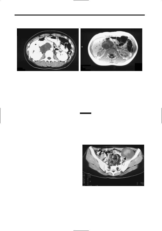
Figure 14.32. A: Abdominal wall hernia containing stomach. The


gastric body and antrum extend anteriorly through a hiatus


(arrow). B: Ventral hernia identified with CT (arrow). The hernia


had been growing and an adenocarcinoma was found during


hernia repair, believed to be secondary to a previously resected

A

colon cancer.

wide hiatus. A barium study detects bowel

others are detected with a small bowel barium

herniation.

study. A retrograde barium study through the

These hernias are somewhat difficult to

stoma is often helpful in outlining these hernias.

repair because of their wide hiatus and because


the surrounding structures tend to be weak. At


times extraperitoneal fixation of a prosthetic

Internal Hernia

mesh to the 12th rib superiorly and iliac crest


inferiorly is necessary.

Patients with an internal hernia range from


those who are asymptomatic, to those who have

Sciatic Foramen Hernia

intermittent nonspecific symptoms, to those


who have small bowel obstruction. Especially

In a sciatic foramen hernia a loop of bowel or

when intermittent, these hernias are difficult to

adjacent tissue herniates through one of the

diagnose with either barium studies or CT.

sciatic foramina posteriorly. Bowel obstruction

At times only a vague point of obstruction,

and strangulation ensue. These hernias are

either partial or complete, is identified with

rare.

imaging, but the etiology of obstruction is not

Sciatic hernias are a cause of chronic pelvic

evident.

pain in women; most are left sided, and a rare

Computed tomography findings of sympto-

one can even contain an ovary and fallopian

matic internal hernias consist of small bowel

tube. Computed tomography or MRI should

obstruction, a focal collection of dilated bowel

identify the hernial content.

displacing adjacent bowel, and stretched and


displaced mesenteric vessels.

Incisional Hernia

Peritoneoceles and enteroceles are discussed


in Chapter 5. Cystoceles are discussed in

Either omentum or part of the stomach or bowel

Chapter 11.

can be part of an incisional hernia (Fig. 14.32).


CT should detect these hernias, except that

Transmesenteric Hernia

some hernias reduce with the patient supine. A


barium study detects herniation if it includes

A transmesenteric hernia is the most common

stomach or bowel.

internal hernia. Although its appearance varies

A parastomal hernia is a type of incisional

considerably, the presence of dilated small

hernia occurring alongside an intestinal stoma

bowel loops close to the abdominal wall without

on the abdominal wall. Some of these hernias

overlying omental fat should suggest this diag-

are evident on physical examination while

nosis (73). Associated small bowel volvulus and

923

PERITONEUM, MESENTERY, AND EXTRAPERITONEAL SOFT TISSUES

ischemia are common complications, and thus surgical correction without undue delay is imperative with these hernias.

Bowel obstruction due to internal hernia is a complication after orthotopic liver transplantation (74); these obstructions are transmesenteric or retroanastomotic in location and are related to the choledochoenteric anastomosis. Similar to other transmesenteric hernias, some of these patients develop a volvulus and bowel ischemia. Computed tomography findings consist of abnormal mesenteric vessels and focal dilated small bowel loops displacing colon from the abdominal wall, yet a prospective diagnosis of internal hernia is rarely made (74).

Foramen of Winslow Hernia

A small bowel herniation into the lesser sac through the foramen of Winslow can lead to both small bowel obstruction and ischemia. A barium study reveals loops of small bowel posterior to the stomach and displacing the stomach anteriorly and inferiorly. A lesser sac hernia can also develop through a defect in the gastrocolic ligament; CT identifies herniated bowel surrounded by the liver, stomach, and pancreas.

Omentum or colon can also occasionally herniate. Ascending colon herniation through the

foramen of Winslow was identified with oral and IV contrast-enhanced CT (75); conventional radiographs revealed this herniated large bowel indenting and displacing the stomach to the left.

Paraduodenal Hernia

A paraduodenal hernia is associated with incomplete midgut rotation. These are also called mesocolic hernias and consist of small bowel, usually jejunum, herniating into a right or left paraduodenal fossa. Clinically, these patients range from asymptomatic to those with a small bowel obstruction.

In a left paraduodenal hernia the distal duodenum and proximal jejunum herniate through a peritoneal defect into a left fossa, posterior to the transverse and descending mesocolon.

Imaging of a typical left paraduodenal hernias reveals a discrete focal collection of jejunal loops posterior to the stomach and close to the pancreatic tail, although findings can be confusing. Noncontrast imaging initially suggests a pancreatic tail tumor, but oral contrast should identify this tumor to consist of jejunal loops located between the stomach and pancreas (Figs. 14.33 and 14.34). Bowel obstruction is inconsistent and often intermittent. The inferior mesenteric vein, which is mesocolic in location, outlines the anterior hernial margin and

A B

Figure 14.33. Left paraduodenal hernia. Transverse (A) and coronal (B) postcontrast CT show a small bowel loop within the left paraduodenal region (arrows). The inferior mesenteric vein (arrowheads) is displaced anteriorly and laterally. The central hypodense region in part A represents mesenteric fat. (Source: Okino Y, Kiyosue H, Mori H, et al. Root of the small bowel mesentery: correlative anatomy and CT features of pathologic conditions. Radiographics 2001;21:1475–1490, with permission from the Radiological Society of North America.)