

Учебники / Gene Therapy of Cochlear Deafness - Present Concepts and Future Aspects Ryan 2009
.pdfRegarding cellor time-specific regulatory sequences, their isolation, identification and verification can be difficult. The core promoter of a gene and proximal enhancers are typically contained within the first few hundred base pairs 5 to the locus at which mRNA transcription begins [16]. They are thus relatively easy to isolate and usually small enough to insert into a gene therapy vector. However enhancers, which most often determine cellular specificity, can be located much further upstream on the 5 side of the gene, or within introns or other transcribed regions of the gene, or 3 to the transcribed region [17, 18].
An initial step in the identification of regulatory elements is cross-species homology analysis. The regions of genes that are translated into protein are often quite homologous across species, because of the constraints imposed by protein function and the conservation of DNA codons. Regulatory regions of genes are also constrained, due to both structural constraints and conservation of DNA motifs that support the binding of TFs. However, the degree of conservation is much less than that seen in coding regions of DNA, since many TF recognition motifs display substantial sequence variation. This is especially true for enhancers and repressors. Even with these constraints, regulatory regions are often conserved across even widely separated species. Most proximal promoters show strong conservation, while about 60% of gene enhancers are also conserved [7]. Computer programs to identify conserved DNA sequences are helpful, but those that identify conserved patterns of TF-binding sites, even if they differ in underlying sequence, are more useful [19]. Of course, conservation provides only candidate regions. These must be cloned, inserted usually upstream from a reporter gene in a gene therapy vector, and expressed. Expression in cell lines can be suggestive, but in vivo expression is the ultimate test of regulatory construct specificity, either in terms of cellular or temporal specificity.
DNA segments extensive enough to contain the relevant regulatory elements may be too large to fit into a gene therapy vector. In this case, the regulatory elements must be identified, and unnecessary DNA edited out. Control of gene expression with such an edited promoter must then be verified. This requires in vivo analysis since in vitro responses can be quite different from those seen in the intact organism [20].
Regarding conditional promoters, binding sites for nuclear receptors can be added to many promoters to make them dependent upon ligands. However, this process does not work for all promoters. The conditional response element may disrupt normal promoter function, or may fail to regulate promoter activity [21]. Again, the function of the conditional promoter must be verified in vivo.
Gene-Regulatory Elements for Use in the Inner Ear
As noted above, the inner ear is a highly complex organ with many distinct cell types that contribute to its structure and function. Moreover, the development of the ear requires exceptional coordination amongst these and other cells in both the temporal and spatial
104 |
Ryan · Mullen · Doherty |
domains [22]. Thus the differential regulation of genes in the inner ear is extraordinarily complex. Even in the adult inner ear, many different genes must be expressed appropriately for the processes of auditory transduction and neural encoding to occur. During development, hundreds if not thousands of genes must be differentially expressed in various cell types and for varying periods of time. It is presumably for this reason that so many mutations have been found to affect the inner ear: more than 400 genetic syndromes include hearing loss as part of the phenotype [23], nearly 150 nonsyndromic deafness loci have been linked, and more than 60 nonsyndromic genes have been identified [http://webh01.ua.ac.be/hhh/]. It is estimated that mutations in more than 300 additional genes may produce inherited hearing loss. The diversity of mutations means that application of gene therapy to the inner ear will be challenging, with the need to produce gene products in many different cell types, and in many instances for restricted periods of time during development. Thus the cellular and temporal targeting of gene therapy may be particularly important for the inner ear.
Of course, not all gene therapy may need to be targeted to a particular cell type. For example, if delivery of a survival gene to hair cells (HCs) or spiral ganglion neurons (SGNs) was desired to protect them from a transient insult such as ototoxicity, it might not matter if the gene were also delivered to the surrounding cells. In contrast, if the gene product alters the function or structure of a cell, it would be more likely to be deleterious if expressed in the wrong cell type.
The utilization of cellor temporally-specific gene promoters to target gene expression is an effective means of controlling gene therapy. However, it should be noted that this is not the only manner in which targeting might be achieved. An alternative is to use a vector that exhibits preferential entry into only certain cell types. For example, some herpes viruses preferentially enter neurons [24], while different isotypes of adeno-associated virus show different cellular tropisms, which can be expanded through molecular engineering [25]. This aspect of targeting is discussed in other articles of this volume. Also, the transduction of cells via viral vectors is temporary, thus delivery at the appropriate time might be achieved by the appropriate timing of delivery. However, when these strategies are not practical, promoter control of gene therapy would be appropriate.
The ideal gene promoter for use in an inner ear cell type would be expressed in that cell type, and in no other cell. In the case of the adult inner ear, it would be expressed constitutively throughout the adult life of the cell. In the case of developmental gene therapy, it would be expressed at the time required for effective treatment of the targeted disorder.
Fortunately, the existence of diverse gene expression patterns provides ample genetic resources with which to target genes to the inner ear. While relatively few genes are expressed in the inner ear and nowhere else in the body, many genes have been shown to have expression patterns that are localized to particular cells within the inner ear. Given that gene therapy vectors can be delivered locally to the labyrinth, the promoters of such genes are good candidates for use in targeting gene delivery to
Targeting Inner Ear Gene Therapy |
105 |
inner ear cells. This is particularly true for inner ear neurons, given that there are many neural-specific genes from which promoters have been isolated.
Potential Cell-Specific Regulators of Inner Ear Gene Expression
Any gene that is selectively expressed in cells of the inner ear contains regulatory elements that could potentially be used to target gene expression. Not all such elements are necessarily suitable for this purpose. As noted above, enhancers/repressors can be located many thousands of bases away from the expressed sequence of a gene, and the identification of targeting regulatory sequences may not always be possible. However, in the majority of genes, enhancers and repressors are close enough to be identified, albeit this process typically requires considerable effort. If the natural sequence is too large for gene therapy vectors it can often be edited. A small number of promoters and enhancers that direct gene expression to cochlear cells have already been identified.
Hair Cells
Arguably the most obvious target for gene therapy of the inner ear is the HC. This highly specialized cell is central to the auditory and vestibular transduction process. Many of the genes responsible for inherited hearing loss are expressed in the HC, and their mutation primarily affects this cell type. Expressing therapeutic sequences in HCs is thus desirable, while expressing HC genes in other cochlear cells could well have deleterious effects on function. Therefore HC-specific promoters are of special interest for inner ear gene therapy.
There are a moderately large number of genes that are expressed in HCs but not in other types of cochlear cells. These are expressed at various periods in the life cycle of the cell, and thus could provide gene therapy at different developmental periods. A list of genes whose promoters could be used for this purpose is provided in table 1. In some cases, the promoters for these genes have been partially isolated and characterized.
The results of attempts at regulatory sequence isolation are informative. For example, the prestin gene is expressed in outer HCs, where it mediates electromotility and the active cochlear amplifier. When DNA fragments from the 5 region of the gene and the first intron were cloned and linked to a reporter in transgenic mice, expression was observed in inner and vestibular HCs, and in SGNs, but not in outer HCs [32].
An analysis of the Myo7A promoter by Boëda et al. [34] found that 118 bp 5 to the transcription initiation site, plus ~2 kb consisting of the first exon and intron of the gene, were sufficient to drive expression that closely mimicked that of Myo7A in the inner ear, specific to HCs after their formation. Neither the 5 118 bp nor the intron alone produced expression, so both regulatory elements were required.
106 |
Ryan · Mullen · Doherty |

Table 1. A selection of hair cell-specific genes
Gene |
Time of expression |
Hair cells |
Regulatory |
References |
|
|
|
sequence isolated |
|
|
|
|
|
|
Atoh1 |
Developmental |
All |
yes |
Bermingham et al. [26], |
|
|
|
|
Lumpkin et al. [27] |
|
|
|
|
|
POU4F3 |
Developmental, |
All |
Yes |
Erkman et al. [28], |
|
adult |
|
|
Ryan et al. [29] |
|
|
|
|
|
Lhx3 |
Developmental, |
All |
No |
Hertzano et al. [30] |
|
adult |
|
|
|
|
|
|
|
|
Prestin |
Adult |
OHCs |
Yes |
Zheng et al. [31], |
|
|
|
|
Tian et al. [32] |
|
|
|
|
|
Myo7A |
Developmental, |
All |
Yes |
Gibson et al. [33], |
|
adult |
|
|
Boëda et al. [34] |
|
|
|
|
|
Myo6 |
Developmental, |
All |
No |
Hasson et al. [35] |
|
adult |
|
|
|
|
|
|
|
|
Oncomod |
Adult |
OHCs |
No |
Sakaguchi et al. [36] |
|
|
|
|
|
α9AchR |
Developmental, |
All |
No |
Park et al. [37] |
|
adult |
|
|
|
|
|
|
|
|
α10AchR |
Developmental, |
All |
No |
Elgoyhen et al. [38] |
|
adult |
|
|
|
|
|
|
|
|
OHCs = Outer hair cells.
A construct consisting of 1.7 kb located 3 to the expressed sequence of the murine atoh1 (math1) gene produces expression in HCs of the inner ear, from fate specification through postnatal development (fig. 3), but not in adult HCs. This was found to be related to two highly conserved enhancer elements within the region. Interestingly, either enhancer could produce HC expression, indicating a high degree of redundancy. Moreover, both enhancers also drove expression in most other tissues that express Atoh1, thus the enhancer is not cell-specific. The enhancer construct also produces some expression in supporting cells, which is not observed in the atoh1 gene.
A construct consisting of 400 bp 5 to the POU4F3 gene does not produce expression in HCs. In contrast, 8.5 kb 5 to the POU4F3 gene produces expression that, in the postnatal mouse inner ear, is limited to HCs [39]. Expression begins shortly after HC fate is adopted, and it is present in all cochlear and vestibular HCs through the first 2 weeks of life. However, this fragment also produces expression in ganglion neurons prenatally, and produces less expression in adult outer HCs than is characteristic of the POU4F3 gene [29]. This presumably reflects the absence of enhancer and repressor elements located elsewhere in the gene.
Targeting Inner Ear Gene Therapy |
107 |
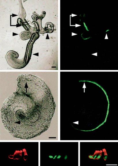
E13.5
utr
sac
crista
coch
a |
|
a` |
E14.5
b |
|
b` |
c |
E17.5 |
MyosinVIIa |
c` |
GFP |
c`` |
Fig. 3. Expression of green fluorescent protein (GFP) driven by regulatory elements from the atoh1 gene, in the inner ear of transgenic mice. a Entire membranous labyrinth at E13.5. The sensory organs of the vestibular system, including the utricle (utr), saccule (sac), and crista, which contain differentiating hair cells (HCs), express GFP. The more immature cochlea (coch), in which HCs have yet to develop, does not. b Dissected cochlea at E14.5. HCs have now developed through 1.5 turns. The arrow indicates the basal region where HC differentiation initiates, arrowhead indicates the leading edge of HC differentiation as shown by the extent of GFP-expressing HCs. Scale bars equal 100 μm. c Confocal image of E176.5 organ of Corti [27]. Reprinted from Gene Expr Patterns 2003;3:389–395, Math 1-driven GFP expression in the developing nervous system of transgenic mice, Lumpkin EA et al., with permission from Elsevier.
108 |
Ryan · Mullen · Doherty |
This illustrates a common problem with enhancer/promoter constructs. They usually do not replicate the expression pattern of the gene with complete fidelity, due to an incomplete complement of regulatory elements, or to potential differences in secondary structure or epigenetic alterations of construct DNA when compared to native DNA. Other post-transcriptional regulatory mechanisms may also generate differences between the gene products of a native gene and a transgene. MicroRNAs targeted to regions of the native transcript may degrade it prior to translation [12] in ways that do not occur in the transcript as encoded by the gene therapy vector, since introns and other untranslated regions are often deleted.
Supporting Cells
Another important target for gene therapy is the supporting cells of the sensory epithelia. The most common inherited form of nonsyndromic deafness is due to mutations in the gene encoding connexin 23 [40], which is highly expressed in adult supporting cells but not in HCs [41]. Other genes expressed selectively in supporting cells include α- and β-tectorin, which are components of the acellular membrane that lie above the inner ear sensory epithelia. The supporting cell antigen is a protein of unknown function that is highly expressed in supporting cells [42]. The cell cycle regulator, p27/Kip1, is expressed in developing supporting cells, but not HCs [43]. A p27/Kip1 construct that directs expression to the inner ear has been generated by recombination in a bacterial artificial chromosome, but the regulatory sequences have not been further localized [44]. FGF receptor 3 is expressed in developing pillar cells and Deiter’s cells of the cochlea [45]. A promoter for this gene has been isolated and defined [46], but not for the inner ear. The LIM-domain protein Islet 1 is expressed in developing HCs and supporting cells, but in adulthood becomes restricted to supporting cells [47]. None of the regulatory elements that direct the expression of these or other genes to supporting cells have been characterized, but given the number of candidates this should certainly be possible.
Inner Ear Ganglion Neurons
The SGNs are the only neurons whose cell bodies are found within the cochlea. Thus a wide variety of genes with expression that is restricted to neurons could supply promoters for use with locally applied gene therapy vectors. This includes a variety of genes encoding neuron-specific structural proteins such as neurofilaments, neurotransmitter receptors such as GluR2 or GluR3. Similarly, many neuron-specific developmental genes are expressed only in the neurons of the inner ear. This includes TFs such as POU4F1 and POU4F2 [28, 48]. Differentiation between type I and type II
Targeting Inner Ear Gene Therapy |
109 |
SGNs could be achieved using the promoter of the peripherin gene [49], which is only expressed in type II neurons [50].
Stria Vascularis
Inherited deafness can be caused by mutation of a number of genes normally expressed in the stria vascularis, due to its critical function as an ion transport epithelium and generator of the endocochlear potential. A number of these genes are preferentially expressed in the stria vascularis, and not in other cochlear tissues. For example, the gene encoding the β2 subunit of Na,K-ATPase is expressed in the marginal, intermediate and basal cells of the stria during late development and adulthood [51]. Bioinformatic analysis of the gene indicates 768 bp of a highly conserved sequence immediately 5 to the CAP site, suggesting promoter/enhancer elements that may control tissue-specific expression [52], although this has not been tested in the inner ear. The genes encoding the KVLQT1 and IsK potassium channels, and ubiquitin A-52, are expressed only in the marginal cells, again in late development and adulthood [53–55]. Promoters for the potassium channel genes have yet to be isolated. However the uba52 promoter region has been partially characterized [56].
Spiral Ligament
Recent observations have supported the concept that the spiral ligament is a very active participant in cochlear function, serving as a site for ion transport between the perilymph and stria vascularis [57] and perhaps serving other functions. This is consistent with the level of glucose metabolism that has been observed in the ligament [58], which equals that of the stria. Genes that are selectively expressed at this site include Crym, found in fibrocytes of the ligament, and collagen type IX, found in type II fibrocytes [55].
Other Promoters
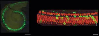
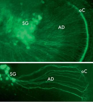
In addition to genes that are normally expressed in tissues, some altered, viral or hybrid promoters have been found to be expressed selectively in cochlear tissues. This is presumably due to the fortuitous presence of binding sites for regulatory factors that are present in cochlear cells. An example of this is the human elongation factor 1α promoter (fig. 4), which for unknown reasons induces expression preferentially in cells of the organ of Corti [59]. Neuron-specific promoters have also been generated artificially. A mutated form of the thy1 promoter has been shown to drive expression in neurons, with integration into different sites producing expression in different subsets of neurons [60], including SGNs (fig. 5).
110 |
Ryan · Mullen · Doherty |
Base
|
|
sc |
pillar |
|
|
|
|
|
Apex |
|
|
|
|
|
ohc |
a |
Midbase |
|
b |
|
|
Fig. 4. Expression of green fluorescent protein (GFP) driven by the human elongation factor 1α (ef1α) promoter in the organ of Corti of the mouse. Plasmid DNA was electroporated into the E11.5 mouse otocyst. The inner ear was fixed and processed for histochemistry at E17.5 [59]. Reprinted from Methods Mol Biol 2009;493:125–139, Electroporation-mediated gene transfer to the developing mouse inner ear, Brigande JV et al., with kind permission of Springer Science+Business Media.
Potential Limitations
The isolation of functional cell-specific regulatory sequences is not always possible due to distant enhancers or lack of sufficient homology for identification. Moreover, many of the regulatory sequences that have been isolated, as listed above, have been characterized in transgenic models in which the promoter is integrated into the genome. However, regulatory sequences may behave differently in a viral vector. This can be due to changes in the conformation of the DNA due to vector DNA. Alternatively, the state of acetylation or methylation of nuclear proteins or DNA within in the regulatory sequence may be different in a viral vector than in native DNA. Artificial DNA introduced into cells has been shown to be associated with histones, but in a manner different to that seen in the genome [61]. Viruses also produce their own histone-like proteins that can be acetylated [62] and that may interact with transgene-regulatory sequences. Interestingly, inhibitors of histone deactylation have been shown to significantly increase viral vector transduction of mammalian cells [63], implicating acetylation as an important factor in viral vector gene expression. This has recently been found to be true for transduction of cells in the organ of Corti by adenovirus vectors [64].
Conclusions
The delivery of gene therapy to specific cochlear cells will be aided by the use of generegulatory mechanisms that occur naturally in cochlear cells. It is clear from the above discussion that many combinations of gene promoters and enhancers are available
Targeting Inner Ear Gene Therapy |
111 |
a
b
Fig. 5. Expression of yellow fluorescent protein, driven by an altered thy1 promoter, in the neurons of the neonatal spiral ganglion of a transgenic mouse. a Neuronal somata in the spiral ganglion (SG) are intensely labeled, as are afferent dendrites (AD) and a dense plexus of nerve terminals underneath the inner hair cells (HCs) in the organ of Corti (oC). b Higher magnification image of SG neurons and their dendrites, with branching terminations under the inner HCs characteristic of this developmental stage.
that could be used to target gene delivery to particular cochlear cells. Additional regulatory sequences are sure to be discovered as the characterization of genes expressed in the inner ear continues. As enhancers that direct expression to particular inner ear cell types are better understood, it will also be possible to engineer vectors that combine different features. Thus it will be possible to use enhancers to direct expression to a cell type, via a promoter that can be separately regulated. The most obvious example would be to combine enhancers that direct expression to a particular cell type, with conditional promoters that can be turned on or off by the application of an endogenous agent such as a Tet-On response element. The promise of regulatory control of gene therapy vectors seems great.
112 |
Ryan · Mullen · Doherty |

References
1 Bazan-Peregrino M, Seymour LW, Harris AL: Gene therapy targeting to tumor endothelium. Cancer Gene Ther 2007;14:117–127.
2Friedmann T: Gene Therapy: Fact and Fiction in Biology’s New Approaches to Disease. Woodbury, CSHL Press, 1994.
3 Lodish H, Berk A, Zipursky S, Lawrence S, Matsudaira P, Baltimore D, Darnell J (eds): Molecular Cell Biology. New York, Freeman, 1999.
 4 Schones DE, Zhao K: Genome-wide approaches to studying chromatin modifications. Nat Rev Genet 2008;9:179–191.
 5 Verdone L, Agricola E, Caserta M, Di Mauro E: Histone acetylation in gene regulation. Brief Funct Genomic Proteomic 2006;5:209–221.
 6 Zhang Y, Reinberg D: Transcription regulation by histone methylation: interplay between different covalent modifications of the core histone tails. Genes Dev 2001;15:2343–2360.
 7 Heintzman ND, Stuart RK, Hon G, Fu Y, Ching CW, Hawkins RD, Barrera LO, Van Calcar S, Qu C, Ching KA, Wang W, Weng Z, Green RD, Crawford GE, Ren B: Distinct and predictive chromatin signatures of transcriptional promoters and enhancers in the human genome. Nat Genet 2007;39:311–318.
 8 Leonhardt H, Rahn HP, Cardoso MC: Functional links between nuclear structure, gene expression, DNA replication, and methylation. Crit Rev Eukaryot Gene Expr 1999;9:345–351.
 9 Haines TR, Rodenhiser DI, Ainsworth PJ: Allelespecific non-CpG methylation of the Nf1 gene during early mouse development. Dev Biol 2001;240: 585–598.
 10 Lobe CG: Transcription factors and mammalian development. Curr Top Dev Biol 1992;27:351–383.
11Davidson EH: The Regulatory Genome: Gene Regulatory Networks in Development and Evolu-
tion. New York, Academic Press, 2006.
 12 Chen K, Rajewsky N: The evolution of gene regulation by transcription factors and microRNAs. Nat Rev Genet 2007;8:93–103.
 13 Hallikas O, Palin K, Sinjushina N, Rautiainen R, Partanen J, Ukkonen E, Taipele J: Genome-wide prediction of mammalian enhancers based on analysis of transcription factor binding affinity. Cell 2006; 24:47–59.
 14 Sipo I, Wang X, Hurtado Picó A, Suckau L, Weger S, Poller W, Fechner H: Tamoxifen-regulated adenoviral E1A chimeras for the control of tumor selective oncolytic adenovirus replication in vitro and in vivo. Gene Ther 2006;13:173–186.
 15 Fechner H, Wang X, Picó AH, Wildner J, Suckau L, Pinkert S, Sipo I, Weger S, Poller W: A bidirectional Tet-dependent promotor construct regulating the expression of E1A for tight control of oncolytic adenovirus replication. J Biotechnol 2007;127:560–574.
 16 Fickett JW, Hatzigeorgiou AG: Eukaryotic promoter recognition. Genome Res 1997;7:861–878.
 17 Visel A, Bristow J, Pennacchio LA: Enhancer identification through comparative genomics. Semin Cell Dev Biol 2007;18:140–152.
 18 Heintzman ND, Ren B: The gateway to transcription: identifying, characterizing and understanding promoters in the eukaryotic genome. Cell Mol Life Sci 2007;64:386–400.
 19 Xie D, Cai J, Chia NY, Ng HH, Zhong S: Cross-species de novo identification of cis-regulatory modules with GibbsModule: application to gene regulation in embryonic stem cells. Genome Res 2008;18:1325– 1335.
 20 Oosterveen T, van Vliet P, Deschamps J, Meijlink F: The direct context of a hox retinoic acid response element is crucial for its activity. J Biol Chem 2003; 278:24103–24107.
 21 Houdebine LM: Methods to generate transgenic animals and to control transgene expression. J Biotechnol 2002;98:145–160.
 22 Friedman LM, Dror AA, Avraham KB: Mouse models to study inner ear development and hereditary hearing loss. Int J Dev Biol 2007;51:609–631.
23 Toriello HV, Reardon W, Gorlin HJ: Hereditary Hearing Loss and Its Syndromes. Oxford, Oxford University Press, 2004.
 24 Frampton AR Jr, Goins WF, Nakano K, Burton EA, Glorioso JC: HSV trafficking and development of gene therapy vectors with applications in the nervous system. Gene Ther 2005;12:891–901.
 25 Kwon I, Schaffer DV: Designer gene delivery vectors: molecular engineering and evolution of adenoassociated viral vectors for enhanced gene transfer. Pharm Res 2008;25:489–499.
 26 Bermingham NA, Hassan BA, Price SD, Vollrath MA, Ben-Arie N, Eatock RA, Bellen HJ, Lysakowski A, Zoghbi HY: Math1:an essential gene for the generationofinnerearhaircells.Science1999;284:1837– 1841.
 27 Lumpkin EA, Collisson T, Parab P, Omer-Abdalla A, Haeberle H, Chen P, Doetzlhofer A, White P, Groves A, Segil N, Johnson JE: Math1-driven GFP expression in the developing nervous system of transgenic mice. Gene Expr Patterns 2003;3:389– 395.
Targeting Inner Ear Gene Therapy |
113 |