

Учебник по НТП
.pdfV. Enhancing Skills In English-Russian Interpretation
Render orally the following text:
The Human Eye
The human eye is wrapped in three layers of tissue. The first is the sclerotic coat which creates the "white" of the eye except in the front where it forms the transparent cornea. The cornea admits light to the interior of the eye and bends the light rays to that they can be brought to a focus. The surface of the cornea is kept moist and dust-
free by secretions from the tear glands.
The second layer, the choroid coat, is deeply pigmented with melanin. The choroid coat forms the iris which, too, is pigmented and is responsible for eye "color" depending on how much melanin is in it. The darker the eye color, the more melanin there is in the iris. So, dark brown eyes have a lot of melanin in them, while the blue eyes have the least of all. Melanin is a pigment that plays a part in protection against the sun, which is why paler-colored eyes have only evolved in cooler, less sunny climates. Sometimes human eyes are different colors, or heterochromia iridium. It’s thought to result from an alteration to one of the genes that control eye color.
The lens is located just behind the iris. It is held in position by zonules extending from an encircling ring of muscle. When this ciliary muscle is relaxed, its diameter increases, the zonules are put under tension, and the lens is flattened; contracted, its diameter is reduced, the zonules relax, and the lens becomes more spherical. These changes enable the eye to adjust its focus between far objects and near objects. If the eyeball is too short or the lens too flat or inflexible, the light rays entering the eye–particularly those from
113

nearby objects–will not be brought to a focus by the time they strike the retina. Eyeglasses with convex lenses can correct the problem. Farsightedness is called hypermetropia. If the eyeball is too long or the lens too spherical, the image of distant objects is brought long or the lens too spherical, the image of distant objects is brought to a focus in front of the retina and is out of focus again before the light strikes the retina. Nearby objects can be seen more easily. Eyeglasses with concave lenses correct this problem by diverging the light rays before they enter the eye. Nearsightedness is called myopia.
The pupil of the eye is variable and under the control of the autonomic nervous system. In dim light (or when danger threatens), the pupil opens wider letting more light into the eye. In bright light the pupil closes down. This not only reduces the amount of light entering the eye but also improves its image-forming ability (as does "stopping down" the iris diaphragm of a camera).
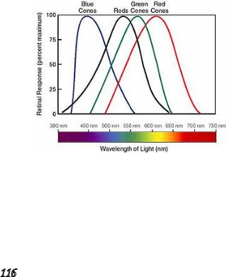
The last but not the least layer is the retina. It contains the light receptors, the rods and three kinds of cones, each "tuned" to absorb light from a portion of the spectrum of visible light. Rods are extremely sensitive to light. A single photon (the minimum unit of light) absorbed by a small cluster of adjacent rods is sufficient to send a signal to the brain. So although rods provide us with a relatively grainy, colorless image, they permit us to detect light that is over a billion times dimmer than what we see on a bright sunny day.
The three types of cones provide us the basis of color vision. Cones are "tuned" to different portions of the visible spectrum: cones that absorb long-wavelength light (red) peaking at 565 nm; cones that absorb middle-wavelength light (green) with a peak absorption at 535 nm; cones that absorb short-wavelength light (blue) with a peak absorption at 440 nm. The response of cones is not all-or-none. Light of a given wavelength (color), say 500 nm (green), stimulates all three types of cones, but the green-absorbing cones will be stimulated most strongly. Like rods, the absorption of light does not trigger action potentials but modulates the membrane potential of the cones.
Most of the 3 million cones in each retina are confined to a small region just opposite the lens called the fovea. So our sharpest and colorful images are limited to a small area of view. Because we can quickly direct our eyes to anything in view that interests us, we tend not to be aware of just how poor our peripheral vision is.
Patient: I always see spots before my eyes.
Doctor: Didn’t the new glasses help?
Patient: Sure, Doctor! Now I see the spots much clearer.
114

Abnormal color insensitivity occasionally occurs in humans, though the term color blindness is something of a misnomer. Very few (~1 in 105) people cannot distinguish colors at all. Most "color-blind" people actually confuse the red and green of traffic lights. As high as 8% of the males in some populations have an inherited defect in their ability to discriminate reds and greens. These defects are found almost exclusively in males because the genes that encode the red-absorbing and greenabsorbing opsins (transmembrane protein) are on the X chromosome.
VI. Enhancing Skills In Russian-English Interpretation
Render orally the following text:
Ночное Зрение
Известно, что глаз человека отличается совершенной оптической системой, позволяющей фиксировать огромный диапазон
видимых световых раздражителей, интенсивность которых может различаться в 10 млрд. раз. Особый интерес для психологов и физиологов представляют закономерности ночного зрения человека. Наиболее интенсивно такие поиски велись в годы второй мировой войны и были нацелены на решение конкретных прикладных военных задач.
В процессе исследований выявлено, что сетчатка глаза состоит из нескольких слоев нервных клеток, заканчивающихся колбочками и палочками (рецепторами света). Колбочки, которых
всетчатке насчитывается около 7 млн., обладают более низкой чувствительностью и представляют собой аппарат дневного света, позволяющий различать цвета. Палочки, количество которых
всетчатке составляет около 130 млн., отличаются высокой чувствительностью к слабым интенсивностям света и являются аппаратами ночного зрения. Повышение световой чувствительности глаз по мере пребывания в темноте получило название темновой адаптации. Установлено, что темновая адаптация начинается с момента погружения глаз в темноту и стабилизируется через 6080 мин. Познание закономерностей ночного видения позволяет существенно ускорить время темновой адаптации глаз. Каковы же эти закономерности?
Во-первых, колбочки и палочки распределены по сетчатке глаза неравномерно. Первые расположены в центре, вторые же находятся на периферии. Отсюда следует, что для обнаружения малозаметных объектов в ночное время их лучше рассматривать периферической частью сетчатки.
Во-вторых, колбочки и палочки заметно отличаются по степени
115
чувствительности к свету с различной длиной волн. Так, адаптированный к темноте глаз наиболее чувствителен к длинам волн порядка 511 миллимикрон и почти не воспринимает волны с длинами выше 620 миллимикрон. Это означает, что при ночном зрении глаз максимально чувствителен к синему цвету и относительно нечувствителен к красному. Другими словами, ночью объекты синего цвета мгновенно обнаруживаются человеком, но при этом возможна так называемая темновая дезадаптация глаз, т.е. снижение их чувствительности к свету, «засветка»).
В годы второй мировой войны в армии США были сконструированы плотно прилегающие красные очки, которые не пропускали лучей длиной волны меньше 620 миллимикрон, но через которые проходило достаточно света для функционирования колбочкового зрения. В таких очках человек мог находиться в хорошо освещенном помещении и одновременно полностью адаптироваться к темноте. Это позволяло практически исключить этап темновой адаптации глаз и мгновенно включаться в деятельность.
В-третьих, установлена закономерность функционирования симпатической нервной системы человека, проявляющаяся в том, что возбуждение одной из систем симпатической иннервации влечет за собой возбуждение прочих симпатических систем. Группа ученых Института психологии АПН РСФСР в 1941-1946 годах исследовала способы улучшения ночного зрения и выделила так называемые «физиологические стимуляторы», позволяющие в короткое время значительно повысить световую чувствительность глаз.
116

К таким стимуляторам отнесены:
а) форсированное дыхание (или гипервентиляция, т.е. углубленное, резкое и частое дыхание, начинающееся с полного выдоха, в течение 1-3 минут);
б) термические раздражители (обтирание лица холодной водой, холодный компресс на затылок);
в) мышечная работа (простые гимнастические упражнения); в) вкусовые раздражители (было отмечено, что после употреб-
ления 10 грамм сахара чувствительность ночного зрения возрастает на 210%, при прослушивании приятной музыки—на 240%. И, напротив, при прослушивании грустной музыки чувствительность к свету снижается на 60% и при употреблении горькой пищи—на 50%).
Проведенные эксперименты до сих пор считаются классическими, а полученные результаты—актуальными. Они свидетельствуют о том, что применение физиологических стимуляторов позволяет повысить чувствительность ночного зрения и слуха на 40-50%. Время темновой адаптации глаз сокращается с 40-50 мин. до 4-5 мин., то есть зрительная чувствительность возрастает в 10 раз быстрее, чем при темновой адаптации без применения стимуляторов. При этом однократное их применение обеспечивает повышение чувствительности на 1-1,5 часа, многократное—на 2-3 часа. Одновременно названные стимуляторы являются надежным и эффективным средством снятия утомления в области кинестатической и сенсорной деятельности.
VII. Solving Translation Problems
English technical, scientific, and medical texts are abundant in attributive noun groups—strings of nouns that can be of 2-5 and even more words long. In these groups, the rightmost (last) noun is called the head-word, and all the other words it precedes in the string serve as its attributes (determinants). In most cases there are no equivalent syntactic patterns for attributive noun groups in English and in Russian, so translation strategies may employ various transformations and additional explanations, for example:
blood testing set—набор для исследования крови;
anaesthesia column housing—прибор для наблюдения за пациентами в операционной;
last cruise control set speed—последняя установленная скорость стабилизации;
Copy out the underlined attributive noun groups from the text below and translate them into Russian. Comment on transformations you’ve made to convey their meaning. Translate the entire text into Russian.
117

Blood Groups And Blood Typing
Experiments with blood transfusions (the transfer of blood or blood components into a person’s blood stream) have been carried out for hundreds of years. It was not until 1901, when the Austrian Karl Landsteiner discovered human blood groups, that blood transfusions became safer. Mixing blood from two individuals can lead to blood clumping or agglutination. The clumped red cells can crack and cause toxic reactions which may lead to fatal consequences. Karl Landsteiner found out that blood clumping was an immunological reaction which occurs when the blood transfusion receiver has antibodies against the donor blood cells. Karl Landsteiner’s work made it possible to determine blood types and thus paved the way for blood transfusions to be carried out safely. For this discovery he was awarded the N0bel Prize
in Physiology or Medicine in 1930.
An adult human has about 4–6 liters of blood circulating in the body. Among other things, blood transports oxygen to various parts of the body. Blood consists of several types of cells floating around in a fluid called plasma. The red blood cells contain hemoglobin, a protein that binds oxygen. Red blood cells transport oxygen to, and remove carbon dioxide from the body tissues. The white blood cells fight infection.
The differences in human blood are due to the presence or absence of certain protein molecules called antigens and antibodies. The antigens are located on the surface of the red blood cells and the antibodies are in the blood plasma. Individuals have different types and combinations of these molecules inherited from their parents. There are more than 20 genetically determined blood group systems known today, but the AB0 and Rh systems are the most important ones used for blood transfusions. According to the AB0 blood typing system there are four different kinds of blood types: A, B, AB or 0 (null). The table shows the four AB0 phenotypes (“blood groups”) present in the human population and the genotypes that give rise to them.
Phenotypes |
Antigens |
Antibodies |
Genotypes |
|
on RBCs |
in serum |
|||
|
|
|||
A |
A |
Anti-B |
AA or A0 |
|
B |
B |
Anti-A |
BB or B0 |
|
AB |
A and B |
Neither |
AB |
|
0 |
Neither |
Anti-A and anti-B |
00 |
118
The blood group 0 (null): no A or B antigens on the surface of the red blood cells but both A and B antibodies are in the blood plasma. The O group is the oldest of the blood groups. Back in the Stone Age, everyone would have been O and today it’s still the most common group in the UK, especially in the N0rth of England. Over in Central and South America and the USA most people are O too. The fact that anyone can receive O blood reflects the fact that all other blood groups are derived from it.
The blood group A: A antigens on the surface of the red blood cells and B antibodies in the blood plasma. Group A is the second oldest blood group, appearing around 25,000–15,000 BC, when larger human settlements first appeared as farming developed. It’s the commonest group in N0rway, Denmark, Austria, Armenia and Japan.
The blood group B: B antigens on the surface of the red blood cells and A antibodies in the blood plasma. In the Chinese or Asian communities around a quarter of all people share the blood group B. It emerged between 15,000 and 10,000 BC as tribes migrated from Africa to Europe, Asia and the Americas and mingled with other populations.
The blood group AB: both A and B antigens on the surface of the red blood cells and no A or B antibodies at all in the blood plasma. The newest and rarest group, AB, only appeared between 1000 and 500 years ago, and is believed to have occurred as a response to the existing blood groups interfusion on a major scale. In Japan, China and Pakistan around 10% of the population boast this rarest of blood groups.
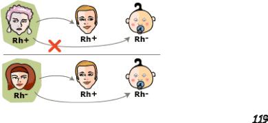
Rh positive and negative antigens are transmembrane proteins with loops exposed at the surface of red blood cells. They appear to be used for the transport of carbon dioxide and/or ammonia across the plasma membrane. They are named for the rhesus monkey in which they were first discovered. The major importance of the Rh system for human health is to avoid the danger of mother-fetus RhD incompatibility. During birth, there is often a leakage of the baby’s red blood cells into the mother’s circulation. If the baby is Rh positive (having inherited the trait from its father) and the mother Rh-negative, these red cells will cause her to develop antibodies against the RhD antigen. The antibodies do
not cause any problems for that child, but can cross the placenta and attack the red cells of a subsequent Rh+ fetus. This destroys the red cells producing the disease, called erythroblastosis fetalis or hemolytic disease of the newborn, which may be so severe as to kill the fetus or even the newborn infant.
119

For a successful blood transfusion, AB0 and Rh blood groups must be compatible between the donor blood and the patient blood. If they are not, the red blood cells from the donated blood will agglutinate and clog blood vessels to stop the blood circulation to various parts of the body. The transfusion will work if a patient to receive blood has a blood group without any antibodies against the donor blood’s antigens. People with blood group 0 are called “universal donors” and people with blood group AB–“universal receivers.”
Blood |
Antigens |
Antibodies |
Can Give |
Can Receive |
Group |
Blood To |
Blood From |
||
AB |
A and B |
None |
AB |
AB, A, B, 0 |
A |
A |
B |
A and AB |
A and 0 |
B |
B |
A |
B and AB |
B and 0 |
0 |
None |
A and B |
AB, A, B, 0 |
0 |
VIII. Mastering English Grammar
Translate the sentences into Russian paying special attention to the equivalent-lacking grammatical structures:
1. Declarative knowledge provides an explicit, consciously accessible record of individual previous experiences and a sense of familiarity about those experiences.
2.An important factor that influences what is stored and how strongly it is stored is whether the action is followed by rewarding or punishing consequences.
3.After years of study, there is much support for the idea that memory involves a persistent change in the relationship between neurons.
4.Another important model for the study of memory is the phenomenon of long-term potentiation (LTP), a long-lasting increase in the strength of a synaptic response following stimulation.
5.Scientists believe that no single brain center stores memory.
6.In non-right handers (which include left-handed and ambidextrous people), language functions are far more likely to involve the right hemisphere.
7.A prominent and influential model is based on studies of patients who have lost speech and language abilities due to stroke, and from behavioral and functional neuroimaging studies of normal people.
120

8.Wernicke’s area is a semantic processing area. It is associated with the short-term memory involved in speech recognition and production, as well as some hearing function and object identification.
9.The amygdala appears to play a prominent role in these memory events.
10.Damage to Wernicke’s area causes Wernicke’s aphasia, a condition in which people can hear language being spoken, but cannot understand it.
IX. Fostering Critical Thinking Skills
Read the text. Find additional material to expand the topic and write a commented essay in Russian on Memory and Language:
Memory And Language
While much is unknown about memory, scientists can recognize certain pieces of the process. The motivation of researchers is twofold: to understand human behavior better—from how we learn to why people have trouble remembering some facts—and to discover ways to prevent or cure many devastating brain disorders. For example, the brain appears to process different kinds of information in separate ways and then store it differently. Procedural knowledge, the knowledge of how to do something, is expressed in skilled behavior and learned habits. Declarative knowledge provides an explicit, consciously accessible record of individual previous experiences and a sense of familiarity about those experiences. Declarative knowledge requires processing in the medial temporal region and parts of the thalamus, while procedural knowledge requires processing by the basal ganglia. Other kinds of memory depend on the amygdala (emotional aspects of memory) and the cerebellum (motor learning where precise timing
is involved).
An important factor that influences what is stored and how strongly it is stored is whether the action is followed by rewarding or punishing consequences. This is an important principle in determining what behaviors an organism will learn and remember. The amygdala appears to play a prominent role in these memory events.
How exactly does memory occur? After years of study, there is much support for the idea that memory involves a persistent change in the relationship between neurons. The stability of long-term memory is conferred by structural modifications within neurons that change the strength and number of synapses. Another important model for the study of memory is the phenomenon of longterm potentiation (LTP), a long-lasting increase in the strength of a synaptic response following stimulation. LTP occurs prominently in the hippocampus, as well as in other brain areas.
121

It is good to rub and polish our brain against that of others. Michel deMontaigne
The more you use your brain, the more brain you will have to use.
George A. Dorsey
Scientists believe that no single brain center stores memory. It most likely is stored in the same, distributed collection of cortical processing systems involved in the perception, processing and analysis of the material being learned. In short, each part of the brain most likely contributes differently to permanent memory storage.
One of the most prominent intellectual activities dependent on memory is language. As is true for every other functional ability, parts of the brain specialize in language. We now know that there are small differences in the sizes of some regions in the the left and the right hemispheres. These differences may form the basis for the first major brain specialization for language—lateralization of language to the left hemisphere.
In about 98 percent of right-handers, the left hemisphere accomplishes most language processing functions. In non-right handers (which include left-handed and ambidextrous people), language functions are far more likely to involve the right hemisphere. There is some evidence that lateralization differs in males and females. There is also evidence that the non-dominant hemisphere is primarily involved in functions that are just one step beyond the essential language functions of relating form to literal meaning. These include determining the emotional state of a speaker from his or her tone of voice, and appreciating humor and metaphor.
A prominent and influential model, based on studies of patients who have lost speech and language abilities due to stroke, and from behavioral and functional neuroimaging studies of normal people, proposes that the underlying structure of speech comprehension arises in Wernicke’s area, a portion of the left hemisphere of the brain. This temporal lobe region is connected with Broca’s area in the frontal lobe where a program for vocal expression is created. This program is then transmitted to a nearby area of the motor cortex that activates the mouth, tongue and larynx.
Wernicke’s area is a semantic processing area. It is associated with the short-term memory involved in speech recognition and production, as well as some hearing function and object identification. Wernicke’s area is most often associated with processing of incoming language, whether it be written, spoken or signed. Damage to Wernicke’s area causes
122