

|
2. NMR spectroscopy of dienes and polyenes |
119 |
||||||
TABLE 25. |
13C (67.5 MHz) and (400 MHz) 1H NMR spectral data of alisamicin |
|||||||
(75) (CDCl3, 303 K)a |
|
|
|
|
|
|
|
|
Position |
υCb |
|
|
1H |
|
|
|
|
|
|
|
υ |
|
HMBC partner |
|||
|
|
|
|
|
|
|
||
|
|
(multiplicity, J in Hz) |
|
2JCH |
3JCH |
|||
1 |
188.63 |
|
— |
|
|
|
|
|
2 |
128.08 |
|
— |
|
|
|
|
|
3 |
126.36 |
7.40 |
(d, 2.6) |
|
C2 |
C1 |
||
4 |
71.20 |
|
— |
|
|
|
|
|
5 |
57.41 |
3.70 |
(dd, 2.6, 3.6) |
|
C4 |
C7 |
||
6 |
52.93 |
3.65 |
(d, 3.6) |
|
C1, C5 |
C2 |
||
7 |
136.29 |
5.86 |
(dd, 14.5, 0.3) |
|
|
C3, C9 |
||
8131.58 6.58 (dd, 11.3, 14.5)
9139.52 6.58 (dd, 14.8, 11.3)
10131.74 6.42 (ddd, 11.2, 14.8, 0.3)
11 |
143.45 |
7.32 |
(dd, 11.2, 14.7) |
|
C13 |
12 |
121.59 |
6.05 |
(d, 14.7) |
C13 |
C10 |
13 |
165.48 |
|
— |
|
|
10 |
165.16 |
|
— |
C10 |
|
20 |
120.95 |
5.84 |
(d, 14.8) |
|
|
30 |
144.16 |
7.22 |
(ddm, 14.8, 10.5) |
C30 |
|
40 |
125.52 |
6.12 |
(dd, 10.5, 15.5) |
C30 |
|
50 |
150.76 |
6.12 |
(m) |
|
|
60 |
41.13 |
2.10 |
(m) |
|
|
70 , 110 |
32.25 |
1.76 |
(m) (eq), |
|
|
80 , 100 |
|
1.13, (m) (ax) |
|
|
|
25.80 |
1.73 |
(m) (eq), |
|
|
|
90 |
|
1.28 |
(m) (ax) |
|
|
26.00 |
1.67 |
(m) (eq), |
|
|
|
100 |
|
1.18 |
(m) (ax) |
|
|
197.39 |
|
— |
|
|
|
200 |
115.01 |
|
— |
|
|
300 |
174.15 |
|
— |
|
|
400 |
32.14 |
2.61 |
(m) |
|
|
500 |
25.65 |
2.53 |
(m) |
|
|
300 -OH |
— |
13.52 |
(s) |
|
|
4-OH |
— |
3.25 |
(s) |
C10 |
C1, C3 |
2-NH |
— |
7.54 |
(s) |
||
13-NH |
— |
7.58 |
(s) |
C13 |
C300 |
aThe 1H and 13C chemical shifts are in ppm from CH3 4Si and CDCl3 as internal standards, respectively.
bThe carbon multiplicities were determined by DEPT-135 experiment.
was obtained from the culture filtrate of an actinomycete identified as Streptomyces gannmycius, and found to be identical with 77 by direct comparison.
The structure was determined by NMR spectral analysis including a variety of twodimensional NMR techniques. The 500-MHz 1H NMR spectrum of 77 taken in CDCl3 (Figure 26) revealed the presence of 5 aromatic protons, 15 olefinic protons, a methoxy (υ3.65), an allylic methyl (υ2.14) and a tertiary methyl group (υ1.33). The 13C NMR spectrum showed signals due to all 34 carbons, which were assigned to 7 quaternary carbons, 23 methines, 1 methylene and 3 methyls by DEPT experiments. The 13C and 1H NMR spectral data are summarized in Table 27.

120 |
Yoshito Takeuchi and Toshio Takayama |
||
|
O |
H |
H |
|
NH |
|
|
|
O |
|
H |
|
O |
H |
H |
H
OH
H
 H
H
H
 H
H
H
 H
H
O
NH
OH  O
O
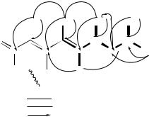
FIGURE 24. NOE network of alisamycin (75). Reproduced by permission of Japan Antibiotics Research Association from Reference 45
|
|
8′ |
|
|
|
7′ |
|
|
|
H3C |
|
|
|
CH3 |
|
||
|
|
|
|
|
|
6′ |
|
|
|
9′ |
|
|
|
|
5′ |
|
|
|
H3C |
|
|
|
|
|
|
|
|
|
4′ |
|
|
|
|
|
|
1′ |
|
|
3′ |
|
|
|
|
|
H3C |
|
|
O |
|
|
|||
|
2′ |
|
|
|
− |
|||
9 |
4 |
|
|
|
|
5 |
+ |
N |
H3C 3 |
4a |
|
|
N |
|
|||
|
|
|
|
|
||||
|
|
|
|
|
|
|
6 |
|
2 |
|
|
|
|
|
|
7 |
|
O |
N |
8a |
|
|
8 |
O |
|
|
|
H |
|
|
|
|
|
|
|
|
|
|
|
|
|
|
|
|
|
|
|
|
O |
|
|
||
|
|
(76) |
|
|
|
|
|
|
2. NMR spectroscopy of dienes and polyenes |
121 |
||
TABLE 26. |
13C and 1H NMR spectra of lagunamycin |
|
|
(76) in CDCl3 |
|
|
|
Atom |
13C |
1H |
|
1 |
|
9.60 (1H, s) |
|
2161.3 (s)
3130.0 (s)
4151.4 (s)
4a |
116.3 (s) |
5173.6 (s)
687.5 (s)
7168.8 (s)
8172.5 (s)
8a |
138.6 |
(s) |
|
|
9 |
14.0 |
(q) |
2.18 |
(3H, s) |
10 |
16.8 |
(q) |
1.90 |
(3H, d, J D 1.3) |
20 |
137.4 |
(s) |
|
(1H, dq, J D 9.4, 1.3) |
30 |
135.0 |
(d) |
4.86 |
|
40 |
30.4 |
(d) |
2.68 |
(1H, m) |
50 |
46.6 |
(t) |
1.19 |
(2H, m) |
60 |
25.9 |
(d) |
1.61 |
(1H, m) |
70 |
22.4 |
(q) |
0.93 |
(3H, d, J D 6.4) |
80 |
23.2 |
(q) |
1.93 |
(3H, d, J D 6.4) |
90 |
20.4 |
(q) |
1.01 |
(3H, d, J D 6.6) |
|
CH3 |
CH3 |
CH3 |
CH3 |
O |
|
|
CH |
HC |
C |
C |
C |
C |
CH3 |
|
|
|
H |
|
HN |
|
H |
|
|
1H−1H COSY NOESY
1H−13C long-range COSY
FIGURE 25. A partial structure of lagunamycin (76) as revealed by 1H – 1H COSY, NOESY and 1H – 13C long-range COSY experiments. Reproduced by permission of Japan Antibiotics Research Association from Reference 46
All one-bond 1H – 13C connectivities were established by a heteronuclear multiplequantum coherence (HMQC) experiment. Partial structures including a tetraene system, a phenyl group and a diol moiety as shown in Figure 27A were determined by a 1H – 1H COSY experiment.
The remaining olefinic methine (C21), which could not be assigned due to overlapping of three olefinic protons (H20, H21 and H22) at υ6.22, was assumed to form another tetraene system together with C16 – C20, C22 and C23 from their chemical shifts. 1H – 13C long-range couplings in the heteronuclear multiple-bond correlation (HMBC) spectrum confirmed this tetraene moiety. As shown in Figure 27B, long-range couplings
122 |
|
|
Yoshito Takeuchi and Toshio Takayama |
|
|
|
|
|||||||||||||
|
HO |
|
26 |
|
|
|
|
|
|
|
|
|
|
|
|
|
|
|
|
|
|
H3C |
9 |
|
|
7 |
5 |
|
|
|
|
|
|
|
|
|
|
||||
27 |
12 |
11 |
|
10 |
8 |
|
6 |
|
|
4 |
O |
|
|
|
|
|
|
|||
|
|
|
|
|
|
|
|
|
||||||||||||
CH3O |
|
|
|
|
|
|
|
|
|
|
|
|
|
|
||||||
|
|
|
|
|
O |
|
|
|
|
|
|
|
|
|
|
|
|
|||
|
13 |
|
14 |
15 |
|
3 |
|
|
1 |
|
NH |
|
|
|
|
|||||
|
|
|
|
|
|
|
|
|
|
|
|
|
|
|||||||
|
|
|
|
|
|
|
|
|
28 |
|
|
|
2 |
|
|
|
|
|
|
|
|
HO |
|
|
|
|
|
|
|
|
|
|
|
|
|
|
30 |
|
|||
|
|
|
|
O 16 |
CH3 |
|
|
|
|
|
|
|
|
|||||||
|
|
|
|
|
|
|
|
24 |
|
25 |
29 |
31 |
||||||||
|
|
|
|
|
|
|
|
|
19 |
21 |
|
|
|
|
|
|
|
|
||
|
|
|
|
|
17 |
|
|
18 |
20 |
|
|
22 |
23 |
34 |
33 |
32 |
||||
|
|
|
|
|
|
|
|
|
|
|
|
|
|
|
||||||
(77)
8 |
7 |
6 |
5 |
4 |
3 |
2 |
1 |
0 |
|
|
|
|
ppm |
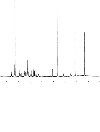
|
|
|
|
FIGURE 26. The 500-MHz 1H NMR spectrum of viridenomycin (77) in CDCl3. Reproduced by permission of Japan Antibiotics Research Association from Reference 47
were observed between H20 (or H21) and the carbon at υ133.7, and H24 and the carbon at υ133.7. Therefore carbon signals at υ134.3, 128.1 and 133.7 were assigned to C20, C21 and C22, respectively.
The connectivities of the partial structures thus obtained were elucidated by observation of the 1H – 13C long-range correlations from H2, H3 and NH25 to a carbonyl carbon (C1,

2. NMR spectroscopy of dienes and polyenes |
123 |
||||
TABLE 27. |
13C and 1H NMR assignments for virideno- |
|
|||
mycin (76) in CDCl3 |
|
|
|
||
No. |
υCa |
|
υH |
(J, Hz)a |
|
1 |
166.2 |
s |
|
|
|
2 |
120.8 |
d |
5.59 |
(d, 11.5) |
|
3 |
134.9 |
d |
6.92 |
(t, 11.5) |
|
4 |
124.6 |
d |
7.57 |
(t, 11.5) |
|
5 |
135.8 |
d |
6.45 |
(t, 11.5) |
|
6 |
126.3 |
d |
6.77 |
(dd, 15.0, 11.5) |
|
7 |
136.3 |
d |
6.46 |
(dd, 15.0, 10.5) |
|
8 |
131.4 |
d |
6.34 |
(dd, 15.0, 10.5) |
|
9 |
141.7 |
d |
5.68 |
(d, 15.0) |
|
10 |
46.8 |
s |
|
|
|
11 |
83.4 |
d |
4.06 |
(d, 6.5) |
|
12 |
85.3 |
d |
4.29 |
(d, 6.5) |
|
13171.1 s
14105.9 s
15167.1 s
16147.7 s
17 |
119.1 |
d |
5.63 |
(d, 11.5) |
18 |
126.3 |
d |
6.17 |
(dd, 14.5, 11.5) |
19 |
133.7 |
d |
5.91 |
(dd, 14.5, 10.0) |
20 |
134.3 |
d |
6.22 |
(br s) |
21 |
128.1 |
d |
6.22 |
(br s) |
22 |
133.7 |
d |
6.22 |
(br s) |
23 |
125.7 |
d |
5.28 |
(ddd, 11.0, 10.0, 6.5) |
24 |
33.7 |
t |
3.12 |
(ddd, 13.5, 10.0, 4.5) |
|
|
|
2.50 |
(ddd, 13.5, 6.5, 3.5) |
25 |
51.5 |
d |
5.49 |
(ddd, 9.5, 4.5, 3.5) |
25-NH |
|
|
5.90 |
(d, 9.5) |
26 |
17.0 |
q |
1.33 |
(s) |
27 |
59.1 |
q |
3.65 |
(s) |
28 |
15.9 |
q |
2.14 |
(s) |
29 |
139.5 s |
|
|
|
30, 34 |
126.3 |
d |
7.28 |
(d, 7.0) |
31, 33 |
128.5 |
d |
7.34 |
(t, 7.0) |
32 |
127.3 |
d |
7.28 |
(d, 7.0) |
as, singlet; d, doublet; t, triplet; q, quartet.
υ166.2), and from H30 and H34 to a methine carbon (C25, υ51.5), thereby showing that the tetraene moiety consisting of C2 to C9 was attached to C25 through an amide linkage, and the phenyl group is connected to C25.
The remaining functional groups including a tertiary methyl, a methoxy, a diol moiety and three quaternary carbons were assembled as shown in Figure 27C by analysis of the HMBC spectral data, which revealed the 1H – 13C long-range couplings from the tertiary methyl (H26) to C9, C10, C11 and C14, from the oxymethine (H12) to C13 and C14, and from the methoxy (H27) to C12. These correlations established a cyclopentene ring structure (C10 – C14) substituted with a methoxy group at C12 and a tetraene moiety at C9. The only remaining carbon (C15) was assignable to an ester from its chemical shift (υ167.1) and an IR absorption at 1700 cm 1. In order to explain the chemical shifts of C13 (υ171.1) and C14 (υ105.9), and a positive ferric chloride reaction for viridenomycin, C13 and C14 must form an enol group conjugated to the ester carbonyl (C15). The ester linkage between C15 and C16 was determined by the chemical shift of C16
(a) |
15.0 |
10.5 |
|
15.0 |
11.5 |
11.5 |
|
11.5 |
11.5 |
|
|
|
|
|
|
|
|
|
|||||||||||
CH |
|
|
|
CH |
CH |
|
CH |
|
|
CH |
|
|
|
CH |
CH |
CH |
|
|
|
|
|
|
|||||||
|
|
|
|
|
|
|
|
|
|
|
|
|
|||||||||||||||||
|
|
|
|
|
|
|
|
|
|
|
|
||||||||||||||||||
5.68 |
6.34 |
6.46 |
|
|
6.77 |
|
|
6.45 |
|
|
7.57 |
6.92 |
|
5.59 |
|
|
|
|
|
|
|
|
|||||||
28 CH3 |
2.14 |
|
|
|
|
|
|
|
|
|
|
|
|
|
|
|
|
5.5 |
2.50 |
3.5 |
|
|
|
||||||
|
|
14.5 |
|
|
|
|
|
|
|
|
|
|
|
|
H |
|
|
|
|||||||||||
|
|
11.5 |
|
|
|
10.0 |
|
|
|
|
|
|
11.0 |
|
|
|
|
|
9.5 |
||||||||||
|
|
|
|
|
|
|
|
|
|
|
|
|
|
|
|
|
|
|
|
|
|
|
|
||||||
16 |
|
|
|
|
18 |
|
|
|
|
|
|
|
20 |
|
|
|
22 |
|
|
|
|
|
25 |
|
|
|
|||
C |
|
|
CH |
CH |
|
|
|
|
CH |
|
|
CH |
|
|
|
CH |
|
CH |
|
|
C CH |
|
|
NH |
|||||
|
|
|
|
|
|
|
|
|
|
|
|
|
|||||||||||||||||
|
|
|
5.63 |
6.17 |
|
|
5.91 |
|
6.22 |
|
|
|
6.22 |
5.25 |
|
|
|
5.49 |
5.90 |
||||||||||
|
|
|
|
|
|
|
|
|
|
|
|
|
|
|
|
|
|
|
|
|
10.0 |
|
H |
4.5 |
|
|
|
||
|
|
|
|
|
|
|
|
|
|
|
|
|
|
|
|
|
|
|
|
|
|
|
|
3.12 |
|
|
|
||
|
|
|
|
|
|
|
|
|
|
|
|
|
|
|
|
|
|
|
|
|
|
|
|
|
|
|
|
||
|
|
|
|
|
|
|
|
|
7.0 |
|
|
|
|
|
|
|
|
|
|
|
|
|
|
|
|
|
|
||
7.287.34
|
|
7.0 |
6.5 |
34 |
|
11 |
12 |
29 |
32 7.28 |
CH |
CH |
|
|
4.06 |
4.29 |
|
|
30 |
|
O |
|
O |
|
|
|
|
|
|
|
(b) |
|
|
24 |
(c) |
|
|
|
|
|
5.68 |
|
|
|
|
|
|
|
|
|
1.33 |
H |
|
|
|
|||
|
28 |
|
H |
|
|
|
|
|
|
|
|
||
|
6.22 |
|
4.06 |
|
OH CH3 |
|
|
|
|||||
O |
CH3 19 |
|
|
|
|
|
|
||||||
21 |
|
|
|
H |
|
11 |
26 |
|
141.7 |
5.63 |
|||
|
H |
H |
|
|
|
|
|
|
9 |
||||
|
|
|
|
|
|
|
|
|
|||||
|
|
|
H 24 |
4.29 H |
|
46.8 |
|
|
|
H |
|||
|
|
134.3 |
|
|
|
|
85.3 |
|
14 |
O |
|
17 |
|
H |
|
|
3.65 |
|
|
171.1 |
147.7 |
||||||
133.7 |
|
133.7 |
|
|
105.9 |
167.1 |
|
||||||
128.1 |
59.1 CH |
O |
|
|
|
119.1 |
|||||||
17 |
|
|
|
|
13 |
|
15 |
|
16 |
|
|||
|
|
H6.22 |
|
27 |
3 |
|
|
|
|
|
|||
|
H |
H 6.22 |
|
|
|
HO |
|
|
O |
|
28 CH3 |
2.14 |
|
|
18 |
20 |
22 |
|
|
|
|
|
|
||||
|
|
|
|
|
|
|
|
|
|
|
|
15.9 |
|
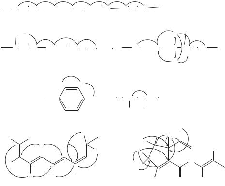
FIGURE 27. Partial structures of viridenomycin. (a): Data from 1H – 1H COSY experiment; (b), (c): The solid line arrows indicate 1H – 13C long-range coupling detected by HMBC
124
2. NMR spectroscopy of dienes and polyenes |
125 |
(υ147.7) and long-range couplings from the allylic methyl (H28) to only two carbons, C16 and C17.
Six of the eight geometries of the two tetraene systems were established to be 2Z,4Z,6E, 8E,18E and 22Z by the coupling constants J 23 D 11.5 Hz, J 45 D 11.5 Hz, J 67 D 15.0 Hz, J 1819 D 14.5 Hz and J 2223 D 11.0 Hz. An upfield chemical shift of C- 28 (υ15.9) and no NOE between H17 and H28 showed the E configuration for C16. The remaining stereochemistry at C20 proved to be 20E by the chemical shifts of C19 (υ133.7) and C22 (υ133.7) observed at a low-field region free from the effects in comparison with C21 (υ128.1) and C23 (υ125.7). NOEs observed from H26 to H12 but not to H11 indicated that the relative configuration of the cyclopentene ring was as shown in Figure 27B. The stereochemistry at C25 remains to be determined.
Thus, the structure of viridenomycin was established except for the absolute configuration. This antibiotic is partially related to hitachimycin, which is a 19-membered lactam antibiotic possessing a phenyl group and a cyclopentene ring, but devoid of the tetraene systems and ester linkage.
Colmenares and coworkers48 reported a 19F NMR study of rhodopsin analogs. 19F NMR spectra of 11-cis and 9-cis isomers of six fluorinated rhodopsin analogs with the label(s) located at the vinylic positions of the polyene chain (8F, 10F, 12F, 14F, 8,12F2, 10,14F2) are reported along with their UV-Vis and CD spectra. The regiospecific F chemical shift data are analyzed in terms of chromophore changes and local perturbation resulting from specific interactions with the protein. Two analogs (11-cis-12-F and 11-cis-8-F) and also 9,11-di-cis-12-F display FOS (fluorine opsin shift) values uniquely different from others. Ab initio 19F NMR chemical shielding calculations of model structures provide support to the assumption that a strong protein perturbation to the 12F position prevails in the binding cavity and that the F8 shift is sensitive to variation of the nearby dihedral angle(s). Possible causes for the broad line width of the F signals of these membrane proteins are discussed.
Freshly reconstituted and concentrated pigment analogs were used for 19F NMR studies for recording the ‘before photoirradiation’ and ‘after irradiation’ spectra. Representative spectra of the 8-F and 14-F monofluoro analogs are shown in Figures 28 and 29. The signal that disappears upon photobleaching is identified as that of the pigment analog, the new signal that appears upon irradiation of the photobleaching product, stereochemistry identified by their F shifts and the HPLC retention time and UV data of the extracted retinal analog. Chemical shifts are listed in Table 28.
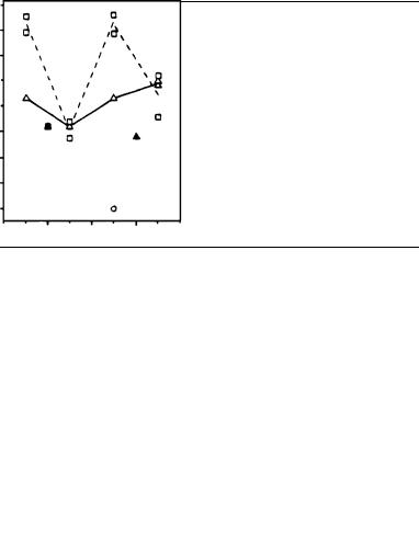
The fluorine chemical shift is very sensitive to changes in the environment. In this study, the F shift of each fluorinated pigment (a protonated Schiff base PSB) is compared to that of the solution value of the corresponding free PSB. The difference in their 19F NMR chemical shifts represents the change imposed by the local environment (the protein binding cavity) on the F probe. This value is now termed the 19F NMR opsin shift (FOS). The FOS values for the 9-cis as well as the 11-cis pigments are listed in Table 28. They have a mean value of 6.6 ppm with 95% of them distributed between 4.6 and 8.6 ppm. Only three FOS values are exceptional, falling outside this range: those of 9,11-di-cis-12- F ( 2.1 ppm), 11-cis-8-F (13.1 ppm) and 11-cis-12-F (13.2 ppm) and, to a lesser degree, also those for the disubstituted 11-cis-8,12-F2 (11.8, 11.7 ppm). The trends are evident in the plot of FOS values vs the F position along the chromophore chain shown in Figure 30.
Li and coworkers49 reported a molecular motion of ˇ-carotene and a carotenoporphyrin dyad (composed of a porphyrin, a trimethylene bridge and a carotenoid polyene) in solution. Internal rotational motions in carotenoid polyenes and porphyrins are of interest because they can mediate energy and electron transfer between these two moieties when the pigments are joined by covalent bonds. Such internal motions can affect the performance of synthetic model systems which mimic photosynthetic antenna function,

126 |
Yoshito Takeuchi and Toshio Takayama |
||
|
(a) |
after hν |
|
|
|
|
|
|
|
F |
CHO |
|
|
||
|
|
||
|
|
|
|
|
|
−103.7 |
before hν |
|
|
|
(excess aldehyde) |
|
|
|
−115.3 |
−100 |
|
−110 |
−120 |
|
|
|
19F δ (ppm) |
|
|
(b) |
|
|
|
|
|
F |
|
−115.1 |
after hν |
|
|
|||
|
(all- trans aldehyde) |
|
||
|
|
|
||
|
|
CHO |
|
|
−99.3 (pigment)104.9 |
|
before hν |
||
|
|
(excess 9-cis aldehyde) |
|
|
−100 |
−110 |
−120 |
|
19F δ (ppm) |
|
FIGURE 28. 283-MHz 19F NMR spectra of isomers of 8-F-rhodopsin in CHAPS before (lower) and after photoirradiation (upper): (a) 11-cis (pulse delay, D5 D 5.0 s, number of acquisitions, NA D 5200, line broadening, LB D 80 Hz); (b) 9-cis (D5 D 50 ms, NA D 160000, LB D 80 Hz). Disappearance of the excess 9-cis aldehyde was due to repeated formation and bleaching of pigment during the irradiation process. Reprinted with permission from Reference 48. Copyright (1996) American Chemical Society
photoprotection and photoinduced electron transfer. Analysis of 13C NMR spin-lattice relaxation times (T1) yields information concerning both overall tumbling of molecules in solution and internal rotations about single bonds. Relaxation time and nuclear Overhauser effect data have been obtained for ˇ-carotene (78) and the related molecules, squalene (82) and carotenoporphyrin (80) which is a zinc meso-tetraphenylporphyrin (79) covalently linked to a carotenoid polyene through a trimethylene bridge. Squalane (81)

2. NMR spectroscopy of dienes and polyenes |
127 |
||
(a) |
−132.7 (all-trans) |
|
|
|
|
−125.4 |
|
|
|
(13-cis) |
|
|
F |
after hν |
|
|
|
||
|
|
||
|
CHO |
|
|
−117.1
before hν
|
−120 |
−130 |
|
|
19F d (ppm) |
(b) |
(all-trans) |
|
|
|
−132.2 |
|
F |
−125.0 |
|
||
|
(13-cis) |
|
|
CHO |
|
|
after hν |
|
|
|
|
|
−123.5 |
−120.3 |
(9-cis pigment) |
before hν |
−120 |
−130 |
19F d (ppm)
FIGURE 29. 283-MHz 19F NMR spectra of 14-F-rhodopsin in CHAPS before and after photoirradiation: (a) 11-cis (D5 D 50 ms, NA D 196136, LB D 80 Hz); (b) 9-cis (D5 D 50 ms, NA D 200000, LB D 80 Hz). The 13-cis isomer was from dark isomerization. Reprinted with permission from Reference 48. Copyright (1996) American Chemical Society
and squalene (82), which lack conjugated double bonds, behave essentially as limp string, with internal rotations at least as rapid as overall isotropic tumbling motions. In contrast, ˇ-carotene reorients as a rigid rod, with internal motions which are too slow to affect relaxation times. Modeling it as an anisotropic rotor yields a rotational diffusion coefficient for motion about the major axis, which is 14 times larger than that for rotation
128 |
Yoshito Takeuchi and Toshio Takayama |
TABLE 28. 19F NMR chemical shifts of retinylidene PSBs, (protonated schiff bases), rhodopsin pigments and corresponding 19F NMR opsin shifts
Analog |
PSB CD2Cl2 |
Pigment (CHAPS)c |
F NMR OSa |
11-cis-8-F |
116.8b |
103.7 |
13.1 |
11-cis-10-F |
112.2b |
107.4 |
4.8 |
11-cis-12-F |
107.8 |
94.6 |
13.2 |
11-cis-14-F |
125.5 |
117.1 |
8.4 |
11-cis-8, 12-F2 |
117.0 |
105.2 |
11.8 |
|
104.6 |
92.9 |
11.7 |
11-cis-10, 14-F2 |
115.3 |
111.8 |
3.5 |
|
122.7 |
117.6 |
5.1 |
9-cis-8-F |
105.9b |
99.3 |
6.6 |
9-cis-10-F |
119.7 |
115.3 |
4.4 |
9-cis-12-F |
120.9 |
114.3 |
6.6 |
9-cis-14-F |
131.4 |
123.5 |
7.9 |
9-cis-10, 14-F2 |
119.1 |
115.4 |
3.7 |
|
128.8 |
121.2 |
7.6 |
9, 11-di-cis-12-F |
108.7 |
110.8 |
2.1 |
9-cis-9-CF3 |
64.6 |
60.2 |
4.4 |
a19F NMR opsin shift D column 3 column 2 in υ (ppm).
bIn CDCl3.
c CHAPS D A membrane protein solubilizing agent: N,N-Dimethyl-N-(3-sulfopropyl)-3-f[ 3˛, 5ˇ, 7˛, 12˛ -3,7,12-trihydroxy-24-oxocholan-24-yl]aminog-1-propanaminium inner salts.
Fluorine opsin shift (ppm)
12
8
4
0
7 |
9 |
11 |
13 |
15 |
Fluorine location
FIGURE 30. Plot of FOS (ppm) for vinyl-F labels at several locations on the chain of the retinyl chromophore in the ( ) 11-cis, dashed line, ( ) 9-cis, solid line, and (°) 9,11-di-cis configurations. Where appropriate, the averaged FOS values from monoand dilabeled analogs were used for connecting lines. Solid symbols are for CF3. Reprinted with permission from Reference 48. Copyright (1996) American Chemical Society
about axes perpendicular to that axis. The porphyrin reorients more nearly isotropically and features internal librational motions about the single bonds to the phenyl groups. The relaxation time data for the carotenoporphyrin are consistent with internal motions similar to those of a medieval military flail, consisting of a rigid, rod-like carotenoid