

|
|
2. NMR spectroscopy of dienes and polyenes |
|
109 |
|||
TABLE 21. |
13C NMR chemical shift values (ppm) of all-E 66 – 72a |
|
|
||||
C |
66 |
67 |
68 |
69 |
70 |
71 |
72 |
|
|
|
|
|
|
|
|
1 |
75.0 |
75.0 |
74.5 |
74.5 |
74.6 |
74.6 |
75.0 |
2 |
43.7 |
43.7 |
39.3 |
39.3 |
40.0 |
39.2 |
43.7 |
3 |
125.3 |
125.2 |
22.1 |
22.1 |
21.3 |
22.1 |
125.6 |
|
|
|
|
|
|
|
(C0.3) |
4 |
137.5 |
137.5 |
40.5 |
40.5 |
37.2 |
40.1 |
137.5 |
5 |
135.2 |
135.0 |
139.2 |
139.4 |
33.5 |
135.4 |
135.7 |
|
|
|
(C4.0) |
(C4.2) |
|
|
(C0.5) |
6 |
130.6 |
130.6 |
125.8 |
125.7 |
40.7 |
124.0 |
130.4 |
|
|
|
( 4.8) |
( 4.9) |
|
|
|
7 |
124.6 |
124.4 |
124.4 |
124.6 |
128.8 |
26.7 |
125.3 |
|
|
|
|
|
(C4.2) |
|
(C0.7) |
8 |
137.5 |
137.5 |
135.4 |
135.4 |
136.1 |
40.2 |
137.5 |
|
|
|
( 2.1) |
( 2.1) |
( 1.4) |
|
|
9 |
135.9 |
135.5 |
135.4 |
136.0 |
135.4 |
139.5 |
137.2 |
|
|
( 0.4) |
( 0.5) |
|
( 0.5) |
(C3.6) |
(C1.3) |
10 |
132.6 |
132.6 |
131.5 |
131.6 |
130.1 |
125.8 |
131.8 |
|
|
|
( 1.1) |
( 1.0) |
( 2.5) |
( 6.8) |
( 0.8) |
11 |
124.9 |
124.4 |
124.4 |
124.9 |
124.8 |
124.9 |
127.0 |
|
|
( 0.5) |
( 0.5) |
|
|
|
(C2.1) |
12 |
138.0 |
138.1 |
137.5 |
137.4 |
137.1 |
135.3 |
135.6 |
|
|
|
( 0.5) |
( 0.6) |
( 0.9) |
( 2.7) |
( 2.4) |
13 |
136.1 |
135.5 |
135.5 |
136.1 |
136.1 |
136.1 |
146.3 |
|
|
( 0.6) |
( 0.6) |
|
|
|
(C10.3)b |
14 |
133.0 |
132.8 |
132.5 |
132.6 |
132.3 |
131.4 |
111.0 |
|
|
|
( 0.5) |
( 0.4) |
( 0.7) |
( 2.6) |
( 22.0) |
15 |
129.4 |
127.3 |
127.3 |
129.5 |
129.4 |
129.5 |
98.3 |
150 |
|
( 2.1) |
(2.1) |
|
|
|
|
130.3 |
130.3 |
130.1 |
130.1 |
130.0 |
129.5 |
97.3 |
|
140 |
|
|
|
|
( 0.3) |
( 0.8) |
|
131.4 |
125.8 |
125.8 |
131.4 |
131.4 |
131.4 |
109.6 |
|
130 |
|
( 5.6) |
( 5.6) |
|
|
|
( 22.7) |
136.6 |
140.3 |
140.2 |
136.5 |
136.4 |
136.1 |
146.3 |
|
120 |
|
(C3.7) |
(C3.6) |
|
|
( 0.5) |
(C9.8)b |
135.2 |
40.2 |
40.2 |
135.3 |
135.3 |
135.3 |
133.0 |
|
110 |
|
|
|
|
|
|
( 2.2) |
125.1 |
26.5 |
26.5 |
125.0 |
125.0 |
124.9 |
127.3 |
|
100 |
|
|
|
|
|
|
(C2.2) |
125.8 |
124.1 |
124.2 |
125.8 |
125.8 |
125.8 |
125.3 |
|
90 |
|
|
|
|
|
|
( 0.5) |
139.7 |
134.9 |
134.9 |
139.7 |
139.6 |
139.5 |
141.5 |
|
80 |
|
|
|
|
|
|
(C1.8) |
40.2 |
39.7 |
39.7 |
40.2 |
40.2 |
40.2 |
40.2 |
|
70 |
26.7 |
26.7 |
26.7 |
26.7 |
26.7 |
26.7 |
26.7 |
60 |
123.9 |
123.7 |
123.8 |
123.8 |
123.8 |
123.9 |
123.7 |
50 |
135.3 |
135.0 |
135.0 |
135.4 |
135.4 |
135.4 |
135.5 |
40 |
39.7 |
39.7 |
39.7 |
39.7 |
39.7 |
39.7 |
39.7 |
30 |
26.6 |
26.6 |
26.6 |
26.6 |
26.6 |
26.6 |
26.5 |
20 |
124.3 |
124.3 |
124.4 |
124.3 |
124.3 |
124.3 |
124.3 |
10 |
131.3 |
131.2 |
131.2 |
131.3 |
131.3 |
131.3 |
131.3 |
1- CH3 2 |
24.9 |
24.9 |
25.0 |
25.0 |
25.0 |
25.0 |
24.9 |
5-CH3 |
13.0 |
13.0 |
16.9 |
16.8 |
19.6 |
15.9 |
13.0 |
|
|
|
C3.9 |
|
C6.6 |
|
|
(continued overleaf )
110 |
|
|
|
Yoshito Takeuchi and Toshio Takayama |
|
|
|
|
|||||
TABLE 21. |
(continued) |
|
|
|
|
|
|
|
|
|
|||
|
|
|
|
|
|
|
|
|
|
|
|
|
|
C |
|
|
66 |
|
67 |
68 |
|
69 |
70 |
|
71 |
72 |
|
|
|
|
|
|
|
|
|
|
|
|
|
|
|
9-CH3 |
|
|
12.9 |
|
12.8 |
12.8 |
12.8 |
13.0 |
|
17.0 |
12.9 |
|
|
|
|
|
|
|
|
|
|
|
|
|
C4.1 |
|
|
13-CH3 |
12.8 |
|
12.6 |
12.7 |
12.9 |
12.8 |
|
12.8 |
15.3 |
|
|||
130 -CH3 |
12.7 |
|
16.9 |
16.8 |
12.8 |
12.8 |
|
12.8 |
15.3 |
|
|||
90 -CH3 |
|
C4.2 |
C4.1 |
|
|
|
|
|
|
||||
17.0 |
|
16.0 |
16.0 |
17.0 |
17.0 |
|
17.0 |
17.1 |
|
||||
50 -CH3 |
16.0 |
|
16.0 |
16.0 |
16.0 |
16.0 |
|
16.0 |
16.0 |
|
|||
10 -CH3 |
25.7 |
|
25.7 |
25.7 |
25.7 |
25.7 |
|
25.7 |
17.7 |
|
|||
(E) |
|
|
|
|
|
|
|
|
|
|
|
|
|
10 -CH3 |
17.7 |
|
17.6 |
17.7 |
17.7 |
17.7 |
|
17.7 |
25.7 |
|
|||
(Z) |
|
|
49.3 |
|
49.3 |
49.1 |
49.1 |
49.1 |
|
49.1 |
49.3 |
|
|
OCH3 |
|
|
|
|
|
||||||||
aIn parentheses relevant chemical shift differences from 66 of more than š0.03 ppm are given. |
|
|
|||||||||||
b Might be interchanged. |
|
|
|
|
|
|
|
|
|
|
|||
1 |
|
|
7 |
9 |
11 |
13 |
15 |
14′ |
12′ |
10′ |
8′ |
Ar |
|
6 |
|
|
|
|
|
|
|
|
7′ |
||||
2 |
|
|
|
|
|
|
|
|
|||||
|
|
8 |
10 |
12 |
14 |
15′ |
13′ |
11′ |
|
9′ |
|
|
|
|
|
|
|
|
|||||||||
|
|
|
|
|
|
||||||||
3 |
|
|
5 |
|
|
|
|
|
|
|
|
|
|
|
|
|
|
|
|
|
|
|
|
|
|
|
|
|
4 |
|
|
|
|
(73) |
|
|
|
|
|
|
|
(a) |
Ar = C6F5, (b) |
Ar = 4-O2NC6H4, (c) |
Ar = 4-(MeO2CC6H4), (d) Ar = 2,4,6-Me3C6H2, |
||||||||||
(e) |
Ar = Ph, (f) Ar = 4-MeOC6H4 |
|
|
|
|
|
|
|
|
||||
The spectrum of the ester 73c is similar to that of the unsubstituted phenyl compound 73e (both not shown). It is noted that the chemical shift of the HC80 reflects the electronwithdrawing properties of the substituents. A combination of 1D and 2D techniques is used to establish the assignments shown, and the chemical shift changes as compared to ˇ,ˇ-carotene are listed in Tables 22 and 23.
Chemical shifts of compounds 73a, 73d and 73f were deduced as described for 73b. Comparison of the data reveals certain trends that were then utilized in the analyses of the spectra of 73c and 73e, for which HMBC spectra were not determined. First, apparent first-order coupling constants of corresponding protons are similar; approximate values are: J(7, 8; 70 , 80 ), ca 16; J(10, 11; 100, 110 ), ca 10 – 12; J(11, 12; 110, 120 ), ca 15; J(14, 15; 140, 150), ca 10; and J(15, 150), ca 14 Hz. Except for J(7, 8), the coupling constants J(11, 12; 110, 120) are considerably larger than the others, so that the doublets due to HC12, HC120 , and HC100 can be identified, even in regions of overlap. Second, for the paired doublets of HC70 /HC80 in compounds 73a, 73b, 73f the chemical shift of HC80 is greater than that of HC70 . Third, for a given pair with nonprimed and corresponding primed C-nuclei,υ (no n-primed > υ(primed) for odd-numbered C-nuclei, whereas the opposite is true for even-numbered C-nuclei. That is, those atoms that bear a
formal positive charge in the resonance structures, i.e. [ C(ˇ υC D C(˛) ]n Arυ , are deshielded; the others are shielded. Fourth, compared with ˇ,ˇ-carotene, Cˇ atoms are in general deshielded (Table 23), while C˛ nuclei are shielded. Both effects decrease in a regular, albeit nonlinear manner similar to the shift changes reported for apo-ˇ-carotenals. Exceptions are the chemical shifts of C70 and C80 , which are subject to anisotropy effects
2. NMR spectroscopy of dienes and polyenes |
111 |
||
(b) |
|
|
|
11′ |
|
|
|
|
|
10 |
|
|
8′ |
7 8 |
|
(a) |
12′ 12 |
10′ |
|
|
7′ |
|
|
11, 11′ |
|
|
|
|
|
|
|
15, 15′ |
|
|
|
|
14′, 14 |
|
|
6.7 |
6.4 |
6.1 |
|
|
1H d (ppm) |
|
|
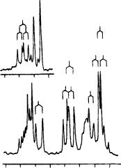
FIGURE 17. 1H NMR spectra of 73d (ca 25 mg ml 1 CDCl3): (a) olefinic region and (b) NOE difference spectrum (irradiation at 2.06 ppm). Reproduced by permission of Neue Schweizerische Chemische Gesellschaft from Reference 43
(of the Ar substituent) that differ from those in ˇ,ˇ-carotene. The fact that all substituents, whether electron-donating or electron-withdrawing in the classical sense, cause shifts in the same direction suggests that in the polyene chain C electron densities are similar. Correlations of 13C chemical shifts and electron densities (AM1 calculation) in these compounds was investigated.
1H NMR data of a minor isomer of 73e are consistent with the (70Z)-structure that is expected on chemical grounds. Thus, the observed highfield shift of one CH3 signal (1.70 vs 2.05 ppm) is expected if Me-C90 lies above the plane of the Ph ring.
Further, the doublets due to HC70 and HC80 in the (E)-isomer (6.57 and 6.90 ppm) are shifted upfield (6.43 and 6.27 ppm) in the (Z)-isomer, as is observed in the spectra of other (E/Z)-isomers.
Yamagishi and coworkers44 reported a structure determination of rumbrin 74, a new cytoprotective substance. Its structure was elucidated by NMR spectral analysis and was found to possess a novel skeleton containing ˛-pyrone, tetraene and pyrrole moieties.
The 1H NMR spectrum of 74 (Figure 21) showed 14 signals, which were attributed to two singlet CH3(υH 1.92 and 2.05), one OCH3(υH 3.95), one imine (υH 11.44) and 10 olefinic methine protons. The 13C NMR spectrum of 74 showed signals for 20 carbons. The distortionless enhancement by polarization transfer (DEPT) experiment assigned them to 3 methyl, 10 sp2 methine and 7 quaternary carbons including one ester carbonyl carbon (C18) and two-oxygenated sp2 carbons (C14 and C16). The 1H – 1H COSY spectrum established a tetraene structure composed of C6 – C12 with E geometrical configurations for the C6 – C7, C8 – C9 and C10 – C11 double bonds, which are apparent from the coupling constants [J 6, 7 D 15.0 Hz, J 8, 9 D 14.5 Hz and J 10, 11 D 14.0 Hz]. HMBC experiment on 74 showed long-range couplings of 19-CH to C13 (υC 124.8) and C14 (υC 159.1), 21CH3 to C16 (υC 165.7), C17 (υC 100.3) and C18 (υC 163.4), 20-OCH3 to
112 |
Yoshito Takeuchi and Toshio Takayama |
ppm
|
7′ 12 |
|
|
15,11,11′,15′ |
10′ |
|
7 10 |
14′ |
|
|
|
|
|
|
|
|
12′ |
14 |
8 |
140
136
132
128
124
6.7 |
6.4 |
6.1 |
|
ppm |
|
12′ |
|
|
6 |
8 |
|
13 |
||
12 |
||
9 |
|
13′ |
10′ |
|
|
9′ |
14′ |
|
|
|
14 |
|
15 |
|
10 |
5 |
15′ |
|
|
|
7 |
|
11 |
11′ |
|
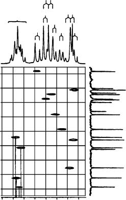
FIGURE 18. Selected portion of a HETCOR plot of 73a (13 mg ml 1 CDCl3). Projections along the axes are 1D spectra obtained at 360 MHz for 1H and 90 MHz for 13C. The spectra contain additional olefinic signals at 141.76(C(80 )), 110.81(C(70 )) and 7.18 (d, H C(80 )) ppm. Reproduced by permission of Neue Schweizerische Chemische Gesellschaft from Reference 43
C16 and H15 to C13, C14 and C17. These correlations established the connectivities of C13 – C18. Taking into consideration the number of oxygen atoms contained in 74 and the chemical shifts of C14 and C18, one oxygen atom must be inserted between C14 and C18. Thus, the existence of an ˛-pyrone unit in 74 was confirmed, as shown in Figure 22.
The HMBC experiment also showed long-range couplings of 19-CH3 to C12 (υC 134.7), C13 (υC 124.8) and C14. Thus, the tetraene and the ˛-pyrone units are linked through C13 (Figure 22). The diagnostic 13C chemical shift for C19 (υC 21.1) and NOE between H12 and 19-CH3 defined the configuration of the C12 – C13 double bond as Z.
In the 1H – 1H COSY spectrum, cross peaks were observed among the two methine protons H2 (υH 6.90, J D 2.6, 3.0 Hz) and H3 (υH 6.13, J D 2.3, 3.0 Hz) and an imine proton (υH 11.44, J D 2.3, 2.6 Hz). In addition, long-range couplings were observed from H3 to C2 (υC 120.2) and C4 (υC 111.5), H2 to C3 (υC 109.0), C4 and C5 (υC 125.9), H6 to C4 and H7 to C5 in the HMBC experiment. These couplings indicated the presence
2. NMR spectroscopy of dienes and polyenes |
113 |
||
o |
|
10 |
|
|
|
|
8 |
15 15′ |
|
7 |
|
|
|
12′ 12 |
|
8′ |
7′ |
10′ |
|
11 |
|
|
|
|
|
|
|
|
|
14′ 14 |
|
|
|
11′ |
|
|
7.3 |
7.0 |
6.7 |
6.4 |
6.1 |
|
|
1H d (ppm) |
|
|
FIGURE 19. Selected portion of the 1H NMR spectrum of 73b (18 mg ml 1 CDCl3). The d due to H C(30 ) and H C(50 ) (8.17 ppm) is not shown. Reproduced by permission of Neue Schweizerische Chemische Gesellschaft from Reference 43
|
|
|
m |
|
o |
8′ |
7′ 10′ |
14 |
|
|
|
|
|
|
|
|
|
|
|||
m |
|
11′ |
|
|
|
|
|
|
||
o |
11 |
|
7′ |
|
|
|
|
125 |
|
|
|
|
|
|
|
|
|||||
|
|
|
|
|
|
|||||
|
|
|
|
|
|
|
|
|
||
|
7 |
|
|
|
|
|
|
|
|
|
|
15′ |
5 |
|
|
|
|
|
130 |
|
|
|
1510 |
|
|
|
|
|
|
|
|
|
|
|
|
|
|
|
|
|
|
|
|
|
14 |
|
|
|
|
|
|
|
|
|
|
14′ |
|
9′ |
|
|
|
|
|
135 |
ppm |
|
|
13′ |
|
|
|
|
|
|||
|
12 10′ |
|
|
9′ |
|
|
|
|||
|
8′ 8 |
13 |
|
6 |
|
|
|
|
|
|
|
|
|
|
|
|
|
|
|
||
|
12′ |
|
|
|
|
|
|
|
140 |
|
|
|
|
IPSO′ |
|
|
|
|
145 |
|
|
|
|
|
P |
|
|
|
|
|
||
|
|
|
|
|
|
|
|
|
||
|
|
8.2 |
|
7.8 |
7.4 |
7.0 |
6.6 |
6.2 |
|
|
|
|
|
|
|
|
|
ppm |
|
|
|
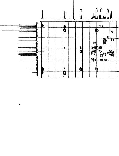
FIGURE 20. Contour plot of a selected portion of the 13C, 1H HMBC spectrum of 73b (20 mg ml 1 CDCl3). Projections along the axes are 1D spectra obtained at 360 MHz for 1H and 90 MHz for 13C. Reproduced by permission of Neue Schweizerische Chemische Gesellschaft from Reference 43
of a 2,3-disubstituted pyrrole ring consisting of C2 – C5, and the linkage to the tetraene unit at C5. Therefore, attachment of the chlorine atom to the quaternary carbon C4 was deduced (Figure 22). Based on all these findings, the total structure of 74 was established to be (1Z, 3E, 5E, 7E)-6-(8-(3-chloro-1H-pyrrol-2-yl)-1,3,5,7-octatetraenyl)-4-methoxy- 3-methyl-2H-pyran-2-one.

114 |
|
Yoshito Takeuchi and Toshio Takayama |
|
|
|||
|
TABLE 22. |
1H NMR chemical shift differences (ppm) of olefinic protons of (all-E)- |
|||||
|
70 -aryl-70 -apo-ˇ-carotens and ˇ,ˇ-carotenea |
|
|
|
|||
|
Hydrogen |
73a |
73b |
73c |
73d |
73e |
73f |
|
|
|
|
|
|
|
|
|
HC15 |
0.05 |
0.07 |
0.03 |
0.01 |
0.00 |
0.00 |
|
HC140 |
0.06 |
0.07 |
0.05 |
0.00 |
0.01 |
0.01 |
|
HC120 |
0.11 |
0.12 |
0.08 |
0.02 |
0.06 |
0.03 |
|
HC110 |
0.01 |
0.00 |
0.00 |
0.01 |
0.00 |
0.01 |
|
HC100 |
0.24 |
0.28 |
0.22 |
0.05 |
0.17 |
0.14 |
|
HC80 |
1.04 |
0.90 |
0.84 |
0.21 |
0.76 |
0.63 |
|
HC70 |
0.19 |
0.38 |
0.38 |
0.37 |
0.37 |
0.34 |
aIn the symmetrical ˇ,ˇ-carotene, υ values of primed and nonprimed atoms are identical. Other chemical shifts of 73a – d were the same (š0.01 ppm) as those reported for ˇ,ˇ-carotene, except for Me-C(90 ): 73a, 2.03; 73b, 2.05; 73c, 2.04; 73d, 2.06; 73e, 2.05; 73f, 2.03; ˇ,ˇ-carotene, 1.98 ppm.
TABLE 23. |
13C NMR chemical shift differences (ppm) of compounds 73a – f (all- |
|||||
E) and ˇ,ˇ-carotenea |
|
|
|
|
|
|
Carbon |
73a |
73b |
73c |
73d |
73e |
73f |
|
|
|
|
|
|
|
C70 |
15.93 |
2.01 |
0.69 |
1.10 |
C0.61 |
C0.22 |
C90 |
1.30 |
1.45 |
1.03 |
0.48 |
0.61 |
0.36 |
C110 |
0.86 |
0.74 |
0.48 |
0.32 |
0.25 |
0.10 |
C130 |
0.50 |
0.48 |
0.34 |
0.19 |
0.18 |
0.07 |
C150 |
0.38 |
0.38 |
0.26 |
0.15 |
0.16 |
0.10 |
C14 |
0.22 |
0.23 |
0.14 |
0.07 |
0.09 |
0.04 |
C12 |
0.22 |
0.25 |
0.17 |
0.09 |
0.13 |
0.09 |
C10 |
0.11 |
0.13 |
0.09 |
0.05 |
0.07 |
0.05 |
C8 |
0.09 |
0.12 |
0.08 |
0.05 |
0.07 |
0.06 |
C6 |
0.14 |
0.17 |
0.14 |
0.09 |
0.15 |
0.14 |
C5 |
0.10 |
0.11 |
0.07 |
0.00 |
0.04 |
C0.02 |
C7 |
0.16 |
0.17 |
0.09 |
0.04 |
0.00 |
0.04 |
C9 |
0.41 |
0.37 |
0.23 |
0.04 |
0.11 |
0.05 |
C11 |
0.40 |
0.43 |
0.26 |
0.04 |
0.10 |
0.02 |
C13 |
0.80 |
0.85 |
0.55 |
0.15 |
0.27 |
0.13 |
C15 |
1.07 |
1.11 |
0.74 |
0.21 |
0.34 |
0.14 |
C140 |
1.83 |
1.91 |
1.30 |
0.41 |
0.69 |
0.39 |
C120 |
3.00 |
2.90 |
1.96 |
0.67 |
0.99 |
0.45 |
C100 |
5.48 |
5.44 |
3.95 |
1.24 |
2.33 |
1.37 |
C80 |
3.94 |
0.34 |
1.66 |
0.71 |
4.17 |
6.09 |
aNegative values indicate upfield shifts, compared to those of ˇ,ˇ-carotene, except that values of C9 and C13 are interchanged.
CH3
H3CO
 O
O
Cl |
O |
CH3
N
H
(74)
2. NMR spectroscopy of dienes and polyenes |
115 |
12 11 ppm
8 |
7 |
6 |
5 |
4 |
3 |
2 |
1 |
|
|
|
|
ppm |
|
|
|
FIGURE 21. The 500 MHz 1H NMR spectrum of rumbrin in DMSO-d6. Reproduced by permission of Japan Antibiotics Research Association from Reference 44
|
|
|
|
|
21 |
|
|
|
|
|
|
|
CH3 |
|
|
|
|
|
|
20 |
17 |
|
O |
|
|
|
H3CO |
|
|||
|
|
|
|
|
|||
|
|
|
|
16 |
|
18 |
|
|
Cl |
|
|
15 |
14 O |
|
|
3 |
4 |
|
|
|
|||
|
|
|
|
|
|
||
|
|
|
|
|
|
|
|
2 |
5 |
7 |
9 |
11 |
|
|
|
|
|
|
|
19 |
|
||
|
|
|
|
13 |
|
||
N |
6 |
8 |
10 |
|
CH3 |
|
|
12 |
|
|
|||||
H |
|
|
|
|
|
|
|
Long-range coupling
NOE
FIGURE 22. 1H – 13C long-range couplings and NOE of rumbrin (74). Reproduced by permission of Japan Antibiotics Research Association from Reference 44
In the 1H – 13C COSY spectrum, the olefinic carbon signals could not be unambiguously assigned because of the overlapping of their proton signals. Therefore, these carbons were assigned and the structure of 74 was confirmed using 1J(CC) information44. The biosynthetic origin of the polyene and ˛-pyrone units was expected to be mainly acetate. Thus, an incorporation experiment with [1,2-13C2]acetate was carried out with a culture of A. umbrium n13. By adding 1 g of sodium [1,2-13C2]acetate 48 hours after the beginning of a 1-liter culture, 4 mg of labeled 74 were obtained. A 2D INADEQUATE experiment using this sample confirmed the structure of 74 and the assignments of all sp2 carbons (Figure 23). The complete carbon and proton assignment is given in Table 24.
Chatterjee and coworkers45 recently reported the taxonomy, production, isolation, structure elucidation and biological properties of a new antibacterial antibiotic alisamycin (75), a new member of the manumycin group of antibiotics obtained by the fermentation of
Streptomyces actuosus.
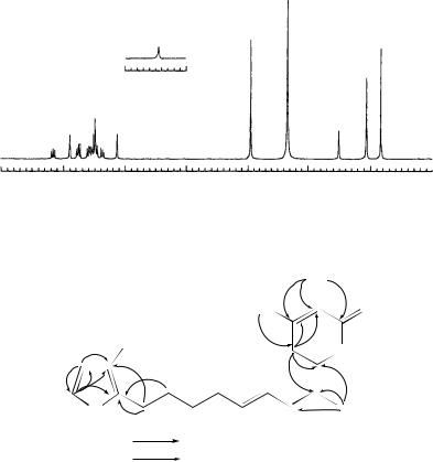
Table 25 summarizes the 1H and 13C NMR spectra of 75. The proton resonances were analyzed by double quantum filtered H H shift-correlated COSY spectrum and
116 |
Yoshito Takeuchi and Toshio Takayama |
FIGURE 23. 2D INADEQUATE spectrum of [1,2-13C2]acetate labeled rumbrin. Reproduced by permission of Japan Antibiotics Research Association from Reference 44
the carbon resonances were assigned by a proton-detected CH shift-correlated multiple quantum coherence (HMQC) NMR experiment. The spectral properties showed strong similarities to those reported for the manumycin group of antibiotics. From the COSY spectrum recorded in CDCl3, four spin systems could be extracted including a conjugated diene moiety attached to a methine multiplet (H60 , υ2.10) being part of a cyclohexane unit, one isolated triene moiety, three signals from the 5-epoxycyclohex-2-enone and two strongly coupled signals representing four protons.
In CDCl3, 75 also revealed the presence of four D2O exchangeable singlets at υ13.52, 7.58, 7.54 and 3.25 corresponding to one enolic hydroxyl, two amides and a hydroxy proton, respectively. On addition of DMSO-d6 as co-solvent, the first three signals underwent large downfield shifts to υ14.00, 9.60 and 8.45, respectively, and the fourth one was not observed. The amide singlet at υ7.54 showed COSY correlation to H20 (υ5.84) and also to the H3 (υ7.40) which, in turn, showed coupling (J D 2.6 Hz) to the epoxy proton H5. All these observations were suggestive of a carboxamide group linking the diene unit to the epoxycyclohexenone. A full confirmation was obtained by a proton-detected longrange CH shift correlation (HMBC)NMR experiment (Table 25). Thus this amide proton

2. NMR spectroscopy of dienes and polyenes |
117 |
|||
TABLE 24. The 500-MHz 1H NMR and 125-MHz 13C |
|
|||
NMR spectral data for rumbrin in DMSO-d6a |
|
|
||
Position |
|
υH |
υC |
|
|
|
|
|
|
1-NH |
11.44 |
(dd 2.3, 2.6) |
|
|
2 |
6.90 |
(dd 2.6, 3.0) |
120.2(d) |
|
3 |
6.13 |
(dd 2.3, 3.0) |
109.0(d) |
|
4 |
|
|
111.5(s) |
|
5 |
|
|
125.9(s) |
|
6 |
6.49 |
(d 15.0) |
120.2(d) |
|
7 |
6.75 |
(dd 11.0, 15.0) |
124.8(d) |
|
8 |
6.59 |
(dd 11.0, 14.5) |
135.9(d) |
|
9 |
6.37 |
(dd 11.3, 14.5) |
131.3(d) |
|
10 |
6.54 |
(dd 11.3, 14.0) |
137.9(d) |
|
11 |
7.16 |
(dd 12.5, 14.0) |
128.9(d) |
|
12 |
6.47 |
(d 12.5) |
134.7(d) |
|
13 |
|
|
124.8(s) |
|
14 |
|
|
159.1(s) |
|
15 |
6.50 |
(s) |
96.2(d) |
|
16 |
|
|
165.7(s) |
|
17 |
|
|
100.3(s) |
|
18 |
|
|
163.4(s) |
|
19 |
2.05 |
(s) |
21.1(q) |
|
20 |
3.95 |
(s) |
56.6(q) |
|
21 |
1.92 |
(s) |
8.5(q) |
|
aCoupling constants in J(Hz) are given in parentheses.
showed 3J(CH) correlation to C3 (υ126.36), C1 (υ188.63); and 2J(CH) correlation to C10 (υ165.16), and could thereby be assigned to the 2-NH proton. The more downfield amide proton showed an exchange cross peak with the enolic proton in the NOESY spectrum and it also showed long-range COSY correlation with the H11 proton (υ7.32).
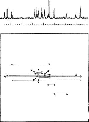
In the HMBC spectrum this NH proton exhibited 3J(CH) correlation to the C300 (υ174.15) and 2J(CH) interaction to the C13 (υ165.48) carbonyl, the latter in turn showing 2J(CH) interaction with H12(υ6.05). Thus it became clear that a carboxamide group linked the conjugated triene and the cyclopentenone unit. A 2J(CH) coupling of the triene terminus H7 to C4 (υH/υC 5.86/71.20) established the point of attachment of the triene unit to C4. An observed NOE interaction between the H7 and H5 lent further support to this attachment and was suggestive of proximal orientation of the trans- 7 bond to the epoxy unit in the most preferred conformation. The absolute configuration at C4 was not established.
The double bond geometries were determined by coupling constant measurements as well as NOE studies. Large coupling constant values (14 – 15 Hz) observed for H12 and H20 established E-configuration of the corresponding double bonds. The olefinic protons H8/H9 and H40 /H50 are isochronous, appearing at υ6.58 and 6.12, respectively, and their coupling constant values could not be measured by simple analysis of the 1H NMR spectrum (Figure 24).
The problem of strong coupling could be resolved by simulating all the olefinic signals with the LAOCOON program and the best fitting values were taken. These values confirmed E-configuration for all the five disubstituted double bonds of 75. Most of the olefinic protons also exhibited long-range couplings (Table 25). The E-configurations of the double bonds were further corroborated by the NOE network (Figure 24) as revealed
118 |
Yoshito Takeuchi and Toshio Takayama |
||||||
|
O |
|
|
|
|
||
|
|
|
|
H |
|
|
2′ |
|
|
|
|
N |
|
|
6′ |
|
1 |
||||||
O |
2 |
|
|
|
|||
|
|
|
|
|
|
||
|
|
|
|
|
|
|
|
5 |
4 |
3 |
O |
||||
|
|
|
|
||||
7 |
OH |
|
10
O 13
13
NH
HO 3″ |
1″ O |
(75)
in a phase-sensitive 2D NOESY spectrum (300 MHz, CDCl3 – DMSO-d6, 500 ms mixing time with 4% random variation).
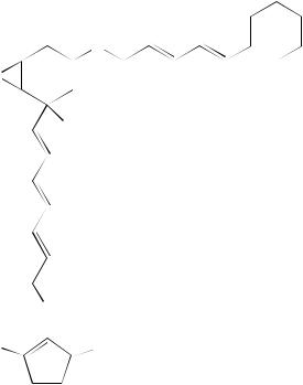
Imai and coworkers46 reported a structural study of lagunamycin (76), a novel 5- lipoxygenase inhibitor which is isolated from the culture filtrate of Streptomyces sp. AA0310 and showed inhibitory activity against 5-lipoxygenases and antibacterial activity against Gram-positive bacteria. The structure of 76 has been elucidated to be 6-diazo-4- [(E)-4,6-dimethyl-2-hepten-2-yl]-3-methyl-2,5,7,8-tetraoxoquinoline by a combination of chemical degradations and NMR studies.
The 13C and 1H NMR data are summarized in Table 26. All one-bond 1H – 13C connectivities were determined by a 13C – 1H COSY experiment. 1H – 1H COSY, NOESY and long-range 13C – 1H COSY experiments indicated a partial structure of C13H21NO containing an amide as depicted in Figure 25. The geometry of the double bond (20 ,30 ) was established as E by measurement of the 3J(CH) value (8.3 Hz) between C10 and H30 in a nondecoupled 13C NMR spectrum.
The lower field 13C NMR signals of 76 suggested a substituted pyridone (υ116.3 s, 130.0 s, 138.6 s, 151.4 s and 161.3 s) and a 2-diazo-3-oxo-1,4-benzoquinone (υ87.5 s, 168.8 s, 172.5 s and 173.6 s) moiety by comparison with the reported values of diazaquinomycin A and 2-diazo-3-oxo-1,4-naphthoquinone, respectively. Similar stabilities of 1- and 2-diazo-3-oxo-1,4-naphthoquinone under acidic conditions indicated the presence of a diazo group in 76. By combining these results, the structure of 76 was assigned.
Seto and coworkers47 reported a study on viridenomycin (77), a novel 24-membered macrocyclic polyene lactam antibiotic. A new antitumor antibiotic, designated AL081,