

|
2. NMR spectroscopy of dienes and polyenes |
89 |
|||||||||||||||||||||||||||||||||||
|
|
|
|
|
|
|
|
|
|
|
|
|
|
|
|
|
|
|
|
|
|
|
|
|
|
|
|
|
|
|
|
|
|
|
|
|
|
|
|
|
|
|
|
|
|
|
|
|
|
|
|
|
|
|
|
|
|
|
|
|
|
|
|
|
|
|
|
|
|
|
|
|
|
|
|
|
|
|
|
|
|
|
|
|
|
|
|
or |
|
|
|
|
|
|
|
|
|
|
|
|
|
|
|
|
|
|
|
||||||
|
|
|
|
|
|
|
|
|
|
|
|
|
|
|
|
|
|
|
|
|
|
|
|
|
|
|
|
|
|
|
|
|
|
|
|
|
|
|
H |
|
W1 |
|
|
|
H |
W2 |
|
||||||||||||||||||||||||||||
|
|
|
|
|
(50) |
|
|
|
|
|
|
|
(51) |
|
|
|
|
|
|
|
|
|
|
||||||||||||||
|
|
|
|
|
|
|
|
|
|
|
|
|
|
|
|||||||||||||||||||||||
|
|
|
Metathesis Catalysts |
|
Ring-forming |
|
|
|
|
|
|
|
|
|
|
||||||||||||||||||||||
|
|
|
|
|
|
|
|
|
|
|
|
|
|
|
|
|
|
|
|
|
|
|
|
|
|
|
|
|
|
|
|
|
|
|
|
|
|
|
|
|
|
|
|
|
|
|
n |
|
|
|
|
|
|
|
|
|
|
|
|
|
|
|
|
|
|
|
|
|
|
n |
|
||||
|
|
|
|
|
|
|
|
|
|
|
|
|
|
|
|
|
|
|
|
|
|
|
|
|
|
|
|
|
|
|
|
||||||
|
|
|
|
|
|
|
|
|
|
|
|
or |
|
|
|
|
|
|
|
|
|
|
|
|
|
|
|
|
|
|
|
|
|
|
|
|
|
|
|
|
|
|
|
|
|
|
|
|
|
|
|
|
|
|
|
|
|
|
|
|
|
|
|
|
|
|
|
|
|
||||||
H |
W1 |
|
|
|
|
|
H |
W2 |
|
||||||||||||||||||||||||||||
(52) |
|
|
|
|
|
|
|
|
|
|
(53) |
|
|
|
|
|
|
|
|
|
|
||||||||||||||||
W1 = CO2(CH2CH2CO)3 |
|
|
|
|
|
|
|
|
|
|
|
|
|
|
|
|
|
|
|
|
|
|
|
|
OMe |
|
|||||||||||
|
|
|
|
|
|
|
|
|
|
|
|
|
|
|
|
|
|
|
|
|
|
|
|
|
|||||||||||||
|
|
|
|
|
|
|
|
|
|
|
|
|
|
|
|
|
|
|
|
|
|
|
|
|
|||||||||||||
|
|
|
|
|
|
|
|
|
|
|
|
|
|
|
|
|
|
|
|
|
|||||||||||||||||
W2 = CO2(CH2CH2CO)3 |
|
|
|
|
|
|
|
|
|
|
|
|
|
|
|
|
|
|
|
|
|
|
|
|
|
|
|
|
|
||||||||
|
|
|
|
|
|
|
|
|
|
N |
|
N |
|
|
|
|
|
|
|
|
|
|
OMe |
||||||||||||||
|
|
|
|
|
|
|
|
|
|
|
|
|
|
|
|
|
|
|
|
|
|
|
|||||||||||||||
|
|
|
|
|
|
|
|
|
|
|
|
|
|
|
|
|
|
|
|
|
|
|
|||||||||||||||
|
|
|
|
|
|
|
|
|
|
|
|
|
|
|
|
|
|
|
|
|
|
|
|
|
|||||||||||||
|
|
|
|
|
|
|
|
|
|
|
|
|
|
|
|
|
|
|
|
|
|
|
|
|
|
|
|
||||||||||
Catalyst: MoCl5, WCl6, PdCl2 |
|
|
|
|
|
|
|
|
|
|
|
|
|
|
|
|
|
|
|
|
|
|
|
|
|||||||||||||
Cocatalyst: (n-Bu)4Sn, EtAlCl2
SCHEME 7
3. Antibiotic polyenes
Ghirlando and coworkers36 reported interactions between a protonated retinal Schiff base and various counterions using two-dimensional NOE NMR. Bacteriorhodopsin (bR) is the protein pigment (a constituent of the purple membrane) of Halobacterium halobium. Its role is to convert light energy directly into a gradient of hydrogen ion concentration across the membrane, which is subsequently used, via a chemiosmotic mechanism, to synthesize adenosine-50-triphosphate. bR consists of a chromophore, all-trans-retinal, covalently bound to the polypeptide backbone through a protonated Schiff base link to the ε-amino group of a lysine. The protonated n-butylamine Schiff base of all-trans-retinal in methanol absorbs at 440 nm, whereas in bR, in its light-adapted form, it has an absorption maximum of 570 nm. This absorption maxima difference between the chromophore in the natural pigment and in methanol is defined as the opsin shift37. A better understanding of the mechanism through which the protein shifts the absorption maximum to longer wavelengths, in addition to accounting for the absorption maxima in various visual pigments ranging from 440 to 625 nm, is of great interest.
At present the opsin shift in bR is interpreted as a result of a combination of different factors: (i) weaker hydrogen bonding in the pigment between the positively charged nitrogen and its counterion relative to the hydrogen bonding existing in methanol solution, (ii) s-trans ring-chain planarity and (iii) interaction of the retinal chromophore with a nonconjugated dipole introduced by the protein.

90 |
Yoshito Takeuchi and Toshio Takayama |
(a)
7.0 |
6.0 |
5.0 |
4.0 |
3.0 |
2.0 |
1.0 |
0.0 |
1H d (ppm)
(b)
7.0 |
6.0 |
5.0 |
4.0 |
3.0 |
2.0 |
1.0 |
1H d (ppm)
FIGURE 8. 1H NMR spectrum of (a): 50 and (b): 52 in CDCl3. Reproduced by permission of Marcel Dekker, Inc. from Reference 35
The importance of the interaction between the protonated retinal Schiff base and its counterion for determining the absorption maxima was pointed out by Blatz and Mohler38, who first noticed a correlation between the type of counterion used and the change in the wavelength of the absorption maximum in aprotic solvents. Excess of trifluoroacetic acid, in methylene chloride as a solvent, introduced a red shift due to the weakening of the electrostatic interaction between the positively charged nitrogen and its counterion. It is therefore important to determine directly the actual spatial location of the counterion along the polyene chain of the all-trans protonated retinal Schiff base in solution. The structure of a retinal Schiff base of t-butylamine is given below.
N
8 |
10 |
12 |
14 |
5

2. NMR spectroscopy of dienes and polyenes |
91 |
(a)Liquid crystal polymers
160 |
140 |
120 |
100 |
80 |
60 |
40 |
20 |
13C d (ppm)
(b)
160 |
140 |
120 |
100 |
80 |
60 |
40 |
13C d (ppm)
FIGURE 9. 13C NMR spectrum of (a): 50 and (b): 52 in CDCl3. Reproduced by permission of Marcel Dekker, Inc. from Reference 35
Blatz and Mohler38 have performed 2D NOE NMR experiments on the protonated t-butylamine Schiff base of all-trans-retinal using different counterions, each carrying at least one nonexchangeable proton. The study has indicated that a proton on the counterion molecule is spatially close, in aprotic solvents, to the protons of the chromophore near the positively charged nitrogen. It has also shown that the ion-pair formation is relaxed in either the presence of excess carboxylic acid (the counterion) or when using methanol as a solvent.
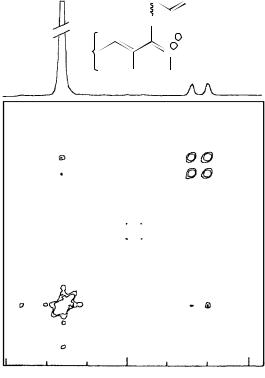
Experiments were performed at 5 °C in order to arrest the cis –trans isomerization of the protonated Schiff base. Spectra with one equivalent of acid and different mixing times showed one NOE cross-peak between H15 of the retinal molecule and the proton on the counterion, as shown for a mixing time of 0.4 s in Figure 10. The strong chemical shift dependence of the H15 resonance on the concentration of the acid dictated the use of less than one equivalent of the protonating formic acid, and therefore an incomplete protonation (>80%) of the retinal, in order to avoid an overlap between the formate and the H15 peaks in the spectrum. This should not affect the observed result since an average chemical shift, between those of H15 of the retinal in its nonprotonated and protonated
92 |
Yoshito Takeuchi and Toshio Takayama |
states, was observed, suggesting a fast exchange. Electrostatic considerations imply that the formate counterion will only interact with the charged protonated retinal Schiff base molecules.
The discrepancy between the intensities of the signals of the formyl proton and of H15 (Figure 10) arises from the large difference in the relaxation times of the two molecules. This has a more pronounced effect on the observed intensity in the 2D NMR projection than in the normal NMR spectrum.
Using two-dimensional NMR spectroscopy, the spatial location of various carboxylate anions relative to the polyene chain of the protonated Schiff base of all-trans-retinal was determined. The observed intermolecular NOE cross-peaks between a proton on the counterion and a proton near the nitrogen atom indicate the existence of ion-pair formation between the protonated retinal Schiff base and various counterions in chloroform. The results suggest that the most likely site of the carboxylate group of the counterion is in the immediate vicinity of the positively charged nitrogen atom of the retinal Schiff base.
|
H |
O |
|
CH3 |
H |
|
|
|
|
O− |
|
13 |
15 |
+ |
|
N |
|||
|
|
H H
H-15
8.7 |
8.4 |
8.1 |
ppm
FIGURE 10. Contour plot of two-dimensional nuclear Overhauser effect 1H NMR (NOESY) of the protonated Schiff base of all-trans-retinal, in chloroform, with formate as the counterion. The intermolecular NOE cross-peak observed between H15 of the retinal and the counterion proton, at a mixing time of 0.4 s, is shown. Top trace: f2 projection of the 2D NOE spectrum. Reproduced by permission of John Wiley & Sons from Reference 36
2. NMR spectroscopy of dienes and polyenes |
93 |
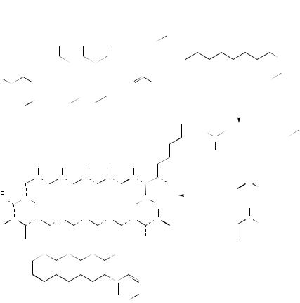
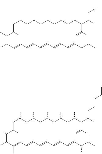
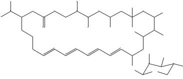
Li and coworkers39 reported the production of new polyene antibiotics. Ethyl (Z)- 16-phenylhexadeca-9-enoate (56), an analog of ethyl oleate (55), was synthesized and added to cultures of Streptomyces cellulosae ATC C12625, which normally produce fungichromin (54) as the principal polyene antibiotic (Figure 11). These cultures showed drastic reduction of fungichromin biosynthesis but afforded four new polyene antibiotics with a truncated four-carbon side chain which are designated as isochainin (58) [an isomer of chainin (57)], 14-hydroxyisochainin (59), 10 -hydroxyisochainin (60) and 10 ,14-dihydroxyisochainin (61). The close correspondence of 13C NMR chemical shifts between these compounds and fungichromin and the coproduction of compounds 58–61 and fungichromin (54) suggest that the stereochemistry at every site is exactly analogous (Table 15).
Recently, the absolute stereochemistry of pentamycin, an antibiotic from Streptomyces pentaticus with the same gross structure as fungichromin (54), has been reported as being either 62a or 62b. Elucidation of the stereochemical relationship between pentamycin (62)
|
|
|
|
|
|
|
|
|
|
|
|
|
6′ |
|
|
|
|
||||
|
|
OH |
OH |
OH |
OH |
OH |
|
|
3′ |
|
|
|
|
||||||||
|
|
|
|
|
|
|
|
|
|
|
|
|
|||||||||
|
|
|
|
|
|
|
|
|
|
|
2 |
|
1′ |
|
|
|
COOEt |
||||
|
12 |
|
|
8 |
|
|
|
4 |
|
|
OH |
|
|
|
|
|
|
||||
|
|
|
|
|
|
|
|
|
|
|
|
|
|||||||||
|
|
|
|
|
|
|
|
|
|
|
|
|
|
|
|
||||||
|
|
|
|
|
|
|
|
|
|
|
|
|
|
|
|
|
|
|
|||
HO |
14 |
OH |
|
|
|
|
|
O |
O |
|
|
|
|
|
|
|
|
|
|||
|
|
|
|
|
|
|
|
|
|
|
|
(55) |
|
|
|||||||
|
|
|
|
|
|
|
|
|
|
|
|
|
|
|
|
|
|
|
|
||
HO 15 |
|
|
18 |
|
|
22 |
|
|
|
26 |
|
28 |
|
|
|
|
|
|
|
|
|
|
|
|
|
|
|
|
|
|
|
|
|
|
|
|
|||||||
|
|
|
|
|
|
|
|
|
|
|
|
|
|
|
|
|
|||||
|
|
|
|
|
|
|
|
|
|
|
|
|
|
|
|
|
|
|
|
||
|
|
|
|
|
|
(54) |
|
|
|
|
OH |
|
|
|
|
|
|
|
|
|
|
|
|
|
|
|
|
|
|
|
|
|
|
|
|
|
|
|
|
|
|||
|
|
|
|
|
|
|
|
|
|
|
|
|
|
|
|
|
|
|
|
|
|
|
|
|
|
|
|
|
|
|
|
|
|
|
|
|
• • |
|
|
|
|
||
|
|
|
|
|
|
|
|
|
|
|
|
|
|
|
|
|
|
|
|
||
|
|
|
• |
• |
• |
• |
• |
|
|
|
|
|
|
• |
Octanoate |
||||||
|
|
|
|
|
|
|
|
|
|
|
• |
|
|||||||||
|
|
|
• |
• |
• |
• |
• |
|
|
|
|
|
|
|
|
|
|||||
|
|
|
|
|
|
|
|
|
|
|
|
|
|
||||||||
|
• |
• • |
OH |
|
|
|
• |
• |
|||||||||||||
|
|
• |
|
|
|
|
|
• |
|
|
|||||||||||
|
|
|
|
|
|
|
|
|
|
|
|
|
• |
|
|||||||
HO |
|
|
|
|
|
|
|
|
|
|
|
|
|
|
|
|
|
Acetate |
|||
|
|
|
|
|
|
|
|
|
|
|
|
|
|
|
|
|
|
|
|
|
|
• |
• |
|
• |
• |
• |
• |
• |
• |
|
|
|
|
|
|
• |
• |
|||||
|
|
|
|
|
|
|
|
|
|
|
OH |
|
|
|
|
|
Propionate |
||||
|
|
|
|
|
|
|
|
|
|
|
|
|
|
|
|
|
|
||||
|
|
|
|
|
|
|
|
|
COOEt |
|
|
|
|
|
|
O2 |
|
||||
|
|
|
|
|
|
|
|
|
|
|
|
|
|
|
|
||||||
|
|
|
|
|
|
|
|
|
|
|
|
|
|
|
|
|
|
Oxygen |
|||
|
|
|
|
|
|
|
|
|
|
|
|
|
|
|
|
|
|
||||
(56)
FIGURE 11. Biosynthetic origin of fungichromic (54) and structure of oleate analog (56). Reproduced by permission of Japan Antibiotics Research Association from Reference 39

94 |
|
Yoshito Takeuchi and Toshio Takayama |
|
|
|
TABLE 15. |
13C chemical shifts (υ) for fungichromin (54), isochainin (58), 14-hydroxyisochainin |
||||
(59), 10 -hydroxyisochainin (60) and 10 ,140 -dihydroisochainin (61)a |
|
|
|||
Carbon |
54 |
58 |
59 |
60 |
61 |
|
|
|
|
|
|
29 |
11.74 |
11.45 |
11.80 |
11.08 |
11.70 |
60 |
14.38 |
— |
— |
— |
— |
28 |
17.96 |
18.29 |
18.30 |
17.95 |
17.91 |
50 |
23.65 |
— |
— |
— |
— |
30 |
26.01 |
23.60 |
23.61 |
19.51 |
19.52 |
40 |
32.88 |
14.21 |
14.25 |
14.23 |
14.23 |
20 |
36.22 |
29.87 |
30.18 |
38.36 |
38.40 |
12 |
39.58 |
42.52 |
39.50 |
41.58 |
39.54 |
4 |
41.38 |
42.70 |
42.33 |
42.86 |
41.34 |
10 |
44.34 |
44.20 |
44.15 |
44.18 |
44.36 |
6 |
45.17 |
44.91 |
44.83 |
45.17 |
45.21 |
8 |
45.33 |
45.11 |
45.16 |
45.26 |
45.36 |
2 |
60.35 |
54.26 |
54.40 |
60.31 |
60.46 |
13 |
70.34 |
67.50 |
70.26 |
67.47 |
70.38 |
11 |
71.45 |
71.00 |
71.35 |
71.12 |
71.46 |
10 |
72.59 |
30.60 |
30.57 |
72.28 |
72.21 |
26 |
73.25 |
73.15 |
73.44 |
73.15 |
73.30 |
3 |
73.41 |
73.29 |
73.55 |
73.60 |
73.30 |
7 |
73.92 |
73.38 |
73.56 |
73.65 |
73.90 |
5 |
74.08 |
73.55 |
73.64 |
73.65 |
74.08 |
9 |
74.20 |
74.24 |
74.02 |
73.91 |
74.17 |
27 |
75.25 |
74.47 |
74.58 |
75.10 |
75.25 |
14 |
78.31 |
45.21 |
78.20 |
45.29 |
78.32 |
15 |
80.43 |
75.63 |
80.32 |
75.83 |
80.50 |
18 |
129.06 |
128.04 |
129.25 |
128.35 |
129.05 |
17 |
129.91 |
129.57 |
129.79 |
129.31 |
129.93 |
24 |
131.97 |
132.43 |
131.99 |
132.25 |
132.03 |
22 |
133.66 |
133.62 |
133.74 |
133.82 |
133.67 |
20 |
134.13 |
134.15 |
133.96 |
134.12 |
134.13 |
23 |
134.21 |
134.19 |
134.32 |
134.12 |
134.17 |
25 |
134.28 |
134.44 |
134.37 |
134.28 |
134.27 |
21 |
134.81 |
134.57 |
134.45 |
134.59 |
134.85 |
19 |
135.36 |
134.68 |
135.18 |
134.96 |
135.41 |
16 |
138.55 |
140.64 |
138.71 |
140.34 |
138.53 |
1 |
172.98 |
175.43 |
175.37 |
173.02 |
173.01 |
a100.6 MHz 13C NMR spectrum in methanol-d4 with solvent reference at 49.00 ppm.
and fungichromin (54) should allow stereochemical assignment of isochainin (58) and its hydroxylated derivatives 59–61 with reasonable confidence.
Sowinski and coworkers40 reported a structure of vacidin A (63), an aromatic heptaene macrolide antibiotic. The constitution of vacidin A, a representative of the aromatic heptaene macrolide antibiotics, was established on the basis of 13C and 1H – 1H double quantum filtered correlated spectroscopy, rotating frame nuclear Overhauser effect spectroscopy, J-resolved 1H as well as 1H – 13C correlation NMR spectra. The geometry of the polyene chromophore was determined as 22E, 24E, 26E, 28Z, 30Z, 32E, 34E.
The 13C NMR spectrum of 64, an amide of 63, showed sixty-two carbon signals of which partial assignments, shown in Table 16, were made based upon distortionless enhancement by polarization transfer(DEPT), 1H – 13C correlation experiments and literature data describing 13C NMR analysis of polyene macrolides.
2. NMR spectroscopy of dienes and polyenes |
95 |
|||||||||||
OH |
OH |
OH |
OH |
OH |
|
|
4′ |
|||||
|
|
|||||||||||
|
|
|
||||||||||
|
1′ |
|||||||||||
|
||||||||||||
|
|
|
|
|
|
|
|
|
2 |
|
||
12 |
|
|
|
8 |
|
|
|
4 |
|
R2 |
||
|
|
|
|
|
|
|
|
|||||
|
|
|
|
|
|
|
|
|
|
|
||
R1 |
1 |
|
14 |
|
|
|
OH |
|
|
|
O |
|
O |
|
|
|
|
|
|
||||||
|
|
|
|
|
|
|
|
26 |
|
|
HO |
15 |
|
|
|
|
|
|
|||
|
|
|
|
|
|
|
|
|||
|
|
18 |
|
22 |
|
|
|
|
|
|
|
|
|
|
|
|
|
|
|||
|
|
|
|
|
|
|
|
|
|
|
|
|
|
Chainin (57) |
R1 = H |
R2 = H |
OH |
||||
|
|
|
||||||||
|
|
|
|
|
|
|||||
|
|
|
Isochainin (58) |
R1 |
= H |
R2 |
= H |
|
|
|
|
(59) |
R1 |
= OH |
R2 |
= H |
|
|
|
||
|
(60) |
R1 |
= H |
R2 |
= OH |
|
|
|
||
|
(61) |
R1 |
= OH |
R2 |
= OH |
|
|
|
||
|
|
|
|
|
|
|
||||
OH |
OH |
OH |
OH |
OH |
 OH
OH
HO 14
14
OH |
O |
O |
HO 15
15
OH
(62a) 14-OH is in a dashed line and 15-OH in a full bond (62b) 14-OH is in a full line and 15-OH in a dashed bond
The data from 1H NMR studies of 63, which included double quantum filtered phase sensitive correlated spectroscopy (DQF-COSY) and rotating frame nuclear Overhauser effect spectroscopy (ROESY) experiments (Figure 12), are collected in Table 17.
The latter, in contrast to nuclear Overhauser enhancement and exchange spectroscopy (NOESY), always feature positive NOEs (negative cross-peaks with respect to diagonal), eliminating known problems of NOEs vanishing or spin diffusion, depending on correlation time, when high field spectrometers are used for measurements of medium-size compounds.
96
|
H3C |
|
OH |
|
37 |
|
41 |
|
38 |
43 |
CH3 |
|
O |
O |
 NH2
NH2
35 |
33 |
31 |
1′ |
5′ |
36 |
30 |
|
|
|
O |
O |
CH3 |
34 |
32 |
|
|
|
2′ |
OH |
|
|
|
27 |
25 |
23 |
21 |
|
|
|
|
|
NH2 |
||||
|
|
|
|
|
|
||
O |
29 |
|
|
|
|
OH |
|
28 |
26 |
24 |
|
22 |
|
|
|
|
|
|
|
1 |
|
|
|
|
|
|
|
|
COR |
|
O |
OH |
OH |
OH |
OH |
|
O |
18 |
|
|
|
|
|||||||
|
|
|
|
|
|
|
|
|
|
|
|
|
|
|
|
|
|
15 |
|
HO |
3 |
5 |
7 |
|
|
13 |
|
|
OH |
|
4 |
6 |
|
|
|
14 |
|
||
|
|
|
|
|
OH |
|
|||
|
|
|
|
|
|
|
|
|
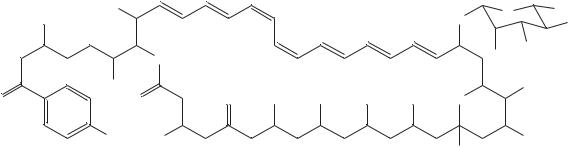
vacidin A (63) |
R = OH |
vacidin A methoxycarbonylmethylamide (64) |
R = NHCH2COOCH3 |
|
|
|
2. NMR spectroscopy of dienes and polyenes |
97 |
||
TABLE 16. |
13C NMR data for vacidin A methoxycarbonylmethylamide (64) |
|
||||
Description |
No. of |
υ (ppm) |
Description |
No. of |
υ (ppm) |
|
|
|
carbon |
|
|
carbon |
|
|
|
atoms |
|
|
atoms |
|
|
|
|
|
|
|
|
CH3 |
|
4 |
13.5, 16.7, 18.6, 52.4 |
C27 |
|
128.58 |
CH2 |
|
13 |
31 – 52 |
C28 |
|
130.91 |
CH: |
|
|
|
C29 |
|
125.53 |
CHCH3 |
|
2 |
34.3, 40.4 |
C30 (C24) |
|
125.28 |
CHNH2 |
|
1 |
57.9 |
C31 |
|
130.84 |
CHCONHR |
1 |
59.3 |
C32 |
|
128.62 |
|
CHOR |
|
13 |
64 – 79 |
C33 (C23) |
|
134.42 |
Acetal |
|
1 |
97.7 |
C34 |
|
133.67 |
CH Ar |
|
4 |
113.7, 131.6 |
C35 |
|
137.88 |
DCH a |
:C22 |
113.67 |
Nonprotonated: |
1 |
98.2 |
|
(olefinic) |
Hemiketal |
|||||
C23 (C33) |
|
130.00 |
C Ar |
2 |
127.0, 171.2 |
|
C24 (C30) |
|
134.80 |
COXR |
3 |
171.8, 174.6, 174.7 |
|
C25 |
|
|
133.10 |
CDO |
2 |
198.1, 208.9 |
C26 |
|
|
135.64 |
|
|
|
aAssignment by H, C-COSY. Interchangeable assignments are shown in parentheses.
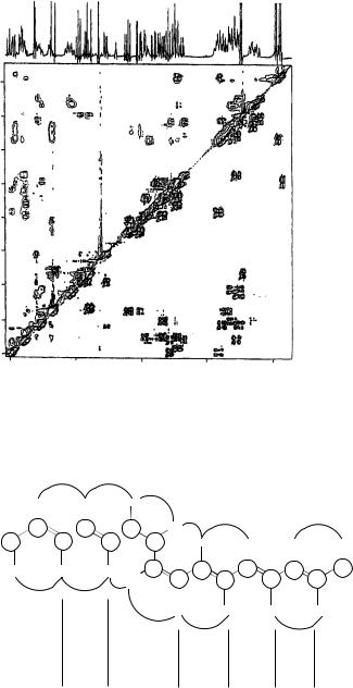
The coupling constants listed in Table 17 were assigned on the basis of the 1D 1H NMR spectrum of 63, but for a few cases the analysis of phase structure of the cross-peaks in the DQF-COSY spectrum was carried out to attribute correct values to the appropriate protons. Also, the analysis of the NOE effects yields the same results (Figure 13).
Hirota and coworkers41 reported a planar structure of new polyene macrolide antibiotic YS-822A (65), which they isolated. 1H and 13C NMR spectra of 65 showed a number of broad and overlapping signals, but the 1H – 1H and 13C – 1H COSY spectra implied the existence of a mycosamine moiety and several other partial structures. The connectivity of these partial structures was established by extensive 2D NMR experiments, including homonuclear Hartmann – Hahn and heteronuclear multiple-bond connectivity measurements, which led to the determination of the gross planar structure of 65.
29 |
|
Me |
OH |
OH
30 |
28 |
1 |
4 |
7 |
Me |
|
|
||
|
|
|
|
|
|
|
O |
OH |
OH |
|
|
23 |
|
|
11
9
O 12
12
15 |
OH |
|
2′ |
O |
O |
|
|
|
1′ |
(65) YS-822A
 OH
OH
31
 COOH
COOH
NH2
 OH
OH
 Me
Me
5′ 6′
Although 1D 1H and 13C NMR spectra of 65 in DMSO-d6 showed a number of broad and overlapping signals, the 13C – 1H COSY spectrum afforded the assignment
98 |
Yoshito Takeuchi and Toshio Takayama |
F2 (ppm)
1.0
2.0
3.0
4.0
5.0
5 |
4 |
3 |
2 |
1 |
F1 (ppm)
FIGURE 12. Spectra of vacidin A methoxycarbonylmethylamide 64. Spectral region 0.72 – 5.06 ppm of 300 MHz ROESY (upper triangle) and DQF-COSY (lower triangle) spectra of VacGlyOMe (15 mg ml 1, pyridine-d5-methanol-d4, 9 : 1) combined along the diagonal. Reproduced by permission of Japan Antibiotics Research Association from Reference 40
|
|
m |
|
|
|
|
m |
|
|
|
|
|
|
|
|
|
|
5.45 |
|
6.24 |
|
|
6.15 |
|
s |
|
|
|
|
||||
|
|
|
|
|
|
|
|
|
|
|||||||
|
137.9 |
15 11 |
134 |
. |
4 |
|
11.5 |
|
. |
|
|
6.63 |
m |
m |
|
s |
9 |
15 |
130 |
8 |
11.5 |
|
|
|
|
||||||||
36 |
|
133.7 |
|
|
|
128.6 |
31 |
|
125.3 |
|
6.95 |
6.37 |
6.24 |
4.83 |
||
|
|
|
|
|
|
|
||||||||||
|
|
|
|
|
|
|
|
|
|
|
30 |
|
|
|
|
|
|
|
|
|
|
|
|
|
|
|
|
11.5 |
|
|
|
|
|
|
|
|
|
|
|
|
|
|
|
29 |
|
128.6 15 11 133.1 |
15 11 130.0 15 9 |
|||
1.81 |
|
6.34 |
|
|
|
7.17 |
6.97 |
125.5 |
11.5 |
|||||||
|
|
|
|
|
130.9 |
11.5 |
135.6 |
134.8 |
137.7 |
|||||||
|
|
|
|
|
|
|
|
|
|
|
|
28 |
|
|
22 |
|
|
|
|
|
|
|
|
|
|
|
|
|
|
|
|
||
|
s |
|
m |
|
|
s |
|
|
|
|
|
|
|
|
|
|
|
|
|
|
|
|
|
|
|
|
|
|
6.51 |
|
6.68 |
6.64 |
6.36 |
|
|
|
|
|
|
|
|
|
|
|
s |
|
|
|
|
|
|
|
|
|
|
|
|
|
|
|
|
|
|
m |
|
w |
|
|
|
3H |
|
|
|
7H |
|
|
|
|
7H, 9H |
11H |
13H |
13H, 19H |
||
FIGURE 13. Polyene part of vacidin A. NOE’s, J(HH) and 1H, and 13C chemical shifts. Reproduced by permission of Japan Antibiotics Research Association from Reference 40