

2. NMR spectroscopy of dienes and polyenes |
159 |
||
simulated |
experimental |
simulated |
|
θ = 40° |
tilt series |
θ = 73° |
|
α= 0°
α= 15°
α= 30°
α= 45°
α= 60°
α= 75°
α= 90°
−80 −40 0 40 80 kHz
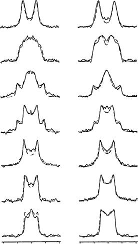
FIGURE 43. Tilt series of 2H NMR spectra from the C19 deuteriomethyl group of retinal in oriented purple membranes, recorded at seven different inclinations (˛) of the sample normal relative to the spectrometer field. Both the experimental data (middle column) and the two simulated series (outer columns) are characterized by an absolute quadrupole splitting of 30 kHz in the zero-tilt spectrum (˛ D 0°, top row). The simulations on the left are based on a methyl group angle of D 40° which corresponds to a positive splitting, while the simulations on the right are based on D 73° with a negative quadrupole splitting. Reprinted with permission from Reference 57. Copyright (1994) American Chemical Society
š1°, since the range around 45° is particularly sensitive as is seen from equation 3. A small change of 1° in would lead to a significant change in the quadrupole splitting of around 2 kHz in the zero-tilt spectrum. When the labeled segment is undergoing any small oscillations with a correlation time of less than 1/ Q, ca 10 5 s, the angle obtained represents the time-averaged orientation of the methyl group.
160 |
Yoshito Takeuchi and Toshio Takayama |
|
|
C18 |
C20 |
α= 0°
α= 15°
α= 30°
α= 45°
α= 60°
α= 75°
α= 90°
−80 −40 |
0 |
40 |
80 |
−80 −40 |
0 |
40 |
80 |
|
kHz |
|
|
|
kHz |
|
|
FIGURE 44. Tilt series of 2H NMR spectra from the deuteriomethyl groups C18 (left column) and C20 (right column) of retinal in oriented purple membranes, at seven different sample inclination (˛) in the spectrometer field. The line-shape simulations are superimposed over the experimental spectra in order to illustrate the good line fit obtained by the prediction method. Reprinted with permission from Reference 57. Copyright (1994) American Chemical Society
Figure 45 illustrates how all the different methyl group orientations with respect to the membrane normal N are accommodated in space by the proposed structure of retinal within bR. This picture is clear from the measured values of , which are indicated as labels to the individual methyl groups. The roughly parallel orientations of the two methyl groups, C18 ( D 37°) and C19 ( D 40°), demonstrate that retinal must have a
2. NMR spectroscopy of dienes and polyenes |
161 |
N
|
+ |
|
|
32° |
|
|
|
cytoplasmic side |
|
|
|
|||||||
|
|
|
|
|
|
|
|
|
||||||||||
|
|
|
20 |
|
|
|
|
|
|
|
|
|
|
|
|
|||
|
N |
15 |
|
|
|
|
|
|
|
|
|
|
|
|
|
|
|
|
|
H |
|
|
|
|
|
|
|
|
|
40° |
|
|
|
|
|
||
|
|
|
|
|
|
|
|
|
|
|
|
|
|
|
||||
|
|
|
|
|
|
|
|
|
|
|
|
|
||||||
|
|
|
13 |
|
|
|
|
|
|
|
|
|
|
|
|
|||
|
|
|
|
|
|
|
|
|
|
|
19 |
|
|
|
|
|
||
|
|
|
|
|
|
|
|
|
|
|
|
|
|
|||||
|
|
|
|
|
|
|
11 |
|
|
|
|
|
|
|
|
|
|
|
|
|
|
|
|
|
|
|
|
|
|
|
|
|
|
|
37° |
||
|
|
|
|
|
|
|
|
|
|
|
|
|
|
|
|
|
||
|
|
|
|
|
|
|
|
|
|
|
|
|
|
|
|
|
||
|
|
|
|
|
|
|
|
|
|
9 |
|
|
|
|
|
|
||
|
|
|
|
|
|
|
|
|
|
|
|
|
|
|
|
|
18 |
|
|
|
|
|
|
|
|
|
|
|
|
|
|
|
|
|
|
||
membrane |
|
|
|
|
|
7 |
|
|
||||||||||
normal |
|
|
|
|
|
|
|
|
|
|
|
|
|
|
|
|
|
|
|
|
|
|
|
|
|
|
|
|
|
|
|
|
|
|
|
|
|
|
|
|
|
|
|
|
6 |
5 |
|
|
||||||||
|
|
|
|
|
|
|
61 |
|
|
4 |
||||||||
|
|
|
|
|
|
|
1 |
|
||||||||||
|
|
|
|
|
|
|
71 |
|
|
|
|
|
||||||
|
|
|
|
|
|
|
2 |
3 |
|
|
||||||||
FIGURE 45. Orientation and conformation of retinal in bR, constructed from the individual methyl group orientations that have been determined by solid-state 2H NMR. The angles of the C CD3 bond vectors with respect to the membrane normal (N) were evaluated for C18 (37°), C19 (40°) and C20 (32°) from the zero-tilt spectra shown in Figure 44 and with the aid of line-shape simulation of the tilt series in Figure 42 and 43. Reprinted with permission from Reference 57. Copyright (1994) American Chemical Society
6s-trans rather than a 6s-cis conformation when bound to bacteriorhodopsin. That is, a 180° rotation around the C6 C7 bond, which has a substantially lower energy barrier compared to the other single bonds of the conjugated system, would produce a structure that is incompatible with the measured angles. This conclusion confirms previous solidstate NMR studies that have proposed a 6s-trans chromophore from comparison with crystalline model compounds. By focussing on the specific angles of the three methyl groups, C18, C19 and C20, along the polyene chain, it is apparent that the chromophore backbone cannot be perfectly straight.
In an undistorted system of conjugated double bonds, the three methyl groups would be expected to be exactly parallel; however, in this case their individual orientations are not the same with respect to the membrane. In particular, the two neighboring methyl groups, C19 ( D 40°) and C20 ( D 32°), show that the carbon framework of the polyene chain must be distorted by an in-plane curvature and possibly an out-of-plane twist. The fact that the two methyl groups, C18 ( D 37°) and C19 ( D 40°), are not entirely parallel may be partially attributed to the additional rotational flexibility around the C6 C7 bond. It thus appears that the observed in-plane curvature, and possibly an out-of-plane twist, relieve the steric crowding of the three methyl groups (C18, C19 and C20) along the retinal chain, as well as the interference of the gem-dimethyl groups (C16 and C17) on the ring with the proton on C8. A more refined picture of the chromophore in terms of the individual bond and torsion angles could be obtained by computer modeling of the molecular framework to the set of geometrical constraints, i.e. to the measured methyl group orientations. However, since the in-plane and out-of-plane distortional modes are interdependent, it is not possible to quantify the contribution of each, and a family of plausible retinal structures which are compatible with the 2H NMR results would emerge.

162 |
Yoshito Takeuchi and Toshio Takayama |
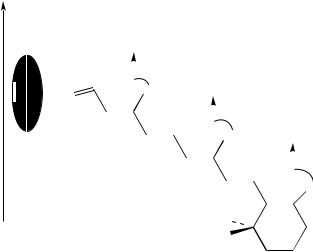
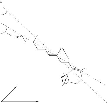
Ulrich and coworkers59 reported the orientation and conformation of the cyclohexene ring of retinal in bacteriorhodopsin of the purple membrane of Halobacterium halobium by solid-state 2H NMR spectroscopy, through the determination of individual chemical bond vectors (Figure 46). The chromophore ([2,4,4,16,16,16,17,17,17,18,18-2H11] retinal) was specifically deuterium-labeled on the cyclohexene ring and incorporated into the protein. A uniaxially oriented sample of purple membrane patches was prepared and measured at a series of inclinations relative to the spectrometer field. Computer simulations were applied in the analysis of the 2H NMR spectrum line shapes. From the deuterium quadrupole splittings, the specific orientations of the three labeled methyl groups on the cyclohexene ring could be calculated. The two adjacent methyl groups (on C1) of the retinal were found to be approximately horizontal to the membrane and make respective angles of 94° š 2° and 75° š 2° with the membrane normal. The third group (on C5) points toward the cytoplasmic side with an angle of 46° š 3°. These intramolecular constraints indicate that the cyclohexene ring lies approximately perpendicular to the membrane surface and that it has a 6s-trans conformation. From the estimated angle of the tilt of the chromophore long axis, it is concluded that the polyene chain is slightly curved downward to the extracellular side of the membrane (Figure 47).
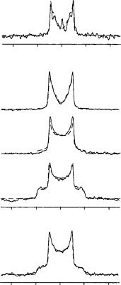
Figure 48 shows representative experimental 2H NMR spectra from the labeled retinal in bR in a dark-adapted PM sample. The line shape simulations that were generated in the data analysis are superimposed on the experimental spectra. The powder pattern [Figure 48(a)] serves as a general reference for the tilt series of spectra recorded at various sample inclinations [Figure 48(b)], because it defines the accessible frequency region over which the spectral intensity can occur. The oriented sample was measured at every 22.5° between 0° and 90°, of which three inclinations are represented in Figure 48(b) with ˛ D 0°, 45° and 90°.
membrane normal |
intracellular side |
|
B |
A |
C |
G |
D |
|
|
F |
E |
Lys
216 N
+H
CD2H
CD3
CD3
glass plate
extracellular side
FIGURE 46. Retinal chromophore in bR is attached via a protonated Schiff base to Lys-216 on helix G and is tilted toward the extracellular side. To determine its detailed structure, retinal was selectively deuteriated on the three methyl groups on the cyclohexene ring and incorporated into bR from H. Halobium. Reprinted with permission from Reference 60. Copyright (1997) American Chemical Society
2. NMR spectroscopy of dienes and polyenes |
163 |
|
z |
|
local |
|
|
tilt |
|
|
avarage |
|
|
chain tilt |
15 |
|
|
|
|
|
N |
|
|
H+ |
13 |
|
14 |
|
|
11 |
|
|
|
|
|
|
12 |
|
|
10 |
|
y |
|
|
|
x |
 9
9  rotation
|
7 |
8 |
CD2H |
|
CD3(b) |
1 |
6 |
5 |
|
|
|
|||
CD3(a) |
|
4 |
||
|
2 |
|||
- |
+ |
3 |
||
|
||||
|
|
|||
|
|
|
||
skew |
|
|
|
FIGURE 47. Three-dimensional structure of the cyclohexene ring of retinal in bR as determined by 2H NMR, relative to the membrane surface in the x – y plane. Analysis of the orientations of the three deuterium labeled methyl groups on the puckered ring (skew around C1 – C6) indicates that the chromophore has a 6s-trans conformation around the C6 – C7 bond. Reprinted with permission from Reference 60. Copyright (1997) American Chemical Society
With the sample aligned horizontally in the magnet, further spectra were recorded over a range of temperatures from 30° down to 120 °C. Representative line shapes are compared in Figure 48 for 21 °C (c) and 60 °C (b). The signal-to-noise ratio improves dramatically with decreasing temperature, and therefore the spectral analysis was based on the set of data acquired at 60 °C. The central resonance line which appears at temperatures above 0 °C is due to HDO. Two components appear to be resolved at 21 °C, but they are found to broaden and merge to give the unresolved line shape at 60 °C. A possible interpretation of this observation could be that some small local fluctuations within the puckered cyclohexene ring are frozen out in a slight glassy disorder, which is commonly found in crystalline retinal derivatives. Nevertheless, the overall line shape does not change significantly over the whole range of temperatures examined, and therefore the structure of the chromophore appears to remain relatively unaffected by freezing.
The characteristic shape of the 2H NMR powder pattern [Figure 48(a)] indicates that the dynamics of the rotating methyl groups is within the fast-motional limit at temperatures down to 120 °C. This assumption is further supported by the increasing signal-to-noise ratio with decreasing temperature, since no loss, but rather a gain, in intensity is observed on cooling. It is also clear that the cyclohexene ring does not undergo significant librational
164 |
Yoshito Takeuchi and Toshio Takayama |
||||
|
(c) |
|
|
|
|
|
|
|
|
|
α = 0° |
|
−80 |
−40 |
0 |
40 |
80 |
|
|
|
kHz |
|
|
|
(b) |
|
|
|
|
|
|
|
|
|
α = 0° |
|
|
|
|
|
α = 45° |
|
|
|
|
|
α = 90° |
|
−80 |
−40 |
0 |
40 |
80 |
|
|
|
kHz |
|
|
|
(a) |
|
|
|
|
|
−80 |
−40 |
0 |
40 |
80 |
|
|
|
kHz |
|
|
FIGURE 48. Representative 2H NMR spectra (full lines) of dark-adapted bR (90 mg) containing deuteriated retinal, with line shape simulations (dashed lines) superimposed. Both the powder spectrum (a) from randomly oriented PM patches and the tilt series (b) over sample inclinations, ˛ D 0°, 45° and 90°, were recorded at 60 °C (number of scans, 1.7 ð 105 , for ˛ D 0°). Spectrum (c) was measured at 21 °C with ˛ D 0° (number of scans, 3 ð 105). Reprinted with permission from Reference 60. Copyright (1997) American Chemical Society
motion within its binding pocket, since the 2H NMR spectra are not time-averaged by this type of random oscillation (with a correlation time c < 10 6 s). The polyene chain of the chromophore has been shown to be completely immobilized on the time scale of 10 9 – 10 2 s, indicating the absence of any rotational freedom.
With a horizontally oriented sample (˛ D 0°), the spectrum of the labeled bR in Figure 48(b) should display three quadrupole splittings corresponding to the three labeled methyl groups on the retinal. It is apparent, however, that the expected three pairs of resonances are not resolved because of spectral overlap of the broadened lines. A computer simulation approach was used to analyze the spectral line shapes despite the overlap, but much qualitative information about the cyclohexene ring can be gained by simple inspection of the experimental data in Figure 48.

2. NMR spectroscopy of dienes and polyenes |
165 |
IV. SPECIAL TOPICS
A. Allenes
Allenes form a unique class of compounds which are dienes (or polyenes), but simultaneously they are the parent compounds for the interesting family of cumulenes. The NMR investigation of these compounds is interesting in view of their unique electronic and structural properties.
A new C15 bromoallene60, dactylallene (103), was isolated from the digestive gland of the anaspidean mollusc Aplysia dactylomela. The structure was established by using mainly oneand two-dimensional NMR techniques, whereas the absolute stereochemistry was determined by X-ray diffractometric analysis. Ichthyotoxicity and antifeedant activity suggests a defensive role of 103 against predators. The structure of 103 is given below together with that of its steroisomer 104.
Br |
|
|
|
|
|
|
|
|
Br |
|
|
|
|
|
|
H |
8 |
|
|
|
|
|
|
H |
|
|
|
|
|
|
|
|
|
|
|
|
Cl |
|
|
|
|
|
Cl |
|
10 |
|
|
7 |
|
|
|
|
|
||||||
|
|
|
|
|
|
|
|
|
||||||
|
|
|
|
|
|
|
|
|
|
|
|
|
|
|
11 |
9 |
|
|
|
|
|
|
|
|
|
|
|
|
|
|
|
|
|
|
|
|
|
|
|
|
|
|
|
|
12 |
|
O |
6 |
|
O |
|
|
|
|
|
||||
|
|
|
|
|
|
|
|
|
|
|
|
|||
13 |
|
5 |
|
|
|
|
|
H |
|
|
|
|
|
H |
14 |
|
4 |
H |
|
|
H |
||||||||
|
|
|
|
|
|
H |
|
|
|
|
|
H |
||
|
|
|
|
|
|
|
|
|
|
|
|
|
||
15 |
O |
|
3 |
|
|
|
|
|
O |
|
|
|
|
|
|
|
|
|
|
|
|
|
|
|
|
||||
|
|
|
|
|
|
|
|
|
|
|
|
|
|
|
|
|
|
2 |
|
• |
|
|
|
|
|
|
• |
|
|
|
|
|
|
|
|
|
|
|
||||||
|
|
|
1 |
|
|
|
|
|
|
|
|
|
|
|
|
|
H |
|
|
|
|
|
Br |
Br |
|
|
|
|
H |
|
(103) |
|
|
|
|
|
|
(104) |
|
|
|
|
|
|
The 1H NMR spectrum displayed signals attributable to a secondary methyl at υ1.33 (H15, d, J D 7.0 Hz), six deshielded methines at υ4.00 (H-6), υ4.19 (H-4), υ4.38 (H-7), υ4.42 (H-14), υ4.48 (H-10), υ4.73 (H-9) and three methylenes at υ1.59 (H-5a, ddd, J D 14.0, 10.7 and 1.9 Hz) and υ1.69 (H-5b, m), υ2.40 (H-8a, m), and υ2.43 (H-8b, m), υ2.43 (H-11a, m) and υ2.89 (H-11b, m), strongly suggesting a nonterpenoid structure containing heteroatoms.
The presence of a bromoallene function was indicated both by two long-range coupled methine signals in the 1H NMR spectrum at υ6.01 (H1, dd, J D 5.8 Hz and 1.7 Hz) and υ5.35 (H3, dd, J D 5.8 and 6.0 Hz) and by the resonances in the 13C NMR spectrum at υ73.66 (C1), υ103.62 (C3) and υ200.99 (C2) (Table 40).
However, dactylallene (103) differs from 104 mainly in the chemical shifts of C14 (υ70.54 in 103, υ61.26 in 104), C4 (υ64.68 in 103, υ76.34 in 104) and C15 (υ14.05 in 103, υ21.19 in 104), suggesting a different relative stereochemistry for C4 or C14. However, the difference between the υ values for C4 and C14 in 103 and 104 is too large to be justified only by a different relative stereochemistry at the chiral center C4 or C14; it is most likely that these assignments in 104 should be reversed. In addition, the sign of [˛]D of 103, opposite to that of 104, indicated a different absolute stereochemistry of the allene residue, which could be predicted as S according to the Lowe – Brewster’s rule61.
Barretta and coworkers62 reported an assignment of the absolute configuration of chiral allenes, which is usually a difficult task. The problem has been solved by suitable chemical

166 |
|
Yoshito Takeuchi and Toshio Takayama |
|
|||
TABLE 40. |
1H and 13C NMR dataa,b for dactylallene (103) |
|
|
|||
Position |
υ1H |
m |
J(Hz) |
υ13C |
mc |
Long-range |
|
|
|
|
|
|
connectivitiesd |
1 |
6.01 |
dd |
5.8 and 1.7 |
73.66 |
d |
H3 |
2 |
|
|
|
200.99 |
s |
H1, H3, H4 |
3 |
5.35 |
dd |
5.8 and 6.0 |
103.62 |
d |
H1, H4, H5a |
4 |
4.19 |
m |
|
64.68 |
d |
H1, H3, H5a, H14 |
5 |
1.59 |
ddd |
14.0, 10.7, 1.9 |
37.05 |
t |
H4 |
|
1.69 |
|
|
|
|
|
6 |
4.00 |
m |
|
76.69 |
d |
H5b, H7, H8, H9 |
7 |
4.38 |
m |
|
62.51 |
d |
H6, H8 |
8 |
2.40 |
m |
|
38.61 |
t |
|
|
2.43 |
|
|
|
|
|
9 |
4.73 |
m |
|
78.26 |
d |
H7, H11 |
10 |
4.48 |
m |
|
49.96 |
d |
H8, H11 |
11 |
2.43 |
m |
|
37.84 |
t |
H9 |
|
2.89 |
|
|
|
|
|
12 |
5.79 |
m |
|
128.23 |
d |
H11, H14, H15 |
13 |
5.72 |
m |
|
128.23 |
d |
H11, H14, H15 |
14 |
4.42 |
m |
|
70.54 |
d |
H4, H12, H13, H15 |
15 |
1.33 |
d |
7.0 |
14.05 |
q |
|
aBruker AMX 500 MHz, CDCl3; υ values are reported in ppm referred to CHCl3 (υH 7.26) and to CDCl3 (υC 77.0).
bAssignments determined by 1H13CHETCOR, 1H1HCOSY, 1H – 1H decoupling experiments. c Determine by DEPT sequence.
dBy HMBC (J D 10 Hz).
correlations of allenes with centrodisymmetric molecules of known absolute configuration or by developing semiempirical rules, which relate the absolute configuration to the sign of the rotatory power or CD bands.
An alternative approach is provided by NMR spectroscopy. Separate NMR signals can be in principle obtained for stable or short-lived diastereomeric derivatives of the enantiomeric mixtures, the intensities of which are correlated with the enantiomeric composition and their relative stereochemistry to the absolute configuration. For this reason, great effort has been continually devoted to the development of new chiral auxiliaries for NMR spectroscopy. The majority of these are dedicated to the chiral assay of molecules having polar functional groups.
Recently, it was found that the commercially available heptakis(2,3,6-tri-O-methyl)- ˇ-cyclodextrin (permethylated ˇ-cyclodextrin, TRIMEB), induced nonequivalence in the 1H NMR spectra, in CD3OD, of enantiomeric mixtures of trisubstituted allenes devoid of polar functional groups, thus affording a simple and general way to determinations of their enantiomeric purity63.
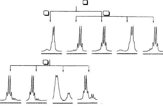
The authors reported that a consistent correlation exists between the absolute configuration of the trisubstituted allenes 105a–e and the permethylated ˇ-cyclodextrin induced shifts of their proton signals. Hence the use of TRIMEB as chiral auxiliary for the rapid and reliable NMR determination of their absolute configuration was proposed. The enantiomeric purities of the samples have been determined by analyzing the 1H NMR spectra of their mixtures with permethylated ˇ-cyclodextrin (with TRIMEB/allene molar ratios of 1 – 2) in CD3OD solutions. In all cases TRIMEB distinguished between the two enantiomers of each of the allenes 105a–e. The corresponding spectral regions relative to the resonances of the allene protons of the substrates in the presence of the cyclodextrin are reported in Figure 49. The bromoallene R -105a showed the major signal at 6.10 ppm, at

2. NMR spectroscopy of dienes and polyenes |
167 |
higher field than the minor signal corresponding to the S-allene at 6.14 ppm. The allenes 105b–e, having S-absolute configuration, generated from the bromoallene 105a, showed a major signal (at 6.12, 6.13, 7.02 and 6.09 ppm for 105b, 105c, 105d and 105e, respectively), which was at lower field than the minor signal due to the R-allene (at 6.09 ppm for 105b–c, 6.95 ppm for 105d and 6.06 ppm for 105e). For the R-allenes 105b–e, it has been also verified that the allene absorption of the R-enantiomers resonates at higher field with respect to the same signal of the S-enantiomer. It is noteworthy that the same kind of correlation between the sense of nonequivalence, i.e., the relative position of the absorption of one enantiomer with respect to the other, and the absolute configuration has been found for the alkyl protons: all the proton signals due to the S-enantiomer are lower field shifted with respect to the corresponding signals due to the R-enantiomer (Table 41).
t-Bu |
|
|
|
H |
C |
|
C |
|
C |
|
|
|||
|
|
|||
Me |
|
|
|
R |
|
(105) |
|
||
(a) R = Br
(b) R = Ph
(c) R = p-FC6H4 (d) R = a-Naph
(e) R = p-MeOC6H4
In conclusion, the most important result is that the use of permethylated cyclodextrin as chiral solvating agent for NMR spectroscopy not only affords a simple and practical way for the determination of the stereochemical purities of trisubstituted allenes, but also allows one to simultaneously determine their absolute configuration. Indeed, TRIMEB induced only positive complexation shifts of all the allene protons, which are greater for the S-enantiomer than for the R-enantiomer, independent of the structure of the allene. This empirical correlation seems to be reliable since it has been satisfied by a large number of trisubstituted allenes.
The method is undoubtedly very attractive from the practical point of view: it only requires the acquisition of a routine NMR spectrum for the suitable allene/TRIMEB mixture.
With the aid of 13C NMR, 6Li NMR and 1H HOESY (heteronuclear Overhauser effect spectroscopy) NMR of ˛-lithiomethoxyallene (106) and 1-lithio-1-ethoxy-3-t-butylallene (107) as well as by ab initio model calculations on monomeric and dimeric ˛-lithiohy- droxyallene, Schleyer and coworkers64 proved that 106 and 107 are dimeric in THF (106 forms a tetramer in diethyl ether) with a nonclassical 1,3-bridged structure. The 13C NMR spectrum of allenyllithium in THF is also in agreement with the allenic-type structure: the chemical shift of C2 (196.4 ppm) resembles that of neutral allene (212.6 ppm), rather than C2 of propyne (82.4 ppm).
The structures of 106 and 107 were also investigated in ether and THF solutions by using IR and NMR methods. Compound 107 was synthesized in order to avoid problems with the rapid rearrangement at > 20 °C of 106 to an alkynyllithium derivative. The lithiomethoxyallene had to be synthesized at ca 78 °C in THF or diethyl ether and it was measured in situ without isolation of the metalated product. Its NMR data are given in Table 42.
But R
C  C
C C
C
168
6.17 6.14 6.11 6.00 ppm |
6.10 6.13 6.10 6.07 ppm |
7.02 8.99 8.98 ppm |
6.12 6.00 6.06 ppm |
105b |
105c |
105d |
105e |
FIGURE 49. 1H NMR (300 MHz, CD3OD) spectra of samples of allenes 105a – e obtained by starting from (S)-105 (ee 89%): Spectral regions corresponding to the allene proton absorptions for mixtures TRIMEB/allene (molar ratio 1 : 1 for 105a – c, e and 2 : 1 for 105d), at 20 °C for 105a – c, e and at 40 °C for 105d. ( ) (R)-105e was obtained by starting from a sample of (S)-105 with lower enantiomeric purity (81%). ( ) (S)-105d – e were obtained by starting from a sample of 105a with lower enantiomeric purity (64%). Reprinted with permission from Reference 62. Copyright (1995) American Chemical Society