

Biomechanics Principles and Applications - Donald R. Peterson & Joseph D. Bronzino
.pdf1-16 |
|
|
|
|
|
|
|
|
|
|
|
|
|
|
|
|
|
|
|
|
|
Biomechanics |
|||
|
|
|
|
|
|
|
|
|
|
|
|
|
|
|
|
|
|
|
|
|
|
|
|
|
|
|
|
10–1 |
1000 |
100 |
10 |
1 |
0.1 |
0.01 |
0.001 |
|
|
||||||||||||||
|
|
|
|
|
|
|
|
|
|
|
|
|
|
(Hz) |
|
|
|
|
|
|
|
|
|
||
|
|
|
Sample dia 1/4 |
|
|
|
|
|
|
|
(c) (compression) |
|
|
|
|||||||||||
|
10–2 |
|
|
|
1/8 |
|
|
|
|
|
|
|
|
|
|
|
|||||||||
|
|
|
|
|
|
|
|
|
|
|
|
Osteons |
(b) Upper bound |
|
|||||||||||
|
|
|
|
|
|
|
|
|
|
|
|
|
|||||||||||||
|
|
|
|
|
|
|
|
|
|
|
|
|
|
|
|
|
|
||||||||
|
|
|
Contributions to the |
|
|
|
|
Lamellae |
|
|
|
|
|
|
|||||||||||
|
|
|
|
|
|
|
300 dia) |
|
|
|
|
|
|
||||||||||||
|
|
|
|
|
|
|
(20 thick) |
|
|
|
|
|
|
||||||||||||
|
10–3 |
loss tangent of |
|
|
|
|
|
|
|
(a) (compression) |
|
||||||||||||||
|
|
|
|
|
|
|
|
|
|
|
|
||||||||||||||
|
|
|
|
|
|
|
|
|
|
|
|
|
|
|
|||||||||||
|
|
tan |
cortical bone |
|
|
|
|
|
|
|
|
|
|
|
|
|
|
|
|
|
Sample dia |
|
|||
|
|
Homogeneous thermoelastic effect |
|
|
|
|
|
|
|
||||||||||||||||
|
|
(a) |
|
|
|
|
|
|
|
1/8 |
|
||||||||||||||
|
10–4 (b) |
Inhomogeneous thermoelastic effect |
|
|
|
|
|
|
|
|
|||||||||||||||
|
|
|
|
|
|
|
|
|
|
||||||||||||||||
|
|
(c) |
Fluid flow effect |
|
|
|
|
|
|
|
|
|
|
|
|
|
|
|
|
||||||
|
|
(d) |
Piezoelectric effect |
|
|
|
|
|
|
|
|
|
|
|
|
|
|
|
|
||||||
|
10–5 |
|
|
|
|
|
|
|
|
|
|
|
|
|
|
|
|
|
|
|
|
|
|
|
|
|
10–6 |
|
|
|
|
|
|
|
(b) |
|
|
|
|
|
|
|
|
|
|
|
|
||||
|
|
|
|
|
|
|
|
|
|
|
|
|
|
|
|
|
|
|
|
|
|
||||
|
|
|
|
|
|
|
|
|
|
|
|
|
|
|
|
|
Haversion canals, 50 dia |
|
|
||||||
|
|
(d), “dry” bone x |
|
|
|
|
|
Lacunae, 14 dia |
|
|
|
|
|
|
|
|
|
||||||||
|
|
|
|
|
|
0.001 |
0.01 |
0.1 |
|
|
1 |
|
|
10 |
|
|
100 |
1000 |
|
||||||
|
|
|
|
|
|
|
|
|
|
|
|
|
|
(sec) |
|
|
|
|
|
|
|
|
|
||
FIGURE 1.10 Contributions of several relaxation mechanisms to the loss tangent of cortical bone. (a) Homogeneous thermoelastic effect. (b) Inhomogeneous thermoelastic effect. (c) Fluid flow effect. (d) Piezoelectric effect [Lakes and Katz, 1984]. (Courtesy CRC Press.)
Following on Katz’s [1976, 1980] adaptation of the Hashin–Rosen hollow fiber composite model [1964], Gottesman and Hashin [1979] presented a viscoelastic calculation using the same major assumptions.
1.7 Related Research
As stated earlier, this chapter has concentrated on the elastic and viscoelastic properties of compact cortical bone and the elastic properties of trabecular bone. At present there is considerable research activity on the fracture properties of the bone. Professor William Bonfield and his associates at Queen Mary and Westfield College, University of London and Professor Dwight Davy and his colleagues at Case Western Reserve University are among those who publish regularly in this area. Review of the literature is necessary in order to become acquainted with the state of bone fracture mechanics.
An excellent introductory monograph that provides a fascinating insight into the structure–property relationships in bones including aspects of the two areas discussed immediately above is Professor John D. Currey’s Bones Structure and Mechanics [2002], the 2nd edition of the book, The Mechanical Adaptations of Bones, Princeton University Press [1984].
Defining Terms
Apatite: Calcium phosphate compound, stoichiometric chemical formula Ca5(PO4)3 · X, where X is OH− (hydroxyapatite), F− (fluorapatite), Cl− (chlorapatite), etc. There are two molecules in the basic crystal unit cell.
Cancellous bone: Also known as porous, spongy, trabecular bone. Found in the regions of the articulating ends of tubular bones, in vertebrae, ribs, etc.
Cortical bone: The dense compact bone found throughout the shafts of long bones such as the femur, tibia, etc., also found in the outer portions of other bones in the body.
Mechanics of Hard Tissue |
1-17 |
Haversian bone: Also called osteonic. The form of bone found in adult humans and mature mammals, consisting mainly of concentric lamellar structures, surrounding a central canal called the haversian canal, plus lamellar remnants of older haversian systems (osteons) called interstitial lamellae.
Interstitial lamellae: See Haversian bone above.
Orthotropic: The symmetrical arrangement of structure in which there are three distinct orthogonal axes of symmetry. In crystals this symmetry is called orthothombic.
Osteons: See Haversian bone above.
Plexiform: Also called laminar. The form of parallel lamellar bone found in younger, immature nonhuman mammals.
Transverse isotropy: The symmetry arrangement of structure in which there is a unique axis perpendicular to a plane in which the other two axes are equivalent. The long bone direction is chosen as the unique axis. In crystals this symmetry is called hexagonal.
References
Ashman R.B. and Rho J.Y. 1988. Elastic modulus of trabecular bone material. J. Biomech. 21: 177.
Black J. and Korostoff E. 1973. Dynamic mechanical properties of viable human cortical bone. J. Biomech. 6: 435.
Bonfield W. and Grynpas M.D. 1977. Anisotropy of Young’s modulus of bone. Nature, London, 270: 453.
Bumrerraj S. and Katz J.L. 2001. Scanning acoustic microscopy study of human cortical and trabecular bone. Ann. Biomed. Eng. 29: 1.
Choi K. and Goldstein S.A. 1992. A comparison of the fatigue behavior of human trabecular and cortical bone tissue. J. Biomech. 25: 1371.
Chung D.H. and Buessem W.R. 1968. In F.W. Vahldiek and S.A. Mersol (Eds.), Anisotropy in Single-Crystal Refractory Compounds, Vol. 2, p. 217. New York, Plenum Press.
Cowin S.C. 1989. Bone Mechanics. Boca Raton, FL, CRC Press.
Cowin S.C. 2001. Bone Mechanics Handbook. Boca Raton, FL, CRC Press.
Cox H.L. 1952. The elasticity and strength of paper and other fibrous materials. Br. Appl. Phys. 3: 72. Crolet, J.M., Aoubiza B., and Meunier A. 1993. Compact bone: numerical simulation of mechanical
characteristics. J. Biomech. 26: 677.
Currey J.D. 1964. Three analogies to explain the mechanical properties of bone. Biorheology: 1.
Currey J.D. 1969. The relationship between the stiffness and the mineral content of bone. J. Biomech.: 477.
Currey J.D. 1984. The Mechanical Adaptations of Bones. New Jersey, Princeton University Press. Currey J.D. 2002. Bone Structure and Mechanics. New Jersey, Princeton University Press.
Gottesman T. and Hashin Z. 1979. Analysis of viscoelastic behavior of bones on the basis of microstructure.
J. Biomech. 13: 89.
Hashin Z. and Rosen B.W. 1964. The elastic moduli of fiber reinforced materials. J. Appl. Mech.: 223.
Hashin Z. and Shtrikman S. 1963. A variational approach to the theory of elastic behavior of multiphase materials. J. Mech. Phys. Solids: 127.
Hastings G.W. and Ducheyne P. (Eds.). 1984. Natural and Living Biomaterials. Boca Raton, FL, CRC Press. Herring G.M. 1977. Methods for the study of the glycoproteins and proteoglycans of bone using bacterial collagenase. Determination of bone sialoprotein and chondroitin sulphate. Calcif. Tiss. Res.: 29.
Hogan H.A. 1992. Micromechanics modeling of haversian cortical bone properties. J. Biomech. 25: 549. Katz J.L. 1971a. Hard tissue as a composite material: I. Bounds on the elastic behavior. J. Biomech. 4:455. Katz J.L. 1971b. Elastic properties of calcified tissues. Isr. J. Med. Sci. 7: 439.
Katz J.L. 1976. Hierarchical modeling of compact haversian bone as a fiber reinforced material. In R.E. Mates and C.R. Smith (Eds.), Advances in Bioengineering, pp. 17–18. New York, American Society of Mechanical Engineers.
1-18 |
Biomechanics |
Katz J.L. 1980. Anisotropy of Young’s modulus of bone. Nature 283: 106.
Katz J.L. 1981. Composite material models for cortical bone. In S.C. Cowin (Ed.), Mechanical Properties of Bone, Vol. 45, pp. 171–184. New York, American Society of Mechanical Engineers.
Katz J.L. and Meunier A. 1987. The elastic anisotropy of bone. J. Biomech. 20: 1063.
Katz J.L. and Meunier A. 1990. A generalized method for characterizing elastic anisotropy in solid living tissues. J. Mat. Sci. Mater. Med. 1: 1.
Katz J.L. and Ukraincik K. 1971. On the anisotropic elastic properties of hydroxyapatite. J. Biomech. 4: 221. Katz J.L. and Ukraincik K. 1972. A fiber-reinforced model for compact haversian bone. Program and
Abstracts of the 16th Annual Meeting of the Biophysical Society, 28a FPM-C15, Toronto.
Keaveny T.M. and Hayes W.C. 1993. A 20-year perspective on the mechanical properties of trabecular bone. J. Biomech. Eng. 115: 535.
Kinney J.H., Pople J.A., Marshall G.W., and Marshall S.J. 2001. Collagen orientation and crystallite size in human dentin: A small angle x-ray scattering study. Calcif. Tissue Inter. 69: 31.
Kinney J.H., Gladden J.R., Marshall G.W., Marshall S.J., So J.H., and Maynard J.D. 2004. Resonant ultrasound spectroscopy measurements of the elastic constants of human dentin. J. Biomech. 37: 437.
Knets I.V. 1978. Mekhanika Polimerov 13: 434.
Laird G.W. and Kingsbury H.B. 1973. Complex viscoelastic moduli of bovine bone. J. Biomech. 6: 59. Lakes R.S. 1993. Materials with structural hierarchy. Nature 361: 511.
Lakes R.S. and Katz J.L. 1974. Interrelationships among the viscoelastic function for anisotropic solids: Application to calcified tissues and related systems. J. Biomech. 7: 259.
Lakes R.S. and Katz J.L. 1979a. Viscoelastic properties and behavior of cortical bone. Part II. Relaxation mechanisms. J. Biomech. 12: 679.
Lakes R.S. and Katz J.L. 1979b. Viscoelastic properties of wet cortical bone: III. A nonlinear constitutive equation. J. Biomech. 12: 689.
Lakes R.S. and Katz J.L. 1984. Viscoelastic properties of bone. In G.W. Hastings and P. Ducheyne (Eds.), Natural and Living Tissues, pp. 1–87. Boca Raton, FL, CRC Press.
Lakes R.S., Katz J.L., and Sternstein S.S. 1979. Viscoelastic properties of wet cortical bone: I. Torsional and biaxial studies. J. Biomech. 12: 657.
Lang S.B. 1969. Elastic coefficients of animal bone. Science 165: 287.
Lipson S.F. and Katz, J.L. 1984. The relationship between elastic properties and microstructure of bovine cortical bone. J. Biomech. 4: 231.
Lugassy A.A. 1968. Mechanical and Viscoelastic Properties of Bone and Dentin in Compression, thesis, Metallurgy and Materials Science, University of Pennsylvania.
Maharidge R. 1984. Ultrasonic properties and microstructure of bovine bone and Haversian bovine bone modeling, thesis, Rensselaer Polytechnic Institute, Troy, NY.
Park J.B. 1979. Biomaterials: An Introduction. New York, Plenum.
Pellegrino E.D. and Biltz R.M. 1965. The composition of human bone in uremia. Medicine 44: 397. Piekarski K. 1973. Analysis of bone as a composite material. Int. J. Eng. Sci. 10: 557.
Reuss A. 1929. Berechnung der fliessgrenze von mischkristallen auf grund der plastizitatsbedingung fur einkristalle, A. Zeitschrift fur Angewandte Mathematik und Mechanik 9: 49–58.
Rho J.Y., Ashman R.B., and Turner C.H. 1993. Young’s modulus of trabecular and cortical bone material; ultrasonic and microtensile measurements. J. Biomech. 26: 111.
Rho J.Y., Roy M.E., Tsui T.Y., and Pharr G.M. 1999. Elastic properties of microstructural components of human bone tissue as measured by indentation. J. Biomed. Mat. Res. 45: 48.
Ryan S.D. and Williams J.L. 1989. Tensile testing of rodlike trabeculae excised from bovine femoral bone.
J. Biomech. 22: 351.
Sedlin E. 1965. A rheological model for cortical bone. Acta Orthop. Scand. 36.
Smith R. and Keiper D. 1965. Dynamic measurement of viscoelastic properties of bone. Am. J. Med. Elec. 4: 156.
Townsend P.R., Rose R.M., and Radin E.L. 1975. Buckling studies of single human trabeculae. J. Biomech. 8: 199.
Mechanics of Hard Tissue |
1-19 |
Turner C.H., Rho J.Y., Takano Y., Tsui T.Y., and Pharr G.M. 1999. The elastic properties of trabecular and cortical bone tissues are simular: results from two microscopic measurement techniques. J. Biomech. 32: 437.
Van Buskirk W.C. and Ashman R.B. 1981. The elastic moduli of bone. In S.C. Cowin (Ed.), Mechanical Properties of Bone AMD, Vol. 45, pp. 131–143. New York, American Society of Mechanical Engineers.
Vejlens L. 1971. Glycosaminoglycans of human bone tissue: I. Pattern of compact bone in relation to age.
Calcif. Tiss. Res. 7: 175.
Voigt W. 1966. Lehrbuch der Kristallphysik Teubner, Leipzig 1910; reprinted (1928) with an additional appendix. Leipzig, Teubner, New York, Johnson Reprint.
Wagner H.D. and Weiner S. 1992. On the relationship between the microstructure of bone and its mechanical stiffness. J. Biomech. 25: 1311.
Wainwright S.A., Briggs W.D., Currey J.D., and Gosline J.M. 1982. Mechanical Design in Organisms. Princeton, NJ, Princeton, University Press.
Weiner S. and Traub W. 1989. Crystal size and organization in bone. Conn. Tissue Res. 21: 259.
Yoon H.S. and Katz J.L. 1976a. Ultrasonic wave propagation in human cortical bone: I. Theoretical considerations of hexagonal symmetry. J. Biomech. 9: 407.
Yoon H.S. and Katz J.L. 1976b. Ultrasonic wave propagation in human cortical bone: II. Measurements of elastic properties and microhardness. J. Biomech. 9: 459.
Further Information
Several societies both in the United States and abroad hold annual meetings during which many presentations, both oral and poster, deal with hard tissue biomechanics. In the United States these societies include the Orthopaedic Research Society, the American Society of Mechanical Engineers, the Biomaterials Society, the American Society of Biomechanics, the Biomedical Engineering Society, and the Society for Bone and Mineral Research. In Europe there are alternate year meetings of the European Society of Biomechanics and the European Society of Biomaterials. Every four years there is a World Congress of Biomechanics; every three years there is a World Congress of Biomaterials. All of these meetings result in documented proceedings; some with extended papers in book form.
The two principal journals in which bone mechanics papers appear frequently are the Journal of Biomechanics published by Elsevier and the Journal of Biomechanical Engineering published by the American Society of Mechanical Engineers. Other society journals that periodically publish papers in the field are the
Journal of Orthopaedic Research published for the Orthopaedic Research Society, the Annals of Biomedical Engineering published for the Biomedical Engineering Society, and the Journal of Bone and Joint Surgery (both American and English issues) for the American Academy of Orthopaedic Surgeons and the British Organization, respectively. Additional papers in the field may be found in the journal Bone and Calcified Tissue International.
The 1984 CRC volume, Natural and Living Biomaterials (Hastings G.W. and Ducheyne P., Eds.) provides a good historical introduction to the field. A recent more advanced book is Bone Mechanics Handbook (Cowin S.C., Ed. 2001), the 2nd edition of Bone Mechanics (Cowin S.C., Ed. 1989).
Many of the biomaterials journals and society meetings will have occasional papers dealing with hard tissue mechanics, especially those dealing with implant–bone interactions.

1-20 |
Biomechanics |
Appendix
The Voigt and Reuss moduli for both transverse isotropic and orthotropic symmetry are given below:
Voigt transverse isotropic
K V = 2(C11 + C12) + 4(C13 + C33)
9
G V = (C11 + C12) − 4C13 + 2C33 + 12(C44 + C66)
30
Reuss transverse isotropic
KR = |
C33(C11 + C12) − 2C132 |
|
|
(C11 + C12 − 4C13 + 2C33) |
|||
|
|||
G R = |
5 C33(C11 + C12) − 2C132 C44C66 |
||
2 C33(C11 + C12) − 2C132 (C44 + C66) + [C44C66(2C11 + C12) + 4C13 + C33]/3 |
|||
Voigt orthotropic
K V = C11 + C22 + C33 + 2(C12 + C13 + C23)
9
G V = [C11 + C22 + C33 + 3(C44 + C55 + C66) − (C12 + C13 + C23)]
15
Reuss orthotropic
KR = |
|
|
− 2(C11C23 + C22C13 + C33C12) |
|
|
||
C11C22 |
+ C22C33 + C33C11 |
||
+ 2(C12C23 + C23C13 + C13C12) − C122 + C132 + C232
GR = 15/(4{(C11C22 + C22C33 + C33C11 + C11C23 + C22C13 + C33C22)
−[C12(C12 + C23) + C23(C23 + C13) + C13(C13 + C12)]}/
+ 3(1/C44 + 1/C55 + 1/C66))
where is given in Equation 1.15.
(1.A1)
(1.A2)
(1.A3)
(1.A4)

Richard L. Lieber
University of California
Thomas J. Burkholder
Georgia Institute of Technology
2
Musculoskeletal Soft Tissue Mechanics
2.1 Structure of Soft Tissues . . . . . . . . . . . . . . . . . . . . . . . . . . . . . . . 2-1
|
Cartilage • |
Tendon and Ligament • |
Muscle |
2.2 |
Material Properties . . . . . . . . . . . . |
. . . . . . . . . . . . . . . . . . . . . . . . 2-4 |
|
|
Cartilage • |
Tendon and Ligament • |
Muscle |
2.3 |
Modeling |
. . . . . . . . . . . . . . . . . . . . . |
. . . . . . . . . . . . . . . . . . . . . . . . 2-6 |
|
Cartilage • |
Tendon and Ligament • |
Muscle |
References . . . . . . |
. . . . . . . . . . . . . . . . . . . . |
. . . . . . . . . . . . . . . . . . . . . . . . 2-13 |
|
Biological soft tissues are nonlinear, anisotropic, fibrous composites, and detailed description of their behavior is the subject of active research. One can separate these tissues based on their mode of loading: cartilage is generally loaded in compression; tendons and ligaments are loaded in tension; and muscles generate active tension. The structure and material properties differ to accommodate the tissue function, and this chapter outlines those features. Practical models of each tissue are described, with particular focus on active force generation by skeletal muscle and application to segmental modeling.
2.1 Structure of Soft Tissues
2.1.1 Cartilage
Articular cartilage is found at the ends of bones, where it serves as a shock absorber and lubricant between bones. It is best described as a hydrated proteoglycan gel supported by a sparse population of chondrocytes, and its composition and properties vary dramatically over its 1- to 2-mm thickness. The bulk composition of articular cartilage consists of approximately 20% collagen, 5% proteoglycan, primarily aggrecan bound to hyaluronic acid, with most of the remaining 75% water [Ker, 1999]. At the articular surface, collagen fibrils are most dense and arranged primarily in parallel with the surface. Proteoglycan content is very low and chondrocytes are rare in this region. At the bony interface, collagen fibrils are oriented perpendicular to the articular surface, chondrocytes are more abundant, but proteoglycan content is low. Proteoglycans are most abundant in the middle zone, where collagen fibrils lack obvious orientation in association with the transition from parallel to perpendicular alignment.
2-1
2-2 |
Biomechanics |
Collagen itself is a fibrous protein composed of tropocollagen molecules. Tropocollagen is a triple-helical protein, which self-assembles into the long collagen fibrils observable at the ultrastructural level. These fibrils, in turn, aggregate and intertwine to form the ground substance of articular cartilage. When cross-linked into a dense network, as in the superficial zone of articular cartilage, collagen has a low permeability to water and helps to maintain the water cushion of the middle and deep zones. Collagen fibrils arranged in a random network, as in the middle zone, structurally immobilize the large proteoglycan (PG) aggregates, creating the solid phase of the composite material.
Proteoglycans consist of a number of negatively charged glycosaminoglycan chains bound to an aggrecan protein core. Aggrecan molecules, in turn, bind to a hyaluronic acid backbone, forming a PG of 50 to 100 MDa, which carries a dense negative charge. This negative charge attracts positively charged ions (Na+) from the extracellular fluid, and the resulting Donnan equilibrium results in rich hydration of the tissue creating an osmotic pressure that enables the tissue to act as a shock absorber.
The overall structure of articular cartilage is analogous to a jelly-filled balloon. The PG-rich middle zone is osmotically pressurized, with fluid restrained from exiting the tissue by the dense collagen network of the superficial zone and the calcified structure of the deep bone. The interaction between the mechanical loading forces and osmotic forces yields the complex material properties of articular cartilage.
2.1.2 Tendon and Ligament
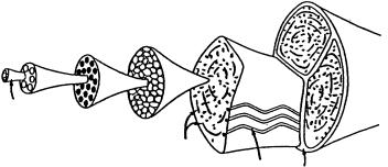
The passive tensile tissues, tendon and ligament, are also composed largely of water and collagen, but contain very little of the PGs that give cartilage its unique mechanical properties. In keeping with the functional role of these tissues, the collagen fibrils are organized primarily in long strands parallel to the axis of loading (Figure 2.1) [Kastelic et al., 1978]. The collagen fibrils, which may be hollow tubes [Gutsmann et al., 2003], combine in a hierarchical structure, with the 20–40-nm fibrils being bundled into 0.2–12-μm fibers. These fibers are birefringent under polarized light, reflecting an underlying wave or crimp structure with a periodicity between 20 and 100 μm. The fibers are bundled into fascicles, supported by fibroblasts or tenocytes, and surrounded by a fascicular membrane. Finally, multiple fascicles are bundled into a complete tendon or ligament encased in a reticular membrane.
As the tendon is loaded, the bending angle of the crimp structure of the collagen fibers can be seen to reversibly decrease, indicating that deformation of this structure is one source of elasticity. Individual collagen fibrils also display some inherent elasticity, and these two features are believed to determine the bulk properties of passive tensile tissues.
Tendon
Microfibril |
Subfibril |
Fibril |
Fascicle |
Collagen
Fibroblasts
Crimp Fascicular
membrane
FIGURE 2.1 Tendons are organized in progressively larger filaments, beginning with molecular tropocollagen, and building to a complete tendon encased in a reticular sheath.
Musculoskeletal Soft Tissue Mechanics |
2-3 |
2.1.3 Muscle
2.1.3.1 Gross Morphology
Muscles are described as running from a proximal origin to a distal insertion. While these attachments are frequently discrete, distributed attachments, and distinctly bifurcated attachments, are also common. Description of the subdomains of a muscle is largely by analogy to the whole body. The mass of muscle fibers can be referred to as the belly. In a muscle with distinctly divided origins, the separate origins are often referred to as heads, and in a muscle with distinctly divided insertions, each mass of fibers terminating on distinct tendons is often referred to as a separate belly.
A muscle generally receives its blood supply from one main artery, which enters the muscle in a single, or sometimes two branches. Likewise, the major innervation is generally by a single nerve, which carries both motor efferents and sensory afferents.
Some muscles are functionally and structurally subdivided into compartments. A separate branch of the principle nerve generally innervates each compartment, and motor units of the compartments do not overlap. Generally, a dense connective tissue, or fascial, plane separates the compartments.
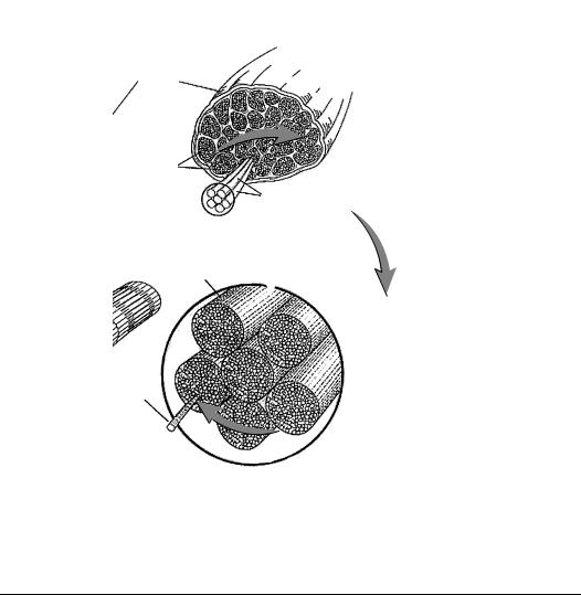
2.1.3.2 Fiber Architecture
Architecture, the arrangement of fibers within a muscle, determines the relationship between whole muscle length changes and force generation. The stereotypical muscle architecture is fusiform, with the muscle originating from a small tendonous attachment, inserting into a discrete tendon, and having fibers running generally parallel to the muscle axis (Figure 2.2). Fibers of unipennate muscles run parallel to each other but at an angle (pennation angle) to the muscle axis. Bipennate muscle fibers run in two distinct directions. Multipennate or fan-like muscles have one distinct attachment and one broad attachment, and pennation angle is different for every fiber. Strap-like muscles have parallel fibers that run from a broad bony origin to a broad insertion. As the length of each of these muscles is changed, the change in length of its fibers depends on fiber architecture. For example, fibers of a strap-like muscle undergo essentially the same length change as the muscle, where the length change of highly pennate fibers is reduced by their angle.
2.1.3.3 Sarcomere
Force generation in skeletal muscle results from the interaction between myosin and actin proteins. These molecules are arranged in antiparallel filaments, a 2- to 3-nm diameter thin filament composed mainly of actin, and a 20-nm diameter thick filament composed mainly of myosin. Myosin filaments are arranged in a hexagonal array, rigidly fixed at the M-line, and are the principal constituents of the A-band (anisotropic, light bending). Actin filaments are arranged in a complimentary hexagonal array and rigidly fixed at the Z-line, comprising the l-band (isotropic, light transmitting). The sarcomere is a nearly crystalline structure, composed of an A-band and two adjacent l-bands, and is the fundamental unit of muscle force generation. Sarcomeres are arranged into arrays of myofibrils, and one muscle cell or myofiber contains many myofibrils. Myofibers themselves are multinucleated syncitia, hundreds of microns in diameter, and may be tens of millimeters in length that are derived during development by the fusion of myoblasts.
The myosin protein occurs in several different isoforms, each with different force-generating characteristics, and each associated with expression of characteristic metabolic and calcium-handling proteins. Broadly, fibers can be characterized as either fast or slow, with slow fibers having a lower rate of actomyosin ATPase activity, slower velocity of shortening, slower calcium dynamics, and greater activity of oxidative metabolic enzymes. The lower ATPase activity makes these fibers more efficient for generating force, while the high oxidative capacity provides a rich energy source, making slow fibers ideal for extended periods of activity. Their relatively slow speed of shortening results in poor performance during fast or ballistic motions.
2-4 |
Biomechanics |
Muscle
Fascicles
Muscle fibers
Muscle
fiber
Sarcomere
Myofibril
FIGURE 2.2 Skeletal muscle is organized in progressively larger filaments, beginning with molecular actin and myosin, arranged as myofibrils. Myofibrils assemble into sarcomeres and myofilaments. Myofilaments are assembled into myofibers, which are organized into the fascicles that form a whole muscle.
2.2 Material Properties
2.2.1 Cartilage
The behavior of cartilage is highly viscoelastic. A compressive load applied to articular cartilage drives the positively charged fluid phase through the densely intermeshed and negatively charged solid phase while deforming the elastic PG-collagen structure. The mobility of the fluid phase is relatively low, and, for rapid changes in load, cartilage responds nearly as a uniform linear elastic solid with a Young’s modulus of approximately 6 MPa [Carter and Wong, 2003].
At lower loading rates, cartilage displays more nonlinear properties. Ker [1999] reports that human limb articular cartilage stiffness can be described as E = E 0(1 + σ 0.366), with E 0 = 3.0 MPa and σ expressed in MPa.
2.2.2 Tendon and Ligament
At rest, the collagen fibrils are significantly crimped or wavy so that initial loading acts primarily to straighten these fibrils. At higher strains, the straightened collagen fibrils must be lengthened. Thus, tendons are more compliant at low loads and less compliant at high loads. The highly nonlinear low load region has been referred to as the “toe” region and occurs up to approximately 3% strain and

Musculoskeletal Soft Tissue Mechanics |
|
|
2-5 |
|||
TABLE 2.1 Tendon Biomechanical Properties |
|
|
|
|||
|
|
|
|
|
|
|
|
|
|
Stress Under |
Strain Under |
Tangent |
|
|
Ultimate |
Ultimate |
Normal Loads Normal Loads |
Modulus |
|
|
Tendon |
Stress (MPa) |
Strain (%) |
(MPa) |
(%) |
(GPa) |
References |
|
|
|
|
|
|
|
Wallaby |
40 |
9 |
15–40 |
|
1.56 |
Bennett et al. [1986] |
Porpoise |
|
|
|
|
1.53 |
Bennett et al. [1986] |
Dolphin |
|
|
|
|
1.43 |
Bennett et al. [1986] |
Deer |
|
|
28–74 |
|
1.59 |
Bennett et al. [1986] |
Sheep |
|
|
|
|
1.65 |
Bennett et al. [1986] |
Donkey |
|
|
22–44 |
|
1.25 |
Bennett et al. [1986] |
Human leg |
|
|
53 |
|
1.0–1.2 |
Bennett et al. [1986] |
Cat leg |
|
|
|
|
1.21 |
Bennett et al. [1986] |
Pig tail |
|
|
|
|
0.9 |
Bennett et al. [1986] |
Rat tail |
|
|
|
|
0.8–1.5 |
Bennett et al. [1986] |
Horse |
|
|
|
4–10 |
|
Ker et al. [1988] |
Dog leg |
|
|
84 |
|
|
Ker et al. [1988] |
Camel ankle |
|
|
18 |
|
|
Ker et al. [1988] |
Human limb |
|
|
|
|
|
McElhaney et al. [1976] |
(various) |
60–120 |
|
|
|
|
|
Human calcaneal |
55 |
9.5 |
|
|
|
McElhaney et al. [1976] |
Human wrist |
52–74 |
11–17 |
3.2–3.3 |
1.5–3.5 |
|
Loren and Lieber [1994] |
|
|
|
|
|
|
|
5 MPa [Butler et al., 1979; Zajac, 1989]. Typically, tendons have nearly linear properties from about 3% strain until ultimate strain, which ranges from 9 to 10% (Table 2.1). The tangent modulus in this linear region is approximately 1.5 GPa. Ultimate tensile stress reported for tendons is approximately 100 MPa [McElhaney et al., 1976]. However, under physiological conditions, tendons operate at stresses of only 5 to 10 MPa (Table 2.1) yielding a typical safety factor of 10.
2.2.3 Muscle
Tension generated by skeletal muscle depends on length, velocity, level of activation, and history. Performance characteristics of a muscle depend on both its intrinsic properties and the extrinsic organization of that tissue. Whole muscle maximum shortening velocity depends both upon the sliding velocity of its component sarcomeres and on the number of those sarcomeres arranged in series. Likewise, maximum isometric tension depends on both the intrinsic tension-generating capacity of the actomyosin crossbridges and on the number of sarcomeres arranged in parallel. The relationship between intrinsic properties and extrinsic function is further complicated by pennation of the fibers. Given the orthotropic nature of the muscle fiber, material properties should be considered relative to the fiber axis. That is, the relevant area for stress determination is not the geometric cross section, but the physiological cross section, perpendicular to the fiber axis. The common form for estimation of the physiological cross sectional area (PCSA) is:
PCSA =
M · cos( )
ρ · FL
where M is muscle mass, is pennation angle, ρ is muscle density (1.06 g/cm3), and FL is fiber length. Likewise, the relevant gage length for strain determination is not muscle length, but fiber length, or fascicle length in muscles composed of serial fibers.
Maximum muscle stress: Maximum active stress, or specific tension, varies somewhat among fiber types and species (Table 2.2) around a generally accepted average of 250 kPa. This specific tension can be determined in any system in which it is possible to measure force and estimate the area of contractile material. Given muscle PCSA, maximum force produced by a muscle can be predicted by multiplying this PCSA by specific tension (Table 2.2). Specific tension can also be calculated for isolated muscle fibers or motor units in which estimates of cross-sectional area have been made.
Maximum muscle contraction velocity: Muscle maximum contraction velocity is primarily dependent on the type and number of sarcomeres in series along the muscle fiber length [Gans, 1982].