

Biomechanics Principles and Applications - Donald R. Peterson & Joseph D. Bronzino
.pdf
The Editors
Donald R. Peterson, Ph.D., M.S., an assistant professor in the Schools of Medicine, Dental Medicine, and Engineering at the University of Connecticut, and director of the Biodynamics Laboratory and the Bioengineering Facility at the University of Connecticut Health Center, offers graduate-level courses in biomedical engineering in the fields of biomechanics, biodynamics, biofluid mechanics, and ergonomics, and teaches in medicine in the subjects of gross anatomy and occupational biomechanics. He earned a B.S. in both aerospace and biomedical engineering from Worcester Polytechnic Institute, a M.S. in mechanical engineering from the University of Connecticut, and a Ph.D. in biomedical engineering also from the University of Connecticut. Dr. Peterson’s current research work is focused on the development of laboratory and field techniques for accurately assessing and modeling human–device interaction and human and/or organism performance, exposure, and response. Recent applications of these protocols model human interactions with existing and developmental devices such as powered and nonpowered tools, spacesuits and spacetools for NASA, surgical and dental instruments, musical instruments, sports equipment, and computer input devices. Other research initiatives focus on cell biomechanics, the acoustics of hearing protection and communication, hand–arm vibration exposure, advanced physiological monitoring methods, advanced vascular imaging techniques, and computational biomechanics.
Joseph D. Bronzino received the B.S.E.E. degree from Worcester Polytechnic Institute, Worcester, MA, in 1959, the M.S.E.E. degree from the Naval Postgraduate School, Monterey, CA, in 1961, and the Ph.D. degree in electrical engineering from Worcester Polytechnic Institute in 1968. He is presently the Vernon Roosa Professor of Applied Science, an endowed chair at Trinity College, Hartford, CT, and president of the Biomedical Engineering Alliance and Consortium (BEACON), which is a nonprofit organization consisting of academic and medical institutions as well as corporations dedicated to the development and commercialization of new medical technologies (for details visit www.beaconalliance.org).
He is the author of over 200 articles and 11 books including the following: Technology for Patient Care (C.V. Mosby, 1977), Computer Applications for Patient Care (Addison-Wesley, 1982), Biomedical Engineering: Basic Concepts and Instrumentation (PWS Publishing Co., 1986), Expert Systems: Basic Concepts (Research Foundation of State University of New York, 1989), Medical Technology and Society: An Interdisciplinary Perspective (MIT Press and McGraw-Hill, 1990), Management of Medical Technology
(Butterworth/Heinemann, 1992), The Biomedical Engineering Handbook (CRC Press, 1st ed., 1995; 2nd ed., 2000; Taylor & Francis, 3rd ed., 2005), Introduction to Biomedical Engineering (Academic Press, 1st ed., 1999; 2nd ed., 2005).
Dr. Bronzino is a fellow of IEEE and the American Institute of Medical and Biological Engineering (AIMBE), an honorary member of the Italian Society of Experimental Biology, past chairman of the Biomedical Engineering Division of the American Society for Engineering Education (ASEE), a charter member and presently vice president of the Connecticut Academy of Science and Engineering (CASE), a charter member of the American College of Clinical Engineering (ACCE), and the Association for the Advancement of Medical Instrumentation (AAMI), past president of the IEEE-Engineering in Medicine
xi
and Biology Society (EMBS), past chairman of the IEEE Health Care Engineering Policy Committee (HCEPC), past chairman of the IEEE Technical Policy Council in Washington, DC, and presently editor-in-chief of Elsevier’s BME Book Series and Taylor & Francis’ Biomedical Engineering Handbook.
Dr. Bronzino is also the recipient of the Millennium Award from IEEE/EMBS in 2000 and the Goddard Award from Worcester Polytechnic Institute for Professional Achievement in June 2004.
xii

Contributors
Kai-Nan An
Biomedical Laboratory
Mayo Clinic
Rochester, Minnesota
Gary J. Baker
Stanford University
Stanford, California
Thomas J. Burkholder
School of Applied Physiology
Georgia Institute of Technology
Atlanta, Georgia
Thomas R. Contield
Argonne National Laboratory
Roy B. Davis, III
Shriner’s Hospital for Children
Peter A. DeLuca
Gait Analysis Laboratory
University of Connecticut
Children’s Medical Center
Hartford, Connecticut
Philip B. Dobrin
Hines VA Hospital and Loyola
University Medical Center
Hines, Illinois
Cathryn R. Dooly
University of Maryland
College Park, Maryland
Jeffrey T. Ellis
Department of Bioengineering
and Bioscience
Georgia Institute of Technology
Atlanta, Georgia
Michael J. Furey
Mechanical Engineering
Department
Virginia Polytechnic Institute
and State University
Blacksburg, Virginia
Wallace Grant
Engineering Science and
Mechanics Department
Virginia Polytechnic Institute
and State University
Blacksburg, Virginia
Alan R. Hargens
Department of Orthopedic
Surgery
University of
California-San Diego
San Diego, California
Robert M. Hochmuth
Department of Mechanical
Engineering
Duke University
Durham, North Carolina
Ben F. Hurley
Department of Kinesiology
College of Health and Human
Performance
University of Maryland
College Park, Maryland
Arthur T. Johnson
Engineering Department
Biological Resource
University of Maryland
College Park, Maryland
J. Lawrence Katz
School of Dentistry
University of
Missouri-Kansas City
Kansas City, Missouri
Kenton R. Kaufman
Biomedical Laboratory
Mayo Clinic
Rochester, Minnesota
Albert I. King
Biomaterials Engineering
Center
Wayne State University
Detroit, Michigan
Jack D. Lemmon
Department of Bioengineering
and Bioscience
Georgia Institute of Technology
Atlanta, Georgia
Baruch B. Lieber
Department of Mechanical and
Aerospace Engineering
State University of
New York-Buffalo
Buffalo, New York
Richard L. Lieber
Departments of Orthopedics
and Bioengineering
University of California
La Jolla, California
xiii
Andrew D. McCulloch
Department of Bioengineering
University of
California-San Diego
La Jolla, California
˜
Sylvia Ounpuu
Center for Motion Analysis University of Connecticut
Children’s Medical Center West Hartford, Connecticut
Donald R. Peterson
University of Connecticut
Health Center
Biodynamics Laboratory
Farmington, Connecticut
Roland N. Pittman
Medical College of Virginia
Richmond, Virginia
Aleksander S. Popel
Department of Biomedical
Engineering
Johns Hopkins University
Baltimore, Maryland
Carl F. Rothe
Department of Physiology
Medical Science
Indiana University
Indianapolis, Indiana
Geert W. Schmid-Schonbein¨
Department of Bioengineering
University of
California-San Diego
La Jolla, California
Artin A. Shoukas
Department of Biomedical
Engineering
Johns Hopkins University
School of Medicine
Baltimore, Maryland
Alexander A. Spector
Biomedical Engineering
Johns Hopkins University
Baltimore, Maryland
Charles R. Steele
Applied Mechanical Division
Stanford University
Stanford, California
Jason A. Tolomeo
Stanford University
Stanford, California
Roger Tran-Son-Tay
University of Florida
Gainesville, Florida
David C. Viano
Wayne State University
Detroit, Michigan
Richard E. Waugh
Department of Pharmacology
and Physiology
University of Rochester
Medical Center
Rochester, New York
Ajit P. Yoganathan
Department of Bioengineering
and Bioscience
Georgia Institute of Technology
Atlanta, Georgia
Deborah E. Zetes-Tolomeo
Stanford University
Stanford, California
xiv

1
Mechanics of Hard Tissue
|
1.1 |
Structure of Bone . . . . . . . . . . . . . . . . . . . . . . . . . . . . . . . . . . . . . |
1-2 |
|
1.2 |
Composition of Bone . . . . . . . . . . . . . . . . . . . . . . . . . . . . . . . . . |
1-4 |
|
1.3 |
Elastic Properties. . . . . . . . . . . . . . . . . . . . . . . . . . . . . . . . . . . . . . |
1-4 |
|
1.4 |
Characterizing Elastic Anisotropy . . . . . . . . . . . . . . . . . . . . . . |
1-9 |
|
1.5 |
Modeling Elastic Behavior . . . . . . . . . . . . . . . . . . . . . . . . . . . . . |
1-12 |
|
1.6 |
Viscoelastic Properties. . . . . . . . . . . . . . . . . . . . . . . . . . . . . . . . . |
1-12 |
|
1.7 |
Related Research . . . . . . . . . . . . . . . . . . . . . . . . . . . . . . . . . . . . . . |
1-16 |
|
Defining Terms . . . . . . . . . . . . . . . . . . . . . . . . . . . . . . . . . . . . . . . . . . . . . |
1-16 |
|
|
References . . . . . . . . . . . . . . . . . . . . . . . . . . . . . . . . . . . . . . . . . . . . . . . . . . |
1-17 |
|
J. Lawrence Katz |
Further Information . . . . . . . . . . . . . . . . . . . . . . . . . . . . . . . . . . . . . . . . |
1-19 |
|
University of Missouri-Kansas City |
Appendix . . . . . . . . . . . . . . . . . . . . . . . . . . . . . . . . . . . . . . . . . . . . . . . . . . . |
1-20 |
|
Hard tissue, mineralized tissue, and calcified tissue are often used as synonyms for bone when describing the structure and properties of bone or tooth. The hard is self-evident in comparison with all other mammalian tissues, which often are referred to as soft tissues. Use of the terms mineralized and calcified arises from the fact that, in addition to the principle protein, collagen, and other proteins, glycoproteins, and protein-polysaccherides, comprising about 50% of the volume, the major constituent of bone is a calcium phosphate (thus the term calcified) in the form of a crystalline carbonate apatite (similar to naturally occurring minerals, thus the term mineralized). Irrespective of its biological function, bone is one of the most interesting materials known in terms of structure–property relationships. Bone is an anisotropic, heterogeneous, inhomogeneous, nonlinear, thermorheologically complex viscoelastic material. It exhibits electromechanical effects, presumed to be due to streaming potentials, both in vivo and in vitro when wet. In the dry state, bone exhibits piezoelectric properties. Because of the complexity of the structure–property relationships in bone, and the space limitation for this chapter, it is necessary to concentrate on one aspect of the mechanics. Currey [1984] states unequivocally that he thinks, “the most important feature of bone material is its stiffness.” This is, of course, the premiere consideration for the weight-bearing long bones. Thus, this chapter will concentrate on the elastic and viscoelastic properties of compact cortical bone and the elastic properties of trabecular bone as exemplar of mineralized tissue mechanics.
1-1

1-2 |
Biomechanics |
1.1 Structure of Bone
The complexity of bone’s properties arises from the complexity in its structure. Thus it is important to have an understanding of the structure of mammalian bone in order to appreciate the related properties. Figure 1.1 is a diagram showing the structure of a human femur at different levels [Park, 1979]. For convenience, the structures shown in Figure 1.1 will be grouped into four levels. A further subdivision of structural organization of mammalian bone is shown in Figure 1.2 [Wainwright et al., 1982]. The individual figures within this diagram can be sorted into one of the appropriate levels of structure shown on Figure 1.1 as described in the following. At the smallest unit of structure we have the tropocollagen molecule and the associated apatite crystallites (abbreviated Ap). The former is approximately 1.5 by 280 nm, made up of three individual left-handed helical polypeptide (alpha) chains coiled into a righthanded triple helix. Ap crystallites have been found to be carbonate-substituted hydroxyapatite, generally thought to be nonstoichiometric. The crystallites appear to be about 4 × 20 × 60 nm in size. This level is denoted the molecular. The next level we denote the ultrastructural. Here, the collagen and Ap are intimately associated and assembled into a microfibrilar composite, several of which are then assembled into fibers from approximately 3 to 5 μm thick. At the next level, the microstructural, these fibers are either randomly arranged (woven bone) or organized into concentric lamellar groups (osteons) or linear lamellar groups (plexiform bone). This is the level of structure we usually mean when we talk about bone tissue properties. In addition to the differences in lamellar organization at this level, there are also two different types of architectural structure. The dense type of bone found, for example, in the shafts of long bone is known as compact or cortical bone. A more porous or spongy type of bone is found, for example, at the articulating ends of long bones. This is called cancellous bone. It is important to note that the material and structural organization of collagen–Ap making up osteonic or haversian bone and plexiform bone are the same as the material comprising cancellous bone.
Articular cartilage
Trabecula
Spongy bone |
|
|
|
Compact bone |
|
|
|
|
Osteon |
|
Collagen fibers |
|
|
|
|
Periostoeum |
|
Concentric |
|
|
|
|
|
|
|
lamella |
Apatite |
Nutrient |
Haversian |
(3–7 m) |
|
artery |
canal |
|
mineral crystals |
|
(200–400 Å long) |
||
Intramedullary |
|
|
|
|
|
|
|
covity |
|
|
|
Line of |
|
|
|
epiphyseal |
|
|
|
fusion |
|
|
|
FIGURE 1.1 Hierarchical levels of structure in a human femur [Park, 1979]. (Courtesy of Plenum Press and Dr. J.B. Park.)
Mechanics of Hard Tissue |
1-3 |
(a)
01 m
(c) |
(b) |
|
10 m |
10 m |
|
|
|
(e) |
(f) |
(g) |
(d)
0.5 m
(i)
(h)
(h)
|
(h) |
1 m |
(h) |
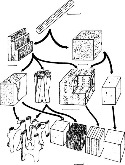
FIGURE 1.2 Diagram showing the structure of mammalian bone at different levels. Bone at the same level is drawn at the same magnification. The arrows show what types may contribute to structures at higher levels [Wainwright et al., 1982]. (Courtesy Princeton University Press.) (a) Collagen fibril with associated mineral crystals. (b) Woven bone. The collagen fibrils are arranged more or less randomly. Osteocytes are not shown. (c) Lamellar bone. There are separate lamellae, and the collagen fibrils are arranged in “domains” of preferred fibrillar orientation in each lamella. Osteocytes are not shown. (d) Woven bone. Blood channels are shown as large black spots. At this level woven bone is indicated by light dotting. (e) Primary lamellar bone. At this level lamellar bone is indicated by fine dashes. (f) Haversian bone. A collection of Haversian systems, each with concentric lamellae round a central blood channel. The large black area represents the cavity formed as a cylinder of bone is eroded away. It will be filled in with concentric lamellae and form a new Haversian system. (g) Laminar bone. Two blood channel networks are exposed. Note how layers of woven and lamellar bone alternate. (h) Compact bone of the types shown at the lower levels. (i) Cancellous bone.
Finally, we have the whole bone itself constructed of osteons and portions of older, partially destroyed osteons (called interstitial lamellae) in the case of humans or of osteons and/or plexiform bone in the case of mammals. This we denote the macrostructural level. The elastic properties of the whole bone results from the hierarchical contribution of each of these levels.

1-4 |
|
|
|
|
|
Biomechanics |
|
|
TABLE 1.1 Composition of Adult Human and Bovine Cortical Bone |
||||||
|
|
|
|
|
|
|
|
|
|
|
|
% Dry Weight |
|
|
|
|
Species % H2O |
Ap |
Collagen |
GAGa |
Reference |
|
|
|
Bovine |
9.1 |
76.4 |
21.5 |
N.D.b |
Herring, 1977 |
|
|
Human |
7.3 |
67.2 |
21.2 |
0.34 |
Pellagrino and Blitz, 1965; Vejlens, 1971 |
|
aGlycosaminoglycan.
bNot determined.
1.2Composition of Bone
The composition of bone depends on a large number of factors: the species, which bone, the location from which the sample is taken, and the age, sex, and type of bone tissue, for example, woven, cancellous, cortical. However, a rough estimate for overall composition by volume is one-third Ap, one-third collagen and other organic components, and one-third H2O. Some data in the literature for the composition of adult human and bovine cortical bone are given in Table 1.1.
1.3 Elastic Properties
Although bone is a viscoelastic material, at the quasi-static strain rates in mechanical testing and even at the ultrasonic frequencies used experimentally, it is a reasonable first approximation to model cortical bone as an anisotropic, linear elastic solid with Hooke’s law as the appropriate constitutive equation. Tensor notation for the equation is written as:
σij = Cijklεkl |
(1.1) |
where σij and εkl are the second-rank stress and infinitesimal second-rank strain tensors, respectively, and Cijkl is the fourth-rank elasticity tenor. Using the reduced notation, we can rewrite Equation 1.1 as
σi = Cij j i, j = 1 to 6 |
(1.2) |
where Cij are the stiffness coefficients (elastic constants). The inverse of the Cij, the Sij, are known as the compliance coefficients.
The anisotropy of cortical bone tissue has been described in two symmetry arrangements. Lang [1969], Katz and Ukraincik [1971], and Yoon and Katz [1976a, b] assumed bone to be transversely isotropic with the bone axis of symmetry (the 3 direction) as the unique axis of symmetry. Any small difference in elastic properties between the radial (1 direction) and transverse (2 direction) axes, due to the apparent gradient in porosity from the periosteal to the endosteal sides of bone, was deemed to be due essentially to the defect and did not alter the basic symmetry. For a transverse isotropic material, the stiffness matrix [Cij] is given by
|
C11 |
C12 |
C13 |
0 |
0 |
0 |
|
|
C12 |
C11 |
C13 |
0 |
0 |
0 |
|
[Cij] = |
C13 |
C13 |
C33 |
0 |
0 |
0 |
(1.3) |
0 |
0 |
0 |
C44 |
0 |
0 |
||
|
0 |
0 |
0 |
0 |
C44 |
0 |
|
|
0 |
0 |
0 |
0 |
0 |
C66 |
|
where C66 = 12 (C11 − C12). Of the 12 nonzero coefficients, only 5 are independent.
However, Van Buskirk and Ashman [1981] used the small differences in elastic properties between the radial and tangential directions to postulate that bone is an orthotropic material; this requires that 9 of
Mechanics of Hard Tissue |
|
|
|
|
|
|
1-5 |
the 12 nonzero elastic constants be independent, that is, |
|
|
|
|
|||
|
C11 |
C12 |
C13 |
0 |
0 |
0 |
|
|
C12 |
C22 |
C23 |
0 |
0 |
0 |
|
[Cij] = |
C13 |
C23 |
C33 |
0 |
0 |
0 |
(1.4) |
0 |
0 |
0 |
C44 |
0 |
0 |
||
|
0 |
0 |
0 |
0 |
C55 |
0 |
|
|
0 |
0 |
0 |
0 |
0 |
C66 |
|
Corresponding matrices can be written for the compliance coefficients, the Sij, based on the inverse equation to Equation 1.2:
εi = Sijσ j i, j = 1 to 6 |
(1.5) |
where the Sijth compliance is obtained by dividing the [Cij] stiffness matrix, minus the ith row and jth column, by the full [Cij] matrix and vice versa to obtain the Cij in terms of the Sij. Thus, although S33 = 1/E 3, where E 3 is Young’s modulus in the bone axis direction, E 3 =C33, since C33 and S33, are not reciprocals of one another even for an isotropic material, let alone for transverse isotropy or orthotropic symmetry.
The relationship between the compliance matrix and the technical constants such as Young’s modulus (Ei) shear modulus (Gi) and Poisson’s ratio (νij) measured in mechanical tests such as uniaxial or pure shear is expressed in Equation 1.6.
|
|
1 |
|
|
−ν21 |
|
−ν31 |
0 |
0 |
0 |
|
||||||||
|
|
|
E 1 |
|
E 2 |
|
E 3 |
||||||||||||
|
|
|
|
|
|
|
|
|
|
|
|
|
|
||||||
|
|
−ν12 |
|
1 |
|
|
−ν32 |
0 |
0 |
0 |
|
||||||||
|
|
|
|
E 2 |
|
E 3 |
|||||||||||||
|
|
|
E 1 |
|
|
|
|
|
|
|
|
|
|
|
|||||
|
|
−ν13 |
|
−ν23 |
|
1 |
|
0 |
0 |
0 |
|
||||||||
|
|
|
|
|
E 3 |
||||||||||||||
[Sij] = |
|
|
E 1 |
|
|
E 2 |
|
|
|
|
|
|
(1.6) |
||||||
|
|
|
|
|
|
|
|
|
|
|
|
1 |
|
|
|||||
|
0 |
|
0 |
|
0 |
|
0 |
0 |
|
||||||||||
|
|
|
|
|
|
|
|||||||||||||
|
|
|
|
|
G 23 |
||||||||||||||
|
|
|
|
|
|
|
|
|
|
|
|
|
|
1 |
|
|
|
||
|
0 |
|
0 |
|
0 |
|
0 |
0 |
|
||||||||||
|
|
|
|
|
|
|
|||||||||||||
|
|
|
|
|
G 31 |
||||||||||||||
|
|
|
|
|
|
|
|
|
|
|
|
|
|
|
|
1 |
|
||
|
0 |
|
0 |
|
0 |
|
0 |
0 |
|
||||||||||
|
|
|
|
|
|
|
|||||||||||||
|
|
|
|
|
G 12 |
||||||||||||||
|
|
|
|
|
|
|
|
|
|
|
|
|
|
|
|
|
|
||
Again, for an orthotropic material, only 9 of the above 12 nonzero terms are independent, due to the symmetry of the Sij tensor:
ν12 |
= |
ν21 |
ν13 |
= |
ν31 |
|
ν23 |
= |
ν32 |
(1.7) |
|||
E 1 |
E 2 |
|
E 1 |
|
E 3 |
|
E 2 |
|
E 3 |
||||
For the transverse isotropic case, Equation 1.5 reduces to only 5 independent coefficients, since
E 1 = E 2 |
ν12 = ν21 ν31 = ν32 = ν13 = ν23 |
|
||
G 23 = G 31 |
G 12 = |
E 1 |
|
(1.8) |
2(1 + ν12) |
||||
In addition to the mechanical tests cited above, ultrasonic wave propagation techniques have been used to measure the anisotropic elastic properties of bone [Lang, 1969; Yoon and Katz, 1976a, b; Van Buskirk and Ashman, 1981]. This is possible, since combining Hooke’s law with Newton’s second law results in a wave