

Biomechanics Principles and Applications - Donald R. Peterson & Joseph D. Bronzino
.pdfThomas R. Canfield
Argonne National Laboratory
Philip B. Dobrin
Hines VA Hospital and Loyola
University Medical Center
11
Mechanics of Blood Vessels
11.1 Assumptions . . . . . . . . . . . . . . . . . . . . . . . . . . . . . . . . . . . . . . . . . . |
11-1 |
Homogeneity of the Vessel Wall • Incompressibility of
the Vessel Wall • Inelasticity of the Vessel Wall
• Residual Stress and Strain
11.2 Vascular Anatomy . . . . . . . . . . . . . . . . . . . . . . . . . . . . . . . . . . . . . 11-2
11.3 Axisymmetric Deformation . . . . . . . . . . . . . . . . . . . . . . . . . . . 11-3
11.4 Experimental Measurements . . . . . . . . . . . . . . . . . . . . . . . . . . 11-4
11.5 Equilibrium . . . . . . . . . . . . . . . . . . . . . . . . . . . . . . . . . . . . . . . . . . 11-5
11.6 Strain Energy Density Functions . . . . . . . . . . . . . . . . . . . . . . . 11-6
Isotropic Blood Vessels • |
Anisotropic Blood Vessels |
References . . . . . . . . . . . . . . . . . |
. . . . . . . . . . . . . . . . . . . . . . . . . . . . . . . . . . 11-12 |
11.1 Assumptions
This chapter is concerned with the mechanical behavior of blood vessels under static loading conditions and the methods required to analyze this behavior. The assumptions underlying this discussion are for ideal blood vessels that are at least regionally homogeneous, incompressible, elastic, and cylindrically orthotropic. Although physiologic systems are nonideal, much understanding of vascular mechanics has been gained through the use of methods based upon these ideal assumptions.
11.1.1 Homogeneity of the Vessel Wall
On visual inspection, blood vessels appear to be fairly homogeneous and distinct from surrounding connective tissue. The inhomogeneity of the vascular wall is realized when one examines the tissue under a low-power microscope, where one can easily identify two distinct structures: the media and adventitia. For this reason the assumption of vessel wall homogeneity is applied cautiously. Such an assumption may be valid only within distinct macroscopic structures. However, few investigators have incorporated macroscopic inhomogeneity into studies of vascular mechanics [1].
11.1.2 Incompressibility of the Vessel Wall
Experimental measurement of wall compressibility of 0.06% at 270 cm of H2O indicates that the vessel can be considered incompressible when subjected to physiologic pressure and load [2]. In terms of the mechanical behavior of blood vessels, this is small relative to the large magnitude of the distortional strains
11-1

11-2 |
Biomechanics |
that occur when blood vessels are deformed under the same conditions. Therefore, vascular compressibility may be important to understanding other physiologic processes related to blood vessels, such as the transport of interstitial fluid.
11.1.3 Inelasticity of the Vessel Wall
That blood vessel walls exhibit inelastic behavior such as length-tension and pressure-diameter hysteresis, stress relaxation, and creep has been reported extensively [3,4]. However, blood vessels are able to maintain stability and contain the pressure and flow of blood under a variety of physiologic conditions. These conditions are dynamic but slowly varying with a large static component.
11.1.4 Residual Stress and Strain
Blood vessels are known to retract both longitudinally and circumferentially after excision. This retraction is caused by the relief of distending forces resulting from internal pressure and longitudinal tractions. The magnitude of retraction is influenced by several factors. Among these factors are growth, aging, and hypertension. Circumferential retraction of medium-caliber blood vessels, such as the carotid, iliac, and bracheal arteries, can exceed 70% following reduction of internal blood pressure to zero. In the case of the carotid artery, the amount of longitudinal retraction tends to increase during growth and to decrease in subsequent aging [5]. It would seem reasonable to assume that blood vessels are in a nearly stress-free state when they are fully retracted and free of external loads. This configuration also seems to be a reasonable choice for the reference configuration. However, this ignores residual stress and strain effects that have been the subject of current research [6–11].
Blood vessels are formed in a dynamic environment that gives rise to imbalances between the forces that tend to extend the diameter and length and the internal forces that tend to resist the extension. This imbalance is thought to stimulate the growth of elastin and collagen and to effectively reduce the stresses in the underlying tissue. Under these conditions it is not surprising that a residual stress state exists when the vessel is fully retracted and free of external tractions. This process has been called remodeling [7]. Striking evidence of this remodeling is found when a cylindrical slice of the fully retracted blood vessel is cut longitudinally through the wall. The cylinder springs open, releasing bending stresses kept in balance by the cylindrical geometry [11].
11.2 Vascular Anatomy
A blood vessel can be divided anatomically into three distinct cylindrical sections when viewed under the optical microscope. Starting at the inside of the vessel, they are the intima, the media, and the adventitia. These structures have distinct functions in terms of the blood vessel physiology and mechanical properties.
The intima consists of a thin monolayer of endothelial cells that line the inner surface of the blood vessel. The endothelial cells have little influence on blood vessel mechanics but do play an important role in hemodynamics and transport phenomena. Because of their anatomical location, these cells are subjected to large variations in stress and strain as a result of pulsatile changes in blood pressure and flow.
The media represents the major portion of the vessel wall and provides most of the mechanical strength necessary to sustain structural integrity. The media is organized into alternating layers of interconnected smooth muscle cells and elastic lamellae. There is evidence of collagen throughout the media. These small collagen fibers are found within the bands of smooth muscle and may participate in the transfer of forces between the smooth muscle cells and the elastic lamellae. The elastic lamellae are composed principally of the fiberous protein elastin. The number of elastic lamellae depends upon the wall thickness and the anatomical location [12]. In the case of the canine carotid, the elastic lamellae account for a major component of the static structural response of the blood vessel [13]. This response is modulated by the smooth-muscle cells, which have the ability to actively change the mechanical characteristics of the wall [14].
Mechanics of Blood Vessels |
11-3 |
The adventitia consists of loose, more disorganized fiberous connective tissue, which may have less influence on mechanics.
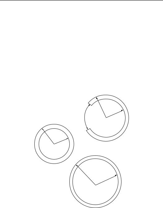
11.3 Axisymmetric Deformation
In the following discussion we will concern ourselves with deformation of cylindrical tubes, see Figure 11.1. Blood vessels tend to be nearly cylindrical in situ and tend to remain cylindrical when a cylindrical section is excised and studied in vitro. Only when the vessel is dissected further does the geometry begin to deviate from cylindrical. For this deformation there is a unique coordinate mapping
(R, , Z) → (r, θ , z) |
(11.1) |
where the undeformed coordinates are given by (R, , Z) and the deformed coordinates are given by (r, θ , z). The deformation is given by a set of restricted functions
r = r (R) |
(11.2) |
θ = β |
(11.3) |
z = μZ + C1 |
(11.4) |
Re
2( –Θo)
Ri
re
ri |
=1 |
=1
re
ri
>1
FIGURE 11.1 Cylindrical geometry of a blood vessel: top: stress-free reference configuration; middle: fully retracted vessel free of external traction; bottom: vessel in situ under longitudinal tether and internal pressurization.

11-4 Biomechanics
where the constants μ and β have been introduced to account for a uniform longitudinal strain and a symmetric residual strain that are both independent of the coordinate .
If β = 1, there is no residual strain. If β =1, residual stresses and strains are present. If β > 1, a longitudinal cut through the wall will cause the blood vessel to open up, and the new cross-section will form a c -shaped section of an annulus with larger internal and external radii. If β < 1, the cylindrical shape is unstable, but a thin section will tend to overlap itself. In Choung and Fung’s formulation, β = π/ o, where the angle o is half the angle spanned by the open annular section [6].
For cylindrical blood vessels there are two assumed constraints. The first assumption is that the longi-
tudinal strain is uniform through the wall and therefore |
|
λz = μ = a constant |
(11.5) |
for any cylindrical configuration. Given this, the principal stretch ratios are computed from the above function as
λr = |
|
dr |
(11.6) |
||||||||
dR |
|
|
|||||||||
λθ = β |
r |
(11.7) |
|||||||||
|
|
||||||||||
R |
|||||||||||
λz |
|
= μ |
(11.8) |
||||||||
The second assumption is wall incompressibility, which can be expressed by |
|
||||||||||
λr λθ λz ≡ 1 |
(11.9) |
||||||||||
or |
|
|
|
|
|
|
|
|
|
|
|
|
r |
|
|
dr |
(11.10) |
||||||
βμ |
|
|
|
|
= 1 |
||||||
R |
dR |
||||||||||
and therefore |
|
|
|
|
|
|
|
|
|
|
|
r dr = |
1 |
|
|
R dR |
(11.11) |
||||||
|
|
|
|||||||||
|
βμ |
||||||||||
Integration of this expression yields the solution
r 2 = |
1 |
R2 + c 2 |
||
βμ |
||||
where |
|
|
|
|
c 2 = re2 − |
1 |
Re2 |
||
|
||||
βμ |
||||
As a result, the principal stretch ratios can be expressed in terms of R as follows:
λr = √ R
βμ(R2 +βμc 2 )
(11.12)
(11.13)
(11.14)
λθ = |
1 |
|
+ |
c 2 |
(11.15) |
βμ |
R2 |
||||
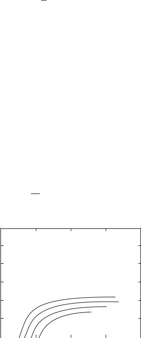
11.4 Experimental Measurements
The basic experimental setup required to measure the mechanical properties of blood vessels in vitro is described in Reference 14. It consists of a temperature-regulated bath of physiologic saline solution to maintain immersed cylindrical blood vessel segments, devices to measure diameter, an apparatus to

Mechanics of Blood Vessels |
11-5 |
mmHg
i p
300
Carotid artery
=1.8
250
200 |
|
|
|
|
|
|
|
=1.6 |
|
|
|
|
|
|
|
|
|
|
|
|
|||
|
|
|
|
|
|
|
|
|
|
|
|
150 |
|
|
|
|
|
|
|
=1.4 |
|
|
|
|
|
|
|
|
|
|
|
|
|
||
100 |
|
|
|
|
|
|
|
|
|
|
|
|
|
|
|
|
|
|
|
|
|||
50 |
|
|
|
|
|
|
=1.2 |
|
|
|
|
|
|
|
|
|
|
|
|
|
|||
0 |
|
|
|
|
|
|
|
|
|
|
|
|
|
|
|
|
|
|
|
|
|
|
|
0.10 |
0.15 |
0.20 |
0.25 |
0.30 |
|||||||
re cm
FIGURE 11.2 Pressure–radius curves for the canine carotid artery at various degrees of longitudinal extension.
|
80 |
|
|
|
|
|
|
|
|
Carotid artery |
|
|
|
|
|
|
70 |
|
|
=1.8 |
|
|
|
|
|
|
|
|
|
||
|
60 |
|
|
|
|
|
|
|
50 |
|
|
|
|
|
|
g |
40 |
|
|
|
|
|
|
|
|
|
|
|
|
|
|
g |
|
|
|
|
|
|
|
f |
30 |
|
|
|
|
|
|
|
|
|
|
|
|
|
|
|
20 |
|
|
|
=1.6 |
|
|
|
10 |
|
|
|
=1.4 |
|
|
|
|
|
|
|
|
|
|
|
0 |
|
|
|
=1.2 |
|
|
|
|
|
|
|
|
|
|
|
0 |
|
|
|
|
|
|
|
0.10 |
0.15 |
0.20 |
0.25 |
0.30 |
||
re cm
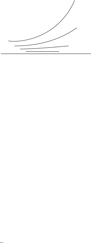
FIGURE 11.3 Longitudinal distending force as a function of radius at various degrees of longitudinal extension.
hold the vessel at a constant longitudinal extension and to measure longitudinal distending force, and a system to deliver and control the internal pressure of the vessel with 100% oxygen. Typical data obtained from this type of experiment are shown in Figure 11.2 and Figure 11.3.
11.5 Equilibrium
When blood vessels are excised, they retract both longitudinally and circumferentially. Restoration to natural dimensions requires the application of internal pressure, pi, and a longitudinal tether force, FT. The internal pressure and longitudinal tether are balanced by the development of forces within the vessel

11-6 |
Biomechanics |
wall. The internal pressure is balanced in the circumferential direction by a wall tension, T . The longitudinal tether force and pressure are balanced by the retractive force of the wall, FR
T = piri |
(11.16) |
FR = FT + piπ ri2 |
(11.17) |
The first equation is the familiar law of Laplace for a cylindrical tube with internal radius ri. It indicates that the force due to internal pressure, pi, must be balanced by a tensile force (per unit length), T , within the wall. This tension is the integral of the circumferentially directed force intensity (or stress, σθ ) across the wall:
re |
|
T = σθ dr = σ¯θ h |
(11.18) |
ri
where σ¯θ is the mean value of the circumferential stress and h is the wall thickness. Similarly, the longitudinal tether force, FT, and extending force due to internal pressure are balanced by a retractive internal force, FR, due to axial stress, σz , in the blood vessel wall:
FR = 2π |
re |
|
σz r dr = σ¯zπ h(re + ri) |
(11.19) |
|
|
ri |
|
where σ¯z is the mean value of this longitudinal stress. The mean stresses are calculated from the above equation as
|
|
ri |
|
|
|
|
(11.20) |
|||
σ¯θ = pi |
|
|
|
|
|
|
|
|||
h |
|
|
|
|
||||||
σ¯z = |
|
|
|
FT |
|
+ |
pi |
|
ri |
(11.21) |
π |
h(re + ri) |
2 h |
||||||||
|
|
|
|
|||||||
The mean stresses are a fairly good approximation for thin-walled tubes where the variations through the wall are small. However, the range of applicability of the thin-wall assumption depends upon the material properties and geometry. In a linear elastic material, the variation in σθ is less than 5% for r /h > 20. When the material is nonlinear or the deformation is large, the variations in stress can be more severe (see Figure 11.10).
The stress distribution is determined by solving the equilibrium equation,
1 d |
σθ |
|
(11.22) |
|||
|
|
|
(r σr ) − |
|
= 0 |
|
r dr |
r |
|||||
This equation governs how the two stresses are related and must change in the cylindrical geometry. For uniform extension and internal pressurization, the stresses must be functions of a single radial coordinate, r, subject to the two boundary conditions for the radial stress:
σr (ri, μ) = − pi |
(11.23) |
σr (re, μ) = 0 |
(11.24) |
11.6 Strain Energy Density Functions
Blood vessels are able to maintain their structural stability and contain steady oscillating internal pressures. This property suggests a strong elastic component, which has been called the pseudoelasticity [4]. This elastic response can be characterized by a single potential function called the strain energy density. It is a scalar
Mechanics of Blood Vessels |
11-7 |
function of the strains that determines the amount of stored elastic energy per unit volume. In the case of a cylindrically orthotropic tube of incompressible material, the strain energy density can be written in the following functional form:
W = W (λr , λθ , λz ) + λr λθ λz p |
(11.25) |
where p is a scalar function of position, R. The stresses are computed from the strain energy by the following:
|
|
|
|
|
|
∂ W |
|
+ p |
(11.26) |
||||||
|
σi = λi |
|
|
|
|||||||||||
|
∂ λi |
|
|||||||||||||
We make the following transformation [15] |
|
|
|
|
|
|
|
|
|
|
|
|
|||
|
λ = |
|
|
|
|
βr |
|
|
|
|
|
(11.27) |
|||
|
|
|
|
|
|
|
|||||||||
|
|
|
|
|
|||||||||||
|
βμ(r 2 |
− c 2) |
|||||||||||||
which upon differentiation gives |
|
|
|
|
|
|
|
|
|
|
|
|
|||
r |
dλ |
|
= |
β−1 |
βλ |
− |
μλ3 |
(11.28) |
|||||||
dr |
|||||||||||||||
|
|
|
|
|
|
|
|
|
|||||||
After these expressions and the stresses in terms of the strain energy density function are introduced into the equilibrium equation, we obtain an ordinary differential equation for p
d p |
= |
β W,λθ |
− W,λr |
− |
dW,λr |
(11.29) |
||||||
|
d |
λ |
βλ |
= |
μλ3 |
|
d |
λ |
||||
|
|
|
|
|
|
|
|
|
||||
subject to the boundary conditions |
|
|
|
|
|
|
|
|
|
|
|
|
|
|
|
|
p(Ri) = pi |
|
|
(11.30) |
|||||
|
|
|
|
p(Re) = 0 |
|
|
|
(11.31) |
||||
11.6.1 Isotropic Blood Vessels
A blood vessel generally exhibits anisotropic behavior when subjected to large variations in internal pressure and distending force. When the degree of anisotropy is small, the blood vessel may be treated as isotropic. For isotropic materials it is convenient to introduce the strain invariants:
I1 |
= λr2 |
+ λθ2 + λ2z |
(11.32) |
I2 |
= λr2 |
λθ2 + λθ2 λ2z + λ2z λr2 |
(11.33) |
I3 |
= λr2 |
λθ2 λ2z |
(11.34) |
These are measures of strain that are independent of the choice of coordinates. If the material is incompressible
I3 = j 2 ≡ 1 |
(11.35) |
and the strain energy density is a function of the first two invariants, then
W = W(I1, I2) |
(11.36) |
11-8 |
Biomechanics |
The least complex form for an incompressible material is the first-order polynomial, which was first proposed by Mooney to characterize rubber:
W = |
G |
[(I1 |
− 3) + k(I2 − 3)] |
(11.37) |
2 |
It involves only two elastic constants. A special case, where k = 0, is the neo-Hookean material, which can be derived from thermodynamics principles for a simple solid. Exact solutions can be obtained for the cylindrical deformation of a thick-walled tube. In the case where there is no residual strain, we have the following:
P = −G (1 + kμ2) |
log λ |
+ |
|
|
1 |
|
+ c 0 |
(11.38) |
|||||||||||
|
|
μ |
|
2μ2λ2 |
|
||||||||||||||
σr = G |
1 |
+ k |
1 |
|
1 |
+ p |
(11.39) |
||||||||||||
|
|
|
|
|
|
+ |
|
|
|||||||||||
|
λ2 |
μ2 |
μ2 |
λ2 |
|||||||||||||||
σθ = G |
λ2 |
+ k |
1 |
|
|
+ λ2μ2 |
+ p |
|
(11.40) |
||||||||||
|
|
|
|
|
|||||||||||||||
μ2 |
|
|
|
||||||||||||||||
σz = G |
μ2 |
+ k |
λ2μ2 + |
1 |
|
|
+ p |
|
(11.41) |
||||||||||
|
|
|
|
||||||||||||||||
|
λ2 |
|
|||||||||||||||||
However, these equations predict stress softening for a vessel subjected to internal pressurization at fixed lengths, rather than the stress stiffening observed in experimental studies on arteries and veins (see Figure 11.4 and Figure 11.5).
An alternative isotropic strain energy density function that can predict the appropriate type of stress stiffening for blood vessels is an exponential where the arguments is a polynomial of the strain invariants. The first-order form is given by
W = |
G 0 |
exp[k1(I1 − 3) + k2(I2 − 3)] |
(11.42) |
2k1 |
mmHg
i p
300
Mooney tube
250G0=0.54 kPa k=21.1
200
150 |
|
|
|
|
|
|
|
|
|
|
=1.8 |
|
|
100 |
|
|
|
=1.6 |
|
|
|
|
|
|
=1.4 |
|
|
|
|
|
|
=1.2 |
|
|
50 |
|
|
|
|
|
|
0 |
|
|
|
|
|
|
0.10 |
0.15 |
0.20 |
0.25 |
0.30 |
||
re cm
FIGURE 11.4 Pressure–radius curves for a Mooney–Rivlin tube with the approximate dimensions of the carotid.

Mechanics of Blood Vessels |
11-9 |
gm
g f
80
70Mooney tube G0=0.54 kPa
60 |
|
|
k=21.1 |
|
|
|
|
|
|
|
|
|
|
|
|
|
|
|
|
|
|
|
|
50 |
|
|
|
|
|
|
|
|
|
|
|
|
|
|
|
|
|
|
|
|
|
|
|
40 |
|
|
|
|
|
|
|
|
|
|
|
|
|
|
|
|
|
|
|
=1.8 |
|
|
|
30 |
|
|
|
|
|
|
|
|
|||
|
|
|
|
|
|
|
|
=1.6 |
|||
|
|
|
|
|
|
|
|
|
|||
20 |
|
|
|
|
|
|
|
|
|
|
|
|
|
|
|
|
|
|
=1.4 |
||||
|
|
|
|
|
|
|
|
||||
10 |
|
|
|
|
|
|
=1.2 |
|
|
|
|
|
|
|
|
|
|
|
|
|
|||
0 |
|
|
|
|
|
|
|
|
|
|
|
–10 |
|
|
|
|
|
|
|
|
|
|
|
|
|
|
|
|
|
|
|
|
|
|
|
0.10 |
0.15 |
0.20 |
0.25 |
0.30 |
|||||||
re cm
FIGURE 11.5 Longitudinal distending force as a function of radius for the Mooney–Rivlin tube.
This requires the determination of only two independent elastic constants. The third, G 0, is introduced to facilitate scaling of the argument of the exponent (see Figure 11.6 and Figure 11.7). This exponential form is attractive for several reasons. It is a natural extension of the observation that biologic tissue stiffness is proportional to the load in simple elongation. This stress stiffening has been attributed to a statistical recruitment and alignment of tangled and disorganized long chains of proteins. The exponential forms resemble statistical distributions derived from these same arguments.
g
i p
300 |
|
|
|
|
|
|
|
W*=G0 exp [k1(11–3)+k3(I2–3)] |
|
|
|
||
|
|
|
|
|
||
250 |
|
G0=16.78 |
|
=1.8 |
|
|
|
|
k1=0.474 |
|
|
|
|
200 |
|
k2=0.008 |
|
=1.6 |
|
|
|
|
|
|
|
||
150 |
|
|
|
|
|
|
100 |
|
|
|
=1.4 |
|
|
|
|
|
|
|
|
|
50 |
|
|
|
=1.2 |
|
|
|
|
|
|
|
|
|
0 |
|
|
|
|
|
|
|
0.15 |
0.20 |
0.25 |
0.30 |
||
0.10 |
||||||
re cm
FIGURE 11.6 Pressure–radius curves for tube with the approximate dimensions of the carotid calculated using an isotropic exponential strain energy density function.
11-10 |
Biomechanics |
mg
g f
80 |
|
|
|
|
|
|
|
|
|
|
|
|
|
|
|
|
|
|
|
|
|
|
|
|
|
|
|
70 |
|
|
W*=G0 exp [k1(I1–3)+k3(I2–3)] |
|
|
=1.8 |
|
|
|
||||
|
|
|
|
|
|
|
|||||||
|
|
|
G0=16.78 |
|
|
|
|
|
|
|
|
|
|
60 |
|
|
k1=0.474 |
|
|
|
|
|
|
|
|
|
|
|
|
|
|
|
|
|
|
|
|
|
|||
50 |
|
|
k2=0.008 |
|
|
|
|
|
|
|
|
|
|
|
|
|
|
|
|
|
|
|
|
|
|
|
|
40 |
|
|
|
|
|
|
|
|
|
=1.6 |
|
|
|
|
|
|
|
|
|
|
|
|
|
|
|
||
30 |
|
|
|
|
|
|
|
|
|
|
|
|
|
|
|
|
|
|
|
|
|
|
|
|
|
|
|
|
|
|
|
|
|
|
|
|
|
|
|
|
|
20 |
|
|
|
|
|
|
|
|
|
=1.4 |
|
|
|
|
|
|
|
|
|
|
|
|
|
|
|
||
10 |
|
|
|
|
|
|
|
|
|
|
|
|
|
|
|
|
|
|
|
|
=1.2 |
|
|
|
|||
0 |
|
|
|
|
|
|
|
|
|
|
|||
|
|
|
|
|
|
|
|
|
|
|
|
|
|
0 |
|
|
|
|
|
|
|
|
|
|
|
|
|
|
|
|
|
|
|
|
|
|
|
|
|
|
|
0.10 |
0.15 |
0.20 |
0.25 |
0.30 |
|||||||||
re cm
FIGURE 11.7 Longitudinal distending force as a function of radius for the isotropic tube.
11.6.2 Anisotropic Blood Vessels
Studies of the orthotropic behavior of blood vessels may employ polynomial or exponential strain energy density functions that include all strain terms or extension ratios. In particular, the strain energy density function can be of the form
W = qn (λr , λθ , λz ) |
(11.43) |
or |
|
W = eqn (λr ,λθ ,λz ) |
(11.44) |
where qn is a polynomial of order n. Since the material is incompressible, the explicit dependence upon λr can be eliminated either by substituting λr = λ−θ 1λ−z 1 or by assuming that the wall is thin and hence that the contribution of these terms is small. Figure 11.8 and Figure 11.9 illustrate how well the experimental data can be fitted to an exponential strain density function whose argument is a polynomial of order n = 3.
Care must be taken to formulate expressions that will lead to stresses that behave properly. For this reason it is convenient to formulate the strain energy density in terms of the Lagrangian strains
ei = 1/2 λi2 − 1 |
(11.45) |
and in this case we can consider polynomials of the lagrangian strains, qn (er , eθ , ez ). Vaishnav et al. [16] proposed using a polynomial of the form
n |
i |
|
W = |
ai j −i eθi − j ezj |
(11.46) |
i =2 |
j =0 |
|
to approximate the behavior of the canine aorta. They found better correlation with order-three polynomials over order-two, but order-four polynomials did not warrant the addition work.
Later, Fung et al. [4] found very good correlation with an expression of the form
W − |
C |
exp |
a1 eθ2 − ez 2 + a2 ez2 − ez 2 + 2a4 eθ ez − eθ ez |
(11.47) |
2 |