

Biomechanics Principles and Applications - Donald R. Peterson & Joseph D. Bronzino
.pdfMechanics of Tissue/Lymphatic Transport |
15-3 |
Arterial |
Pc |
Capillary |
Venous |
|
|
||
|
|
Pv |
|
Pa |
P.S. |
Pt c t |
|
|
|
|
|
|
|
|
To |
|
Pl |
|
thoracic |
|
|
|
duct and |
|
|
|
vena cava |
Initial lymphatic vessel
FIGURE 15.1 Starling pressures that regulate transcapillary fluid balance. Pressure parameters that determine direction and magnitude of transcapillary exchange include capillary blood pressure Pc, interstitial fluid pressure Pt (directed into capillary when positive or directed into tissue when negative), plasma colloidal osmotic pressure πc, and interstitial fluid colloidal osmotic pressure πt. Precapillary sphincters (P.S.) regulate Pc, capillary flow, and capillary surface area A. It is generally agreed that a hydrostatic pressure gradient (Pt > lymph pressure Pl) drains off excess interstitial fluid under conditions of net filtration. Relative magnitudes of pressures are depicted by the size of arrows. (From Hargens A.R. 1986. In R. Skalak and S. Chien (Eds.), Handbook of Bioengineering, vol. 19, pp. 1–35, New York, McGraw-Hill. With permission.)
Therefore, normally at the heart level, Pc is approximately 30 mmHg. However, during upright posture, Pc at foot level is about 90 mmHg and only about 25 mmHg at head level [Parazynski et al., 1991]. Differences in Pc between capillaries of the head and feet are due to gravitational variation of the blood pressure such that the pressure p = ρgh. For this reason, volumes of transcapillary filtration and lymph flows are generally higher in tissues of the lower body as compared to those of the upper body. Moreover, one might expect much more sparse distribution of lymphatic vessels in upper body tissues. The brain has no lymphatics, but most other vascular tissues have lymphatics. In fact, tissues of the lower body of humans and other tall animals have efficient skeletal muscle pumps, prominent lymphatic systems, and noncompliant skin and fascial boundaries to prevent dependent edema [Hargens et al., 1987].
Other pressure parameters in the Starling–Landis equation such as Pt, πc, and πt are not as sensitive to changes in body posture as is Pc. Typical values for Pt range from −2 to 10 mmHg depending on the tissue or organ under investigation [Wiig, 1990]. However, during movement, Pt in skeletal muscle increases to 150 mmHg or higher [Murthy et al., 1994], providing a mechanism to promote lymphatic flow and venous return via the skeletal pump (Figure 15.2). Blood colloid osmotic pressure πc usually ranges between 25 and 35 mmHg and is the other major force for retaining plasma within the vascular system and preventing edema. Interstitial πt depends on the reflection coefficient of the capillary wall (σp ranges from 0.5 to 0.9 for different tissues) as well as washout of interstitial proteins during high filtration rates [Aukland and Reed, 1993]. Typically πt ranges between 8 and 15 mmHg with higher values in upper body tissues compared to those in the lower body [Parazynski et al., 1991; Aukland and Reed, 1993]. Precapillary sphincter activity (see Figure 15.1) also decreases blood flow, decreases capillary filtration area A, and reduces Pc in dependent tissues of the body to help prevent edema during upright posture [Aratow et al., 1991].

15-4 |
Biomechanics |
Soleuspressure |
(mmHg) |
anteriorpressure |
(mmHg) |
Tibialis |
|
160
120
80
40
0 |
2 |
4 |
6 |
8 |
10 |
12 |
160
120
80
40
0 |
2 |
4 |
6 |
8 |
10 |
12 |
Time (sec)
FIGURE 15.2 Simultaneous intramuscular pressure oscillations in the soleus (top panel) and the tibialis anterior (bottom panel) muscles during plantarand dorsiflexion exercise. Soleus muscle is an integral part of the calf muscle pump. (From Murthy, G., Watenpaugh, D.E., Ballard, R.E. et al., 1994. J. Appl. Physiol. 76: 2742. With permission.)
15.2.3 Interstitial Fluid Transport
Interstitial flow of proteins and other macromolecules occurs by two mechanisms, diffusion and convection. During simple diffusion according to Fick’s equation:
J p = − D |
∂ cp |
(15.5) |
∂ x |
where J p is the one-dimensional protein flux, D is diffusion coefficient, and ∂ cp/∂ x is the concentration gradient of protein through interstitial space.
For most macromolecules such as proteins, the diffusional transport is limited. It serves to disperse molecules, but it does not effectively serve to transport large molecules especially if their diffusion is restricted by interstitial matrix proteins, membrane barriers, or other structures that limit their free thermal motion. Instead, both experimental and theoretical evidence highlights the dependence of volume and solute flows on hydrostatic and osmotic pressure gradients [Hargens and Akeson, 1986; Hammel, 1994] and suggests that convective flow plays the dominating role in interstitial flow and transport of nutrients to tissue cells. For example, in the presence of osmotic or hydrostatic pressure gradients, protein transport J p is coupled to fluid transport according to:
Jp = c¯p J v |
(15.6) |
where c¯p is the average protein concentration and Jv is the volume flow of fluid.
Transport of interstitial fluid toward the lymphatics requires convective flow, since it needs to be focused on relatively few channels in the interstitium. Diffusion cannot serve such a purpose because diffusion merely disperses fluid and proteins. Lymph formation and flow greatly depend upon tissue movement or activity related to muscle contraction and tissue deformations. It is also generally agreed that formation of initial lymph depends solely on the composition of nearby interstitial fluid and pressure gradients across the interstitial/lymphatic boundary [Zweifach and Lipowsky, 1984; Hargens, 1986]. For this reason, lymph formation and flow can be quantified by measuring disappearance of isotope-labeled albumin from subcutaneous tissue or skeletal muscle [Reed et al., 1985].

Mechanics of Tissue/Lymphatic Transport |
15-5 |
Arteriole |
Venule |
|
1 mm
Arteriole |
Lymphatic |
|
FIGURE 15.3 Tracing of a typical lymphatic channel (bottom panel) in rat spinotrapezius muscle after injection with a micropipette of a carbon contrast suspension. All lymphatics are of the initial type and are closely associated with the arcade arterioles. Few lymphatics follow the path of the arcade venules, or their side branches, the collecting venules or the transverse arterioles. (From Skalak et al., 1986. In A.R. Hargens (Ed.), Tissue Nutrition and Viability, pp. 243–262, Springer-Verlag, New York. With permission.)
15.2.4 Lymphatic Architecture
To understand lymph transport in engineering terms it is paramount that we develop a detailed picture of the lymphatic network topology and vessel morphology. This task is facilitated by a number of morphological and ultrastructural studies from past decades that give a general picture of the morphology and location of lymphatic vessels in different tissues. Lymphatics are studied by injections of macroscopic and microscopic contrast media and by light and electron microscopic sections. The display of the lymphatics is organ specific and there are many variations in lymphatic architecture [Schmid-Schonbein,¨ 1990]. In this chapter, we will focus our discussion predominantly on skeletal muscle, intestine, and skin. However, the mechanisms outlined below may in part be also relevant to other tissues and organs.
In skeletal muscle, lymphatics are positioned in immediate proximity of the arterioles [Skalak et al., 1984]. The majority of feeder arteries in skeletal muscle and most, but not all, of the arcade arterioles are closely accompanied by a lymphatic vessel (Figure 15.3). Lymphatics can be traced along the entire length of the arcade arterioles, but they can be traced only over relatively short distances (less than about 50 μm) into the side branches of the arcades, the transverse (terminal) arterioles, which supply the blood into the capillary network. Systematic reconstructions of the lymphatics in skeletal muscle have yielded little evidence for lymphatic channels that enter into the capillary network per se [Skalak et al., 1984]. Thus, the network density of lymphatics is quite low compared to the high density of the capillary network in muscle, a characteristic feature of lymphatics in most organs [Skalak et al., 1986]. The close association between lymphatics and the vasculature is also present in skin [Ikomi and Schmid-Schonbein,¨ 1995] and in other organs and may extend into the central vasculature. Recently, Saharinen et al. [2004] reviewed lymphatic vasculature development and molecular regulation in tumor metastasis and inflammation. It is apparent that current understandings of lymphatic growth factors and strategies to limit lymphatic vessel growth may allow manipulation of lymphatic growth in disease.
15-6 |
Biomechanics |
15.2.5 Lymphatic Morphology
Histological sections of the lymphatics permit the classification into two distinct subsets, initial lymphatics and collecting lymphatics. The initial lymphatics (sometimes also denoted as terminal or capillary lymphatics) form a set of blind endings in the tissue that feed into the collecting lymphatics, and that in turn, are the conduits into the lymph nodes. While both initial and collecting lymphatics are lined by a highly attenuated endothelium, only the collecting lymphatics have smooth muscle in their media. In accordance, contractile lymphatics exhibit spontaneous narrowing of their lumen, while there is no evidence for contractility (in the sense of a smooth muscle contraction) in the initial lymphatics. Contractile lymphatics are capable of peristaltic smooth muscle contractions that, in conjunction with periodic opening and closing of intraluminal valves, permit unidirectional fluid transport. The lymphatic smooth muscle has adrenergic innervation [Ohhashi et al., 1982], it exhibits myogenic contraction [Hargens and Zweifach, 1977; Mizuno et al., 1997], and reacts to a variety of vasoactive stimuli [Ohhashi et al., 1978; Benoit, 1997], including signals that involve nitric oxide [Ohhashi and Takahashi, 1991; Bohlen and Lash, 1992; Yokoyama and Ohhashi, 1993]. None of these contractile features has been documented in initial lymphatics.
The lymphatic endothelium has a number of similarities with vascular endothelium. It forms a continuous lining and has typical cytoskeletal fibers such as microtubules, intermediate fibers, and actin in both fiber bundle form and matric form. There are numerous caveolae, Weibel-Palade bodies, but lymphatic endothelium has fewer interendothelial adhesion complexes and a discontinuous basement membrane. The residues of the basement membrane are attached to interstitial collagen via anchoring filaments [Leak and Burke, 1968] that provide relatively firm attachment of the endothelium to interstitial structures.
15.2.6 Lymphatic Network Display
One of the interesting aspects regarding lymphatic transport in skeletal muscle is the fact that all lymphatics inside the muscle parenchyma are of the noncontractile, initial type [Skalak et al., 1984]. Collecting lymphatics can only be observed outside the muscle fibers as conduits to adjacent lymph nodes. The fact that all lymphatics inside the tissue parenchyma are of the initial type is not unique to skeletal muscle, but has been demonstrated in other organs [Unthank and Bohlen, 1988; Yamanaka et al., 1995]. The initial lymphatics are positioned in the adventitia of the arcade arterioles surrounded by collagen fibers (Figure 15.4). Thus, the initial lymphatics are in immediate proximity to the arteriolar smooth muscle, and adjacent to myelinated nerves fibers and a set of mast cells that accompany the arterioles. The initial lymphatics are frequently sandwiched between arteriolar smooth muscle and their paired venules, and they in turn are embedded between the skeletal muscle fibers [Skalak et al., 1984]. The initial lymphatics are firmly attached to the adjacent basement membrane and collagen fibers via anchoring filaments [Leak and Burke, 1968]. The basement membrane of the lymphatic endothelium is discontinuous, especially at the interendothelial junctions, so that macromolecules and even cells and particles enter the initial lymphatics [Casley-Smith, 1962; Bach and Lewis, 1973; Strand and Persson, 1979; Bollinger et al., 1981; Ikomi et al., 1996].
The lumen cross section of initial lymphatics is highly irregular in contrast to the overall circular cross section of collecting lymphatics (Figure 15.4). Luminal cross sections of initial lymphatics are partially or completely collapsed and may frequently span around the arcade arteriole. In fact, we have documented cases in which the arcade arteriole is completely surrounded by an initial lymphatic channel, highlighting the fact that the activity of the lymphatics is closely linked to that of the arterioles [Ikomi and SchmidSchonbein,¨ 1995].
15.2.7 The Intraluminal (Secondary) Lymphatic Valves
Initial lymphatics in skeletal muscle have intraluminal valves that consist of bileaflets and a funnel structure [Mazzoni et al., 1987]. The leaflets are flexible structures and are opened and closed by a viscous pressure drop along the valve funnel. In closed position, these leaflets can support considerable pressures

Mechanics of Tissue/Lymphatic Transport |
15-7 |
(a)
FIGURE 15.4 Histological cross sections of lymphatics (LYM) in rat skeletal muscle before (a) and after (b) contraction of the paired arcade arterioles (ART). The lymphatic channel is of the initial type with a single attenuated endothelial layer (curved arrows). Note, that in the dilated arteriole, the lymphatic is essentially compressed (a) while the lymphatic is expanded after arteriolar contraction (b), which is noticeable by the folded endothelial cells in the arteriolar lumen. In both cases, the lumen cross-sectional shape of the initial lymphatic channels is highly irregular. All lymphatics in skeletal muscle have these characteristic features. (From Skalak T.C., Schmid-Schonbein¨ G.W., and Zweifach B.W. 1984. Microvasc. Res. 28: 95.)
[Eisenhoffer et al., 1995; Ikomi et al., 1997]. This arrangement preserves normal valve function even in initial lymphatics with irregularly shaped lumen cross sections.
15.2.8 The Primary Lymphatic Valves
The lymphatic endothelial cells are attenuated and have many of the morphological characteristics of vascular endothelium, including expression of P-selectin, von Willebrand factor [Di Nucci et al., 1996], and factor VIII [Schmid-Schonbein,¨ 1990]. An important difference between vascular and lymphatic endothelium lies in the arrangement of the endothelial junctions. In the initial lymphatics, the endothelial cells lack tight junctions [Schneeberger and Lynch, 1984] and are frequently encountered in an overlapping

15-8 |
Biomechanics |
(b)
FIGURE 15.4 (Continued.)
but open position, so that proteins, large macromolecules, and even chylomicron particles can readily pass through the junctions [Casley-Smith, 1962, 1964; Leak, 1970]. Examination of the junctions with scanning electron microscopy shows that there exists a periodic interdigitating arrangement of endothelial extensions. Individual extensions are attached via anchoring filaments to the underlying basement membrane and connective tissue, but the two extensions of adjacent endothelial cells resting on top of each other are not attached by interendothelial adhesion complexes. Mild mechanical stretching of the initial lymphatics shows that the endothelial extensions can be separated in part from each other, indicating that the membranes of two neighboring lymphatic endothelial cells are not attached to each other, but are firmly attached to the underlying basement membrane [Castenholz, 1984]. Lymphatic endothelium does not exhibit continuous junctional complexes, and instead has a “streak and dot” like immunostaining pattern of VE-cadherin and associated intracellular proteins desmoplakin and plakoglobulin [Schmelz et al., 1994]. However the staining pattern is not uniform for all lymphatics, and in larger lymphatics a more continuous pattern is present. This highly specialized arrangement has been referred to as the lymphatic endothelial microvalves [Schmid-Schonbein,¨ 1990] or primary lymphatic valves. They are “primary” because fluid from the interstitium must first pass across these valves before entering the lymphatic lumen and then pass across the intraluminal, that is, secondary, valves. Particles deposited into the interstitial
Mechanics of Tissue/Lymphatic Transport |
15-9 |
space adjacent to initial lymphatics pass across the endothelium of the initial lymphatics. However once the particles are inside the initial lymphatic lumen, they cannot return back into the interstitial space unless the endothelium is injured. Indeed, the endothelial junctions of the initial lymphatics serve as a functional valve system [Trzewik et al., 2001].
15.2.9 Mechanics of Lymphatic Valves
In contrast to the central large valves in the heart that are closed by inertial fluid forces, the lymphatic valves are small and the fluid Reynolds number is almost zero. Thus, because no inertial forces are available to open and close these valves, unique valve morphology has evolved in these small valves. The valves form long funnel-shaped channels, which are inserted into the lymph conduits and attached at their base. The funnel is prevented from inversion by attachment via a buttress to the lymphatic wall. The valve wall structure consists of a collagen layer sandwiched between two endothelial layers, and the entire structure is quite deformable under mild physiological fluid pressures. The funnel structure allows a viscous pressure gradient that is sufficient to generate a pressure drop during forward fluid motion to open, and upon flow reversal to close the valves [Mazzoni et al., 1987]. The primary lymphatic valves also open as passive structures at the peripheral endothelial cell extensions. They require sites where they are free to bend into the lumen of the initial lymphatics and where they are not attached by anchoring filaments to the adjacent extracellular matrix [Mendoza et al., 2003].
15.2.10 Lymph Formation and Pump Mechanisms
One of the important questions fundamental to lymphology is: How do fluid and large particles in the interstitium find their way into initial lymphatics? In light of the relative sparse existence of initial lymphatics, a directed convective transport is required that can be provided by either a hydrostatic or a colloid osmotic pressure drop [Zweifach and Silberberg, 1979]. However, the exact mechanism of this unidirectional flow has remained an elusive target. Several proposals have been advanced and these are discussed in detail in Schmid-Schonbein¨ [1990]. Briefly, a number of authors have postulated that there exists a constant pressure drop from the interstitium into the initial lymph, which may support a steady fluid flow into the lymphatics. Nevertheless, repeated measurements with different techniques have uniformly failed to provide supporting evidence for a steady pressure drop to transport fluid into the initial lymphatics [Zweifach and Prather, 1975; Clough and Smaje, 1978]. Under steady-state conditions, no steady pressure drop exists in the vicinity of the initial lymphatics in skeletal muscle within the resolution of the measurement technique (about 0.2 cm H2O) [Skalak et al., 1984]. An order of magnitude estimate of the pressure drop expected at the relatively slow flow rates of the lymphatics shows, however, that the pressure drop from the interstitium may be significantly lower [Schmid-Schonbein,¨ 1990]. Furthermore, the assumption of a steady pressure drop is not in agreement with the substantial evidence that lymph flow rate is enhanced under unsteady conditions (see below). Some investigators have postulated an osmotic pressure in the lymphatics to aspirate fluid into the initial lymphatics [Casley-Smith, 1972] due to ultrafiltration across the lymphatic endothelium, a mechanism referred to as “bootstrap effect” [Perl, 1975]. Critical tests of this hypothesis, such as the microinjection of hyperosmotic protein solutions, have not led to a uniformly accepted hypothesis for lymph formation involving an osmotic mechanism. Others have suggested a retrograde aspiration mechanism, such that the recoil in the collecting lymphatics serves to lower the pressure in the initial lymphatics upstream of the collecting lymphatics [Reddy, 1986; Reddy and Patel, 1995], or an electric charge difference across lymphatic endothelium [O’Morchoe et al., 1984].
15.2.11 Tissue Mechanical Motion and Lymphatic Pumping
An intriguing feature of lymphatic pressure is that lymphatic flow rates depend on tissue motion. In a resting tissue, the lymph flow rate is relatively small. However, different forms of tissue motion serve to
15-10 |
Biomechanics |
enhance lymph flow. This was originally demonstrated for pulsatile pressures in the rabbit ear. Perfusion of the ear with steady pressure (even at the same mean pressure) stops lymph transport, while pulsatile pressures promote lymph transport [Parsons and McMaster, 1938]. In light of the paired arrangement of the arterioles and lymphatics, periodic expansion of the arterioles compresses adjacent lymphatics, and vice versa, a reduction of arteriolar diameter during the pressure reduction phase expands adjacent lymphatics [Skalak et al., 1984] (Figure 15.4). Vasomotion, associated with a slower contraction of the arterioles, but with a larger amplitude than pulsatile pressure, increases lymph formation [Intaglietta and Gross, 1982; Colantuoni et al., 1984]. In addition, muscle contractions, simple walking [Olszewski and Engeset, 1980], respiration, intestinal peristalsis, skin compression [Ohhashi et al., 1991], and other tissue motions are associated with increased lymph flow rates. Periodic tissue motions are significantly more effective to enhance the lymph flow than elevation of the venous pressure [Ikomi et al., 1996], which is also associated with enhanced fluid filtration [Renkin et al., 1977].
A requirement for lymph fluid flow is the periodic expansion and compression of the initial lymphatics. Because initial lymphatics do not have their own smooth muscle, the expansion and compression of initial lymphatics depend on the motion of tissue in which they are embedded. In skeletal muscle, the strategic location of the initial lymphatics in the adventitia of the arterioles provides the milieu for expansion and compression via several mechanisms: arteriolar pressure pulsations or vasomotion, active or passive skeletal muscle contractions, or external muscle compression. Direct measurements of the cross-sectional area of the initial lymphatics during arteriolar contractions or during skeletal muscle shortening support this hypothesis [Skalak et al., 1984; Mazzoni et al., 1990] (Figure 15.5). The different lymph pump mechanisms are additive. Resting skeletal muscle has much lower lymph flow rates (provided largely by the arteriolar pressure pulsation and vasomotion) than skeletal muscle during exercise (produced by a combination of intramuscular pressure pulsations and skeletal muscle shortening) [Ballard et al., 1998].
Measurements of lymph flow rates in an afferent lymph vessel (diameter about 300 to 500 μm, proximal to the popliteal node) in the hind leg [Ikomi and Schmid-Schonbein,¨ 1996] demonstrate that lymph fluid formation is influenced by passive or active motion of the surrounding tissue. Lymphatics in this tissue region drain muscle and skin of the hind leg, and the majority is of the initial type, whereas collecting lymphatics are detected outside the tissue parenchyma in the fascia proximal to the node. Without whole leg rotation, lymph flow remains at low but nonzero values. If the pulse pressure is stopped, lymph flow falls to values below detectable limits (less than about 10% of the values during
|
40 |
|
|
|
40 |
|
|
|
|
|
Dilated arterioles |
|
Contracted arterioles |
||||
(%) |
30 |
|
|
|
30 |
|
|
|
|
|
|
|
|
|
|
|
|
Frequency |
10 |
|
|
|
10 |
|
|
|
|
20 |
|
|
|
20 |
|
|
|
|
0 |
|
|
|
0 |
|
|
|
|
0 |
1000 |
2000 |
3000 |
0 |
1000 |
2000 |
3000 |
|
|
Lymphatic cross-sectional area ( m2) |
|
|||||
Normalized lymphatic cross-sectional area
–2.0
1.5
1.0
 Lymphatics
0.5
Incompressible
material
0.0
0.00.5 1.0 1.5 2.0
Normalized skeletal muscle length
FIGURE 15.5 Histograms of initial lymphatic cross-sectional area in rat spinotrapezius muscle before (left) and after (middle) contraction of the paired arteriole with norepinephrine. Lymphatic cross-sectional area as a function of muscle length during active contraction or passive stretch (right). Cross-sectional area and muscle length are normalized with respect to the values in vivo in resting muscle. Note the expansion of the initial lymphatics with contraction of the arterioles or muscle stretch. (From Skalak T.C., Schmid-Schonbein¨ G.W., and Zweifach B.W. 1984. Microvasc. Res. 28: 95; Mazzoni M.C., Skalak T.C., and Schmid-Schonbein¨ G.W. 1990. Am. J. Physiol. 259: H1860.)
Mechanics of Tissue/Lymphatic Transport |
15-11 |
(a) |
|
0.10 |
|
|
|
rate |
(ml/h) |
|
|
|
|
flowLymph |
0.05 |
|
|
|
|
|
|
|
|
|
|
|
|
|
|
Massage width |
|
|
|
|
|
|
: 1.0 cm |
|
|
0 |
|
|
: 0.5 cm |
|
|
0 |
1 |
2 |
3 |
|
|
|
Massage frequency (Hz) |
||
(b) |
|
0.10 |
|
|
|
rate |
(ml/h) |
|
|
|
|
flowLymph |
0.05 |
|
|
|
|
|
|
|
|
|
|
|
|
0 |
|
|
|
|
|
0 |
0.03 |
0.3 |
3.0 |
|
|
Massage frequency (Hz) |
|||
(c) |
|
0.10 |
rate |
(ml/h) |
|
flowLymph |
0.05 |
|
|
|
|
|
|
0 |
(d) |
|
0.10 |
rate |
(ml/h) |
|
flowLymph |
0.05 |
|
|
|
|
|
|
0 |
Venous pressure=40 mmHg
Massage width=1.0 cm
0 |
1 |
2 |
3 |
|
Massage frequency (Hz) |
|
|
0 |
0.03 |
0.3 |
3.0 |
Massage frequency (Hz)
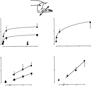
FIGURE 15.6 Lymph flow rates in a prenodal afferent lymphatic draining the hind leg as a function of the frequency of a periodic surface shear motion (massage) without (panels a, b) and with (panels c, d) elevation of the venous pressure by placement of a cuff. Zero frequency refers to a resting leg with a lymph flow rate, which depends on pulse pressure. The amplitudes of the tangential skin shear motion were 1 and 0.5 cm (panels a, b) and 1 cm in the presence of the elevated venous pressure (panels c, d). Note that the ordinates in panels c and d are larger than those in panels a and b. (From Ikomi F., Hunt J., Hanna G. et al. 1996. J. Appl. Physiol. 81: 2060.)
pulse pressure). Introduction of whole leg passive movement causes strong, frequency-dependent lymph flow rates that increase linearly with the logarithm of frequency between 0.03 and 1.0 Hz (Figure 15.6). Elevation of venous pressure, which enhances fluid filtration from the vasculature and elevates the flow rates, does not significantly alter the dependency of lymph flow on periodic tissue motion [Ikomi et al., 1996].
Similarly, application of passive tissue compression on the skin elevates lymph flow rate in a frequencydependent manner. Lymph flow rates are determined to a significant degree by the local action of the lymph pump, because arrest of the heartbeat and reduction of the central blood pressure to zero does not stop lymph flow. Instead, cardiac arrest reduces lymph flow rate only about 50% during continued leg motion or application of periodic shear stress to the skin for several hours [Ikomi and Schmid-Schonbein,¨ 1996]. Periodic compression of the initial lymphatics also enhances proteins and lymphocyte counts in the lymphatics [Ikomi et al., 1996] (Figure 15.7). Thus either arteriolar smooth muscle or parenchymal skeletal muscle activity expands and compresses the initial lymphatics in skeletal muscle. These mechanisms serve to adjust lymph flow rates according to organ activity such that a resting skeletal muscle has a very low lymph flow rate. During normal daily activity or mild or strenuous exercise, lymph flow rates as well as protein and cell transport into the lymphatics increases [Olszewski et al., 1977].
