

Biomechanics Principles and Applications - Donald R. Peterson & Joseph D. Bronzino
.pdfCochlear Mechanics |
17-11 |
expansion of the cell over a narrow frequency band, which is related to the place for the cell along the cochlea. Khanna and coworkers [1989] observe a similar tonic displacement of the whole organ of Corti.
17.5 Active Models
De Boer [1991], Geisler [1993], and Hubbard [1993] discuss models in which the electromotility of the outer hair cells feeds energy into the basilar membrane. The partition stiffness K is expanded from Equation 17.3 into a transfer function, containing a number of parameters and delay times. These are classed as phenomenological models, for which the physiological basis of the parameters is not of primary concern. The displacement gain may be defined as the ratio of ciliary shearing displacement to cell expansion. For these models, the gain used is larger by orders of magnitude than the maximum found in laboratory measurements of isolated hair cells.
Another approach [Steele and colleagues (1993)], which is physiologically based, appears promising. The outer hair cells are inclined in the propagation direction. Thus the shearing of the cilia at the distance x causes a force from the hair cells acting on the basilar membrane at the distance x + x. This “feed-forward” law can be expressed in terms of the pressure as
pohc(x + x) = αp(x) = α[2 pf(x) + pohc(x)] |
(17.16) |
where p, the total pressure acting on the basilar membrane, consists of the effective pressure acting on the basilar membrane from the hair cells pohc and the pressure from the fluid pf. The coefficient α is the force gain supplied by the outer hair cells. With this law, Equation 17.11 is replaced by:
(1 − ein x )n tanh n H = 2 |
ρω2 |
(17.17) |
AK H |
from which the local wave number n must be computed numerically. Only two new parameters are needed, the gain α and the spacing x. With physiologically reasonable gain α = 0.18 and spacing x = 20 μm, the result is an increase of the response of the basilar membrane for higher frequencies by a factor of 102 in a narrow sector, apical to the passive peak. The simple feed-forward addition in Equation 17.17 enhances a narrow band of wave lengths without a closed control loop. At this time, it appears that much of the elaborate structure of the organ of Corti is for the purpose of such a “feed-forward.” This approach is also found to work well for the 1-D model by Geisler and Sang [1995]. The results from the 3-D model for the effect of adding some feed-forward are shown in Figure 17.3. One defect of the current feed-forward results is that the shift of the maximum response point is about one octave, as seen in Figure 17.3, rather than one-half octave consistently shown in normal cochlear measurements of mechanical and neural response.
The significant nonlinear effect is the saturation of the active process at high amplitudes. This can be computed by letting the gain α be a function of the amplitude.
In the normal, active cochlea, it was first observed by Khanna and colleagues [1989] and subsequently by Gummer and colleagues [1996], that the tectorial membrane (TM in Figure 17.1) has a substantially higher amplitude than the basilar membrane. Presumably, the electromotile expansion of the outer hair cells encounters less resistance from the tectorial membrane than the basilar membrane. This shows the importance of getting the correct stiffness and geometry into a model.
17.6 Fluid Streaming
Bek´esy´ [1960] and many others have observed significant fluid streaming in the actual cochlea and in experimental models. Particularly for the high frequencies, it is tempting to seek a component of steady streaming as the significant mechanical stimulation of the inner hair cells [Lighthill, 1992]. Passive models indicated that such streaming occurs only at high sound intensity. Among the many open questions is whether or not the enhancement of amplitude provided by the feed-forward of energy by the outer hair

17-12 |
Biomechanics |
cells and the mechanical nonlinearity at low amplitudes of displacement provided by the ciliary gating can trigger significant streaming. It is clear from Figure 17.1 that the motion and corresponding pressure in the fluid of the inner sulcus is the primary source of excitation for the inner hair cells. The DC pressure associated with DC streaming would be an important effect.
17.7 Clinical Possibilities
A better understanding of the cochlear mechanisms would be of clinical value. Auditory pathology related to the inner ear is discussed by Pickles [1988] and Gulick and colleagues [1989]. The spontaneous and stimulated emissions from the cochlea raise the possibility of diagnosing local inner ear problems, which is being pursued at many centers around the world. The capability for more accurate, physically realistic modeling of the cochlea should assist in this process. A significant step is provided by Zweig and Shera [1995], who find that a random distribution of irregularities in the properties along the cochlea explains much of the emissions.
A patient with a completely nonfunctioning cochlea is referred to as having “nerve deafness.” In fact, there is evidence that in many cases the nerves may be intact, while the receptor cells and organ of Corti are defective. For such patients, a goal is to restore hearing with cochlear electrode implants to stimulate the nerve endings directly. Significant progress has been made. However, despite electrode stimulation of nerves at the correct place along the cochlea for a high frequency, the perception of high frequency has not been achieved. So, although substantial advance in cochlear physiology has been made in the recent past, several such waves of progress may be needed to adequately understand the functioning of the cochlea.
References
Allen J.B. and Neely S.T. 1992. Micromechanical models of the cochlea. Phys. Today 45: 40–47.
Ashmore J.F. 1987. A fast motile response in guinea-pig outer hair cells: the cellular basis of the cochlear amplifier. J. Physiol. 388: 323–347.
Assad J.A. and Corey D.P. 1992. An active motor model for adaptation by vertebrate hair cells. J. Neurosci. 12(9): 3291–3309.
Bek´esy´ G. von. 1960. Experiments in Hearing. McGraw-Hill, New York.
Bohnke¨ F., von Mikusch-Buchberg J., and Arnold W. 1996. 3D Finite Elemente Modell des cochlearen¨ Verstarkers¨. Biomedizinische Technik. 42: 311–312.
Brownell W.E., Bader C.R., Bertrand D., and de Ribaupierre Y. 1985. Evoked mechanical responses of isolated cochlear outer hair cells. Science 227: 194–196.
Canlon B., Brundlin L., and Flock A. 1988. Acoustic stimulation causes tonotopic alterations in the length of isolated outer hair cells from the guinea pig hearing organ. Proc. Natl. Acad. Sci. USA 85: 7033–7035.
Cabezudo L.M. 1978. The ultrastructure of the basilar membrane in the cat. Acta Otolaryngol. 86: 160–175. Dallos P. 1992. The active cochlea. J. Neurosci. 12(12): 4575–4585.
De Boer E. 1991. Auditory physics. Physical principles in hearing theory. III. Phys. Rep. 203(3): 126–231. Freeman D.M. and Weiss T.F. 1990. Hydrodynamic analysis of a two-dimensional model for microme-
chanical resonance of free-standing hair bundles. Hearing Res. 48: 37–68.
Fuhrmann E., Schneider W., and Schultz M. 1987. Wave propagation in the cochlea (inner ear): effects of Reissner’s membrane and non-rectangular cross-section. Acta Mech. 70: 15–30.
Furness D.N., Zetes D.E., Hackney C.M., and Steele C.R. 1997. Kinematic analysis of shear displacement as a means for operating mechanotransduction channels in the contact region between adjacent stereocilia of mammalian cochlear hair cells. Proc. R. Soc. Lond. B 264: 45–51.
Geisler C.D. 1993. A realizable cochlear model using feedback from motile outer hair cells. Hearing Res. 68: 253–262.
Geisler C.D. and Sang C. 1995. A cochlear model using feed-forword outer-hair-cell forces, Hearing Res. 85: 132–146.
Cochlear Mechanics |
17-13 |
Gulick W.L., Gescheider G.A., and Fresina R.D. 1989. Hearing: Physiological Acoustics, Neural Coding, and Psychoacoustics. Oxford University Press, London.
Gummer A.W., Johnston B.M., and Armstrong N.J. 1981. Direct measurements of basilar membrane stiffness in the guinea pig. J. Acoust. Soc. Am. 70: 1298–1309.
Gummer A.W., Hemmert W., and Zenner H.P. 1996. Resonant tectorial membrane motion in the inner ear: its crucial role in frequency tuning. Proc. Natl. Acad. Sci. USA 93: 8727–8732.
Hackney C.M., Furness D.N., and Katori Y. 1996. Stereociliary ultrastructure in relation to mechanotransduction: tip links and the contact region. Diversity in Auditory Mechanics. University of California, Berkeley, pp. 173–180.
Hemmert W., Schauz C., Zenner H.P., and Gummer A.W. 1996. Force generation and mechanical impedance of outer hair cells. Diversity in Auditory Mechanics. University of California, Berkeley, pp. 189–196.
Holley M.D. 1990. Cell biology of hair cells. Semin. Neurosci. 2: 41–47.
Hubbard A.E. 1993. A traveling wave-amplifier model of the cochlea. Science 259: 68–71. Hudspeth A.J. 1989. How the ears work. Nature 34: 397–404.
Iwasa K.H. and Chadwick R.S. 1992. Elasticity and active force generation of cochlear outer hair cells.
J. Acoust. Soc. Am. 92: 3169–3173.
Jen D.H. and Steele C.R. 1987. Electrokinetic model of cochlear hair cell motility. J. Acoust. Soc. Am. 82: 1667–1678.
Kalinec F., Holley M.C., Iwasa K.H., Lim D., and Kachar B. 1992. A membrane-based force generation mechanism in auditory sensory cells. Proc. Natl. Acad. Sci. USA 89: 8671–8675.
Keidel W.D. and Neff W.D., Eds. 1976. Handbook of Sensory Physiology, Volume V: Auditory System. SpringerVerlag, Berlin.
Kemp D.T. 1978. Stimulated acoustic emissions from within the human auditory system. J. Acoust. Soc. Am. 64: 1386–1391.
Khanna S.M., Flock A., and Ulfendahl M. 1989. Comparison of the tuning of outer hair cells and the basilar membrane in the isolated cochlea. Acta Otolaryngol. [Suppl] Stockholm 467: 141–156.
Kolston P.J. and Ashmore J.F. 1996. Finite element micromechanical modeling of the cochlea in three dimensions. J. Acoust. Soc. Am. 99: 455–467.
Lighthill J. 1991. Biomechanics of hearing sensitivity. J. Vibr. Acoust. 113: 1–13. Lighthill J. 1992. Acoustic streaming in the ear itself. J. Fluid Mech. 239: 551–606.
Miller C.E. 1985. Structural implications of basilar membrane compliance measurements. J. Acoust. Soc. Am. 77: 1465–1474.
Nobili R., Mommano F., and Ashmore J. 1998. How well do we understand the cochlea? TINS 21(4): 159–166.
Olson E.S. and Mountain D.C. 1994. Mapping the cochlear partition’s stiffness to its cellular architecture.
J. Acoust. Soc. Am. 95(1): 395–400.
Olson E.S. 1998. Observing middle and inner ear mechanics with novel intracochlear pressure sensors. J. Acoust. Soc. Am. 103(6): 3445–3463.
Pickles J.O. 1988. An Introduction to the Physiology of Hearing, 2nd ed. Academic Press, London.
Preyer S., Renz S., Hemmert W., Zenner H., and Gummer A. 1996. Receptor potential of outer hair cells isolated from base to apex of the adult guinea-pig cochlea: implications for cochlear tuning mechanisms. Auditory Neurosci. 2: 145–157.
Probst R. 1990. Otoacoustic emissions: an overview. Adv. Oto-rhino-laryngol. 44: 1–9.
Raftenberg M.N. 1990. Flow of endolymph in the inner spiral sulcus and the subtectorial space. J. Acoust. Soc. Am. 87(6): 2606–2620.
Ranke O.F. 1950. Theory of operation of the cochlea: a contribution to the hydrodynamics of the cochlea.
J. Acoust. Soc. Am. 22: 772–777.
Rhode W.S. 1971. Observations of the vibration of the basilar membrane in squirrel monkeys using the Mossbauer¨ technique. J. Acoust. Soc. Am. 49: 1218–1231.
Ruggero M.A. 1993. Distortion in those good vibrations. Curr. Biol. 3(11): 755–758.
17-14 |
Biomechanics |
Russell I.J. and Sellick P.M. 1977. Tuning properties of cochlear hair cells. Nature 267: 858–860.
Siebert W.M. 1974. Ranke revisited — a simple short-wave cochlear model. J. Acoust. Soc. Am. 56(2): 594–600.
Steele C.R. 1987. Cochlear mechanics. In Handbook of Bioengineering, R. Skalak and S. Chien, Eds., pp. 30.11–30.22, McGraw-Hill, New York.
Steele C.R. 1992. Electroelastic behavior of auditory receptor cells. Biomimetics 1(1): 3–22.
Steele C.R., Baker G, Tolomeo J.A., and Zetes D.E. 1993. Electro-mechanical models of the outer hair cell, Biophysics of Hair Cell Sensory Systems, In H. Duifhuis, J.W. Horst, P. van Dijk, and S.M. van Netten, Eds., World Scientific.
Strelioff D. and Flock A. 1984. Stiffness of sensory-cell hair bundles in the isolated guinea pig cochlea.
Hearing Res. 15: 19–28.
Taber L.A. and Steele C.R. 1979. Comparison of “WKB” and experimental results for three-dimensional cochlear models. J. Acoust. Soc. Am. 65: 1007–1018.
Tolomeo J.A. and Steele C.R. 1995. Orthotropic piezoelectric propeties of the cochlear outer hair cell wall.
J. Acoust. Soc. Am. 95(5): 3006–3011.
Tolomeo J.A., Steele C.R., and Holley M.C. 1996. Mechanical properties of the lateral cortex of mammalian auditory outer hair cells. Biophys. J. 71: 421–429.
Watts L. 1993. Cochlear Mechanics: Analysis and Analog VLSI. Ph.D. Thesis, California Institute of Technology.
West C.D. 1985. The relationship of the spiral turns of the cochlea and the length of the basilar membrane to the range of audible frequencies in ground dwelling mammals. J. Acoust. Soc. Am. 77(3): 1091–1101.
Zhang L., Mountain D.C., and Hubbard A.E. 1996. Shape changes from base to apex cannot predict characteristic frequency changes. Diversity in Auditory Mechanics. University of California, Berkeley, pp. 611–618.
Zhou G., Bintz L., Anderson D.Z., and Bright K.E. 1994. A life-sized physical model of the human cochlea with optical holographic readout. J. Acoust. Soc. Am. 93(3): 1516–1523.
Zweig G. and Shera C.A. 1995. The origin of periodicity in the spectrum of evoked otoacoustic emissions.
J. Acoust. Soc. Am. 98(4): 2018–2047.
Zwislocki J.J. and Cefaratti L.K. 1989. Tectorial membrane II: stiffness measurements in vivo. Hearing Res. 42: 211–227.
Further Information
The following are workshop proceedings that document many of the developments. De Boer E., and Viergever M.A., Eds. 1983. Mechanics of Hearing. Nijhoff, The Hague.
Allen J.B., Hall J.L., Hubbard A., Neely S.T., and Tubis A., Eds. 1985. Peripheral Auditory Mechanisms. Springer, Berlin.
Wilson J.P. and Kemp D.T., Eds. 1988. Cochlear Mechanisms: Structure, Function, and Models. Plenum, New York.
Dallos P., Geisler C.D., Matthews J.W., Ruggero M.A., and Steele C.R., Eds. 1990. The Mechanics and Biophysics of Hearing. Springer, Berlin.
Duifhuis H., Horst J.W., van Kijk P., and van Netten S.M., Eds. 1993. Biophysics of Hair Cell Sensory Systems. World Scientific, Singapore.
Lewis E.R., Long G.R., Lyon R.F., Narins P.M., Steele C.R., and Hecht-Poinar E., Eds. 1997. Diversity in Auditory Mechanics. World Scientific, Singapore.


Vestibular Mechanics |
18-3 |
(a) |
|
(b) |
dx |
Endolymph |
|
|
y |
||
|
|
|
o |
fluid layer |
|
|
|
b |
Otoconial |
|
|
|
layer |
|
|
|
|
|
|
|
dA |
b |
|
Gel |
|
|
|
||
|
|
|
|
|
|
|
|
|
layer |
|
|
0 |
|
x |
|
|
|
|
|
|
|
|
0 |
Sensory tissue |
|
|
|
|
layer and skull |
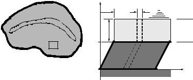
FIGURE 18.1 Schematic of the otolith organ: (a) Top view showing the peripheral region with differential area d A where the model is developed, and the striolar area that makes an arch through the center of the otolith. (b) Cross section showing the layered structure where dx is the width of the differential area d A shown in the top view.
V |
= a characteristic velocity of the skull in the problem (e.g., magnitude of a step change) |
ρo |
= density of the otoconial layer |
ρf |
= density of the endolymph fluid |
τg |
= gel shear stress in the x-direction |
μg |
= viscosity of the gel material |
μf |
= viscosity of the endolymph fluid |
G |
= shear modulus of the gel material |
b |
= gel layer and otoconial layer thickness (assumed equal) |
gx |
= gravity component in the x-direction |
In the equation of motion for the endolymph fluid, the force τfdA acts on the fluid at the fluid–otoconial layer interface (Figure 18.2). This shear stress τf is responsible for driving the fluid flow. The linear NavierStokes equation for an incompressible fluid are used to describe this endolymph flow. Expressions for the pressure gradient, the flow velocity of the fluid measured with respect to an inertial reference frame, and the force due to gravity (body force) are substituted into the Navier-Stokes equation for flow in the x-direction yielding
∂ u |
= μf |
∂ 2u |
(18.1) |
|
ρf |
|
|
||
∂ t |
∂ yf |
|||
with boundary and initial conditions: u(0, t) = v(t); u(∞, t) = 0; and u(yf, 0) = 0.
The gel layer is treated as a Kelvin–Voight viscoelastic material where the gel shear is acting in parallel. This viscoelastic material stress has both an elastic and viscous component acting in parallel. This viscoelastic material model was substituted into the momentum equation and the resulting gel layer equation of
motion is |
|
|
|
|
|
|
|
|
∂ w |
|
t ∂ 2w |
∂ 2w |
(18.2) |
||
ρf |
|
= G |
|
|
dt + μf |
|
|
∂ t |
|
∂ y2 |
∂ y2 |
||||
|
|
0 |
|
f |
|
f |
|
with boundary and initial conditions: w (b, t) = v(t); w (0, t) = 0; w (yg, 0) = 0; and δg(yg, 0) = 0. The elastic term in the equation is written in terms of the integral of velocity with respect to time, instead of displacement, so the equation is in terms of a single dependent variable, the velocity.

18-4 |
Biomechanics |
(a) |
yf |
|
|
|
|
|
u (y,t) |
|
0 |
|
u |
|
0 |
f dA |
|
|
|
||
(b) |
y |
|
|
|
|
o |
|
|
|
|
f dA |
b |
Bx |
v (t) |
|
Wx |
|||
b |
|
||
|
|
||
|
g dA |
|
|
0 |
|
x |
|
|
|
||
(c) yg |
g dA |
|
|
|
|
||
|
|
|
|
b |
|
|
|
|
|
w (y,t) |
|
0 |
|
|
|
|
|
|
FIGURE 18.2 The free-body diagrams of each layer of the otolith with the forces that act on each layer. The interfaces are coupled by shear stresses of equal magnitudes that act in opposite directions at each surface. The τg shear stress acts between the gel–otoconial layer and the τf acts between the fluid–otoconial layer. The forces acting at these interfaces are the product of shear stress τ and area dA. The Bx and Wx forces are the components of the buoyant and weight forces acting in the plane of the otoconial layer, respectively. See the nomenclature table for definitions of other variables.
(a) Endolymph fluid layer: The spatial coordinate in the vertical direction is yf and the velocity of the endolymph fluid is u(yf, t), a function of the fluid depth yf and time t. (b) Otoconial layer: The otoconial layer with thickness b and at a height b above the gel layer vertical coordinate origin. The velocity of the otoconial layer is v(t), which is a function of time only. (c) Gel layer: Gel layer of thickness b, vertical coordinate yg, and horizontal coordinate δg the gel deflection. The gel deflection is a function of both yg and time t. The velocity of the gel is w (yg, t), a function of yg and t.
The otoconial layer equation was developed using Newton’s second law of motion, equating the forces that act on the otoconial layer: fluid shear, gel shear, buoyant, and weight to the product of mass and inertial acceleration. The resulting otoconial layer equation is
|
∂ v |
|
∂ Vs |
|
|
∂ u |
|
|
|
t |
∂ w |
|
|
∂ w |
|
ρob |
+ (ρo − ρf) |
− g x |
= μ f |
|
− G |
|
|
|
dt + μg |
|
|||||
∂ t |
∂ t |
∂ yf |
yf =0 |
0 |
|
∂ yg |
yg =b |
∂ yg |
yg =b |
||||||
|
|
|
|
|
|
|
|
|
|
|
|
|
(18.3)

Vestibular Mechanics |
18-5 |
with the following initial conditions v(0) = 0. The otoconial layer displacement can be calculated by integrating the velocity of this layer with respect to time
|
|
t |
|
δ = |
0 |
v dt |
(18.4) |
Displacement of the otoconial layer is the variable of importance, it is proportional to acceleration, and is the variable transduced into a neural signal.
18.4 Nondimensionalization of the Otolith Motion Equations
The equations of motion are then nondimensionalized to reduce the number of physical and dimensional parameters, and to combine them into some useful nondimensional numbers. The following nondimensional variables, which are indicated by overbars, are introduced into the motion equations:
y¯f = |
yf |
y¯g = |
yg |
¯ |
|
|
μf |
t u¯ = |
u |
v¯ = |
v |
w¯ = |
w |
(18.5) |
b |
b |
t |
= |
ρob2 |
V |
V |
V |
|||||||
Several nondimensional parameters occur naturally as a part of the nondimensionalization process; these parameters are
R = |
ρf |
ε = |
G b2ρo |
M = |
μg |
g¯x = |
ρob2 |
g x |
(18.6) |
ρo |
μf2 |
μf |
V μf |
These parameters represent the following: R is the density ratio and represents the system nondimentional mass, ε is a nondimensional elastic parameter and represents the system elasticity, M is the viscosity ratio and represents the system damping, and g¯x is the nondimensional gravity.
The governing equations of motion in nondimensional form are then as follows:
|
|
Endolymph Fluid Layer |
|
|
R |
∂ u¯ |
= |
|
∂ 2u¯ |
|
|
|
|
|
(18.7) |
|||||||||
|
|
|
|
∂ t¯ |
|
∂ y¯f2 |
|
|
|
|
|
|||||||||||||
|
|
Boundary Conditions |
u¯ |
¯ |
|
|
¯ |
|
u¯ |
|
|
|
¯ |
|
|
|
|
|
||||||
|
|
(0, t) |
= v¯(t) |
|
(∞, t) = 0 |
|
|
|
||||||||||||||||
|
|
Initial Conditions |
u¯(y¯f, 0) = 0 |
|
|
|
|
|
|
|
|
|
|
|
|
|||||||||
|
∂ v¯ |
¯ |
|
|
|
|
∂ u¯ |
|
|
|
|
|
t¯ |
|
∂ w¯ |
|
∂ w¯ |
|||||||
|
|
∂ Vs |
− g¯x |
|
|
|
|
|
|
|
|
|
|
|
||||||||||
Otoconial Layer |
|
+ (1 − R) |
|
|
|
|
= |
|
|
|
|
− ε |
|
|
|
|
|
|
dt − M |
|
|
|||
∂ t¯ |
|
∂ t¯ |
|
∂ y¯f |
0 |
0 |
|
|
∂ y¯g 1 |
∂ y¯g 1 |
||||||||||||||
|
|
|
|
|
|
|
|
|
|
|
|
|
|
|
|
|
|
|
|
|
|
|
(18.8) |
|
|
Initial Conditions |
v¯(0) = 0 |
|
|
|
|
|
|
|
|
|
|
|
|
|
|
|
|||||||
|
|
|
∂ w¯ |
|
|
¯ |
|
∂ 2w¯ |
|
|
|
|
|
|
∂ 2w¯ |
|
|
|
|
|||||
|
Gel Layer R |
= ε |
t |
|
dt |
+ M |
|
|
|
(18.9) |
||||||||||||||
|
∂ t¯ |
0 |
|
∂ y¯ |
2 |
|
|
|
∂ y¯ |
2 |
|
|
|
|||||||||||
|
|
|
|
|
|
|
|
|
g |
|
|
|
|
|
|
g |
|
|
|
|||||
|
Boundary Conditions |
w¯ |
¯ |
|
|
¯ |
|
w¯ |
¯ |
|
|
|
|
|
|
|||||||||
|
(1, t) |
= v¯(t) |
|
(0, t) = 0 |
|
|
|
|||||||||||||||||
|
Initial Conditions |
|
w¯ (y¯g, 0) = 0 |
|
¯ |
|
|
|
|
|
|
|
|
|
|
|||||||||
|
|
|
δg(y¯g, 0) = 0 |
|
|
|
||||||||||||||||||
