


2
Biomolecules and Cells on Surfaces – Fundamental Concepts
Kristi L. Hanson, Luisa Filipponi, and Dan V. Nicolau
2.1 Introduction
In microarray technology, surfaces must be designed and prepared to optimize the immobilization of probe biomolecules and/or cells, but also to resist non-specific binding of target species. Further, the surface and type of immobilization technique selected will a ect the concentration, bioactivity and target–binding ability of bound species. For any given probe molecule, there is likely to be an optimal surface and/or technique which will allow for attachment at the highest possible concentration and with preservation of required activity. However, for multi–probe array formats requiring a variety of probe molecules to be bound to the same type of surface, di culties are encountered selecting a surface and immobilization method able to generate su cient probe concentration, resolution and bioactivity for all probes. The resulting variability in probe concentration and activity within the array also leads to signal variability, causing di culty in data interpretation. Thus, appropriate attachment methods are critical to the success of any array technology.
The aim of this chapter is to summarize the general knowledge and fundamental concepts underlying DNA, protein, small biomolecule and cell attachment to surfaces, and to highlight issues arising in the field of microarray fabrication. The section will provide background knowledge for the reader not familiar with general biomolecule immobilization techniques, while more specific protocols used in microarray technology will be discussed further in Chap. 3.
2.2 Types of Immobilization
Biomolecule attachment is dependent on the properties of the biomolecular surface, the solid surface, and the liquid medium. In most cases, the biomolecular surface will display a higher level of complexity than the attachment surface or the liquid medium, as biomolecules and cells exhibit not only an
24 Kristi L. Hanson et al.
overall charge and hydrophobicity, but also a heterogeneous distribution of these, depending on the types and distributions of surface-exposed groups.
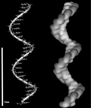
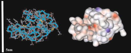
The biomolecules of interest can be broadly grouped into nucleic acids (DNA, RNA, PNA), proteins (antibodies, enzymes, receptors, a bodies), small molecules (e.g. peptides, metabolites) and other biomolecules (e.g. carbohydrates, lipids), of which the first two classes have been by far the most studied with respect to microarray applications. Figures 2.1 and 2.2 depict the distribution of charges and hydrophobicity on a single stranded oligonucleotide and a protein (lysozyme), respectively. The oligonucleotide shows more ordered and predictable patterns, with regularly-spaced negativelycharged phosphate groups in the backbone region, and hydrophobic base pairing regions. In contrast, proteins are characterized by both heterogeneous and irregular regions of positive charge, negative charge, and hydrophobicity. As we will see in the next section, the relative structural simplicity of DNA, as compared to protein, results in more predictable and controllable patterns of surface attachment.
Fig. 2.1. Structure of a single stranded oligonucleotide (left) and the 3D map of the electrostatic potential (darker patches = negative charges)
2 Biomolecules and Cells on Surfaces – Fundamental Concepts |
25 |
Fig. 2.2. Structure of a simple protein (lysozyme, left) and the 3D map of the electrostatic potential (red indicates negative charges; blue indicates positive charges)
Mechanisms of immobilization can be divided into two major categories:
(i) adsorption, which relies on non-covalent interactions (mainly electrostatic, van der Waals, and dehydration of hydrophobic interfaces) and (ii) covalent binding of specific functional groups on the biomolecule to functionalized surfaces. The first mechanism is of a purely physical nature and therefore displays varying levels of reversibility, whereas covalent binding, by definition, involves the formation of essentially irreversible chemical bonds between biomolecule and surface.
2.2.1 Adsorption
In general, the extent of adsorption of any species at the solid–liquid interface will be the net result of several attractive and repulsive forces. For biomolecules, the most important of these include electrostatic interactions, van der Waals forces, energetically favorable dehydration of hydrophobic surfaces, structure rearrangement, and lateral interactions [1, 2].
Electrostatic interactions result from the overlap of the electrical double layers around a charged protein molecule and a charged surface. These interactions generally depend on the net charge of the surface and the molecule, but heterogeneous surface charges distributed around a protein molecule (see Fig. 2.2) can also produce a dipole moment, thereby contributing to overall electrostatic interaction. The relatively weak character of these interactions renders them less appropriate for microarray technology, where strong and irreversible attachment is generally required. However, the possibility of charge control on the surface (e.g., using electrodes) and on the biomolecular surface (e.g. by variation of pH) make these interactions more versatile. For instance, control of surface charge of an electrode allows for the possibility of ‘reusable’ microarrays, where bound species can be desorbed, rinsed and re-arrayed.
Van der Waals forces can be broadly defined as other weak attractive forces contributing to intermolecular attraction, including dipole–dipole in-
26 Kristi L. Hanson et al.
teractions, hydrogen bonding, and dispersion (London) forces. Where electrostatic interactions might be unfavorable due to like charges on molecule and surface, adsorption may still occur due to the strong e ect of van der Waals forces at close range.
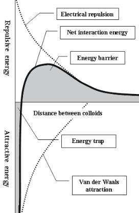
Classic DLVO theory [3, 4] models colloidal or protein interactions and stability based on the balance between the above forces (i.e. electrostatic repulsion and van der Waals attraction). In general, electrostatic forces are felt at longer distances than van der Waals forces, but both forces increase as molecules are brought closer together. At short distances, van der Waals attraction increases more rapidly than electrostatic repulsion, leading to adsorption (in the case of proteins and surfaces) or flocculation (in the case of colloidal particles in solution). Thus, in order for adsorption to occur, likecharged particles must have su cient kinetic energy to overcome the energy barrier, which is dictated by the point of maximum repulsive energy on the net interaction curve (Fig. 2.3).
DLVO theory predicts strong adhesion between hydrophobic particles or molecules, due to the strong e ect of van der Waals interactions at close range. These interactions are sometimes therefore referred to as hydrophobic interactions, but the driving force is considered to be the energetically favorable displacement of water molecules between two hydrophobic surfaces. Regardless of the details of theoretical explanation, there is no doubt that attractive interactions between proteins and hydrophobic surfaces are often very important, and in many cases dominate all other driving forces [1]. The application of these attractive forces to microarrays is complicated by the fact that hydrophobic interactions are often associated with conformational changes in molecular structure, as the hydrophobic interior of the biomolecule ‘unfolds’ to position itself against the hydrophobic interface.
Finally, of particular importance to microarray technology, but usually poorly characterized, lateral interactions will a ect the density of surfacebound biomolecules. These interactions can result from either (1) electrostatic repulsion between molecules with like charges, or (2) dipole–dipole interactions, which can be repulsive or attractive, depending on the alignment and ordering of molecules on the surface.
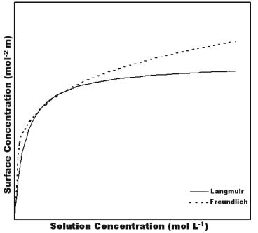
In practice, it is di cult to predict or model the overall e ects of the above interactions, and the nature of biomolecule adsorption on a particular surface is often investigated by determination of relevant adsorption isotherms (Fig. 2.4). Adsorption isotherms relate the quantity of adsorbed protein (relative to available surface area) to the concentration in solution at equilibrium. Typically, the amount of adsorbed protein increases sharply at low solution concentrations, and then eventually approaches a limiting value indicative of the saturated, or maximum possible loading. In the simplest case, the relationship can be modelled by the Langmuir equation, which assumes a single equilibrium constant for the reaction between adsorbed and dissolved protein. An alternative model, known as the Freundlich model, can be derived assuming a certain distribution function for multiple binding sites having di erent
2 Biomolecules and Cells on Surfaces – Fundamental Concepts |
27 |
Fig. 2.3. Net interaction curve formed by subtracting the attraction curve (due to van der Waals forces) from the repulsion curve (due to electrostatic repulsion of like-charged particles)
equilibrium constants. In most cases, one or both of these models can be fit to protein adsorption data.
Evaluation of such isotherms is particularly useful when comparing different adsorption strategies, and can provide insight into maximum possible protein loading concentration, binding geometries and lateral interactions. For example, if the dimensions of the biomolecule are known, the maximum surface coverage achieved can be compared to theoretical monolayer coverage in all possible binding geometries, thereby allowing inference of attachment density and/or attachment orientation.
2.2.2 Covalent Attachment
The covalent binding of biomolecules allows for very strong attachment and in certain instances a positional linking at one end of a biomolecule. A variety
28 Kristi L. Hanson et al.
Fig. 2.4. Typical patterns of Langmuir (solid line) and Freundlich (dashed line) adsorption isotherms
of side groups are easily used for covalent binding, most commonly amino, carboxy, hydroxy, and thiol groups. Consequently similar groups on the surface are needed for a covalent interaction, and in many instances the covalent binding must be enabled by a functionalization of the surface and/or the biomolecule. Quite often the process is ‘standardized’ through the use of crosslinkers and associated protocols, many of these being reviewed in Chap. 3.
2.3 DNA Immobilization on Surfaces
Although DNA immobilization at the solid/liquid interface is not fully understood, especially with respect to molecular conformations at the surface, a wide variety of techniques have been successfully used for probe attachment.
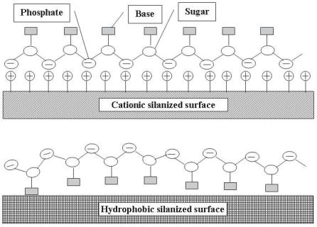
At neutral pH, DNA molecules are charged negatively (Fig. 2.1), and the pattern of charges suggests that phosphates in the DNA backbone would be expected to bind strongly to a positively charged surface, leaving the bases facing towards the solution. As such, positively charged surfaces (e.g., aminopropyltriethoxylsilane [APTES] or poly–L–lysine coated glass) have commonly been used as DNA hybridization sensors [5, 6]. In contrast, hydrophobic and van der Waals interactions would be expected to bind base pairing regions to
2 Biomolecules and Cells on Surfaces – Fundamental Concepts |
29 |
the hydrophobic surface, thus reducing the level of target hybridization in microarray format. These two possible conformations are illustrated in Fig. 2.5 (reprinted from [7]).
Despite the fact that in theory, DNA adsorption to a hydrophobic surface should not allow for e cient hybridization of DNA target, nitrocellulose and nylon supports have been widely used for many years as standard substrates for DNA hybridization [8]. It is interesting to note that both single stranded and double stranded DNA are able to bind by hydrophobic interactions [9], despite the fact that hydrophobic regions of double stranded DNA are presumably buried within the center of the helical structure. As a result, adsorbed double stranded DNA molecules overlap and superimpose through sticky end cohesions, forming complex lattices that are unstable and desorb easily from the surface. When a positive potential is applied to the surface, these lattices form coiled fibers with greatly increased stability due to the electrostatic attraction of phosphate backbone to the positively charged surface, but the DNA duplex is destabilized and stretched as a result of charge–charge repulsion on the unbound side of the DNA helix. Subsequent reorientation of the molecule forces DNA bases from inside the helix to be more exposed to solution. These processes demonstrate the relative simplicity of oligonucleotide
Fig. 2.5. Possible conformations of the DNA/oligonucleotide–surface complex on hydrophobic and cationic surfaces (Reprinted with permission from [7]. Copyright 1998 Academic Press Inc Elsevier Science)
30 Kristi L. Hanson et al.
behavior, as it appears to be dominated by electrostatic, van der Waals and hydrophobic interactions in a quasi–predictable manner.
For microarray applications, it appears that electrostatic interactions between negatively charged DNA and a positively charged surface will produce both higher concentrations of surface-bound probe DNA and more favorable orientation of the probe with respect to hybridization potential [7]. Moreover, the electrostatic adhesion has been found to result in significantly lower surface di usion, which would be advantageous for maintaining high contrast areas of probe attachment.
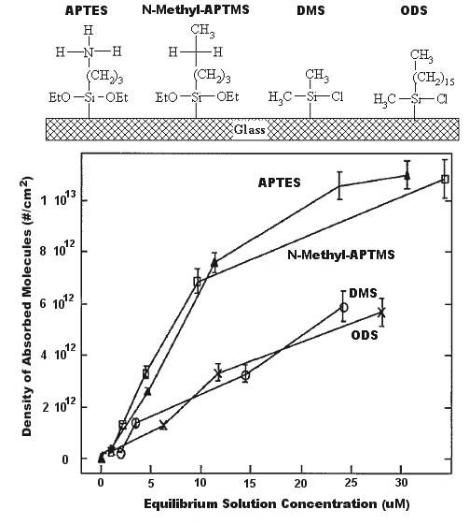
Figure 2.6 shows patterns of oligonucleotide adsorption on hydrophobic and ionic substrates [7]. The chemical structures of the functionalized silanes coupled to the glass surface are shown along with adsorption isotherms for equilibrium oligonucleotide concentrations on the surfaces. Maximum adsorption densities reached > 1 × 10¹³ molecules cm−2 on cationic surfaces, approximately two times higher than on hydrophobic surfaces. The e ect of such densities on hybridization signal were not evaluated in this study, but another study has specifically assessed the e ects of array spot concentration on hybridization signal [10] by direct comparison of spot concentration to hybridization e ciency. With maximum hybridization signals (300–400 a.u.) were observed using a spot concentration of 0.25–1 ng nL−1.
The simplicity of physical adsorption for DNA immobilization can be counterbalanced by several advantages of covalent binding, many strategies for which are specifically described in Chap. 3. Whatever the covalent binding method, the non-covalent interactions precede it and are responsible for the build–up of a high local concentration of molecules near the surface. This high local concentration is needed to achieve a high rate of reaction. However, the very processes responsible for the build–up of the local concentration (in particular electrostatics) can interact with the covalent binding e ciency. X-ray photoelectron spectrometry and cyclic voltametry were used to probe the impact of the terminal functionality of a SAM on the e ectiveness of covalent binding of DNA to SAM-covered electrodes, shedding light on the interaction between electrostatic adsorption and covalent binding [10]. While the ratios of total immobilized DNA on hydroxyl-, carboxyland amino-terminated SAMs was (3–3.5):(1–1.5):1, respectively, the proportion of covalently immobilized DNA was found to be approximately 85%, 93%, and 25%, respectively. These results suggest that protonization of amino groups on the surface resulted in electrostatically driven adsorption of negatively charged DNA, inhibiting the less energetically favorable condensation reaction between the 5 phosphate end of the DNA and the exposed amine group. Attachment to carboxyl-terminated surfaces showed the opposite e ect, with electrostatic repulsion between like negatively charged DNA molecules and surface functional groups inhibiting adsorptive attachment, but higher covalent binding yields. However, the total amount of immobilized DNA on the carboxyl-terminated surfaces was low, due to inhibition of DNA movement towards the reactive surface by electrostatic repulsion. Optimal total attachment, with a high per-
2 Biomolecules and Cells on Surfaces – Fundamental Concepts |
31 |
Fig. 2.6. Concentration of DNA molecules as a function of surface chemistry (Adapted from [7]. Copyright 1998 Academic Press Inc Elsevier Science)
centage of covalently bound DNA, was achieved on neutral or slightly negative hydroxyl-terminated surfaces.
Whatever the method of immobilization, the key performance criterion is the e ciency of hybridization. In theory, this should depend on (i) the surface characteristics, (ii) the surface density of probe molecules, (iii) probe orientation on the surface, and (iv) factors controlling transport of target molecules to the surface. While the DNA density can be controlled, the DNA conformation on the surface is more di cult to modulate. Studies discussed above [6, 7, 9]
32 Kristi L. Hanson et al.
suggest that electrostatically driven DNA adsorption results in orientation of the molecule’s backbone parallel to, rather than perpendicular to, the surface, but with base pairing sites exposed to the liquid medium. While this orientation is conducive to target hybridization, it does not allow for dense probe coverage on the surface, and will therefore limit both the sensitivity and spatial resolution of associated microarrays. That said, simple adsorptive attachment of DNA has been found to be su cient for many microarray applications.
Where higher sensitivity and resolution are required, covalent binding can not only produce higher densities and tighter immobilization of DNA, but also control the orientation of molecular immobilization at either the 3 –hydroxyl or 5 –phosphate end of the DNA chain. However, increased probe density will also a ect intra–strand interactions, surface interactions and charge density at the surface, which can in turn result in substantially di erent ionic strength, pH, and dielectric constant at the surface than in bulk electrolyte solution. It is likely that these di erences will also impinge on the availability of immobilized strands for hybridization. For instance, the standard enthalpy change for the thermal denaturation of target bound DNA was found to be 2–3 times lower for immobilized DNA than for DNA in the bulk solution, and the melting temperature (Tm) was decreased by 6–10◦C [11]. The lower melting temperature suggests that interstrand bonding is weaker on a surface than in solution, depending on bound strand density. Thus, depending on the ability to control immobilization density, there may be variations in sensitivity from case to case. Another study [12] suggests that this e ect is likely to be more pronounced for shorter strands, which were shown to produce lower hybridization signals when spotted at the same concentration as longer molecules. Hybridization signals for shorter strands could, however, be improved by addition of a poly(A)tail. This would be expected if smaller target molecules are held more tightly and closer to the surface, thus being more a ected by surface interactions. In contrast, longer molecules are likely to contain more free loops and ends available for hybridization further away from the surface. This e ect has been observed elsewhere [13], where hybridization was found to be directly dependent on the length of immobilized strands.
2.4 Protein Immobilization on Surfaces
The fundamentals of protein attachment on surfaces have been widely reviewed (e.g., [14–16]), but the extreme diversity and complexity of proteins still make any prediction regarding attachment di cult. Technologies used for DNA microarray application have, to some degree, been adapted to protein microarrays, but the broader use of protein microarray technology is still limited, primarily due to the fact that surfaces and technology allowing uniform and global attachment of a wide variety of proteins are not currently available. This lag in technology stems from the fundamental structural dif-
2 Biomolecules and Cells on Surfaces – Fundamental Concepts |
33 |
ference between proteins and DNA. DNA is (i) uniformly structured with an ordered hydrophilic backbone, (ii) stable, (iii) does not lose binding activity easily, and (iv) has only one interaction site and geometry with target DNA. In contrast, proteins have (i) many di erent structures, (ii) contain heterogeneous hydrophobic and charged domains, (iii) are extremely fragile with activity dependent on retention of three–dimensional structure, and (iv) can have multiple interaction sites.
Additional complications arise with respect to microarray technology, where functional conservation and su cient concentration of bound protein are critical to the success of the technology. Correct orientation of the bound protein is required to increase the exposure of functional domains to solvent/target, but protein adsorption mechanisms often result in random (or widely distributed) orientations on a surface.
In theory, random attachment is not likely to result in a high percentage of protein functional sites in the proper orientation for binding, but successful attachment and target detection have been achieved with random adsorptive attachment techniques (e.g., [17]). Further, a recent study indicates that there may be minimal e ect on functionality between proteins immobilized by directed or random attachment [18]. In contrast, oriented attachment has also been found to increase array sensitivity up to 10–fold [19].
Overall, the need for directed orientation and choice of technique will depend on the specific proteins being used, and no single method is likely to work in all situations. It is clear, however, that as protein arrays become more comprehensive and as the number of proteins in a single array increases, the need for a technology that can accomplish immobilization across a wide range of proteins, or even an entire proteome, will become more desirable. Strategies used thus far can be broadly classified based on adsorptive or covalent binding mechanisms, and then further subdivided into methods resulting in random vs. directed orientation of the molecule.
2.4.1 Random Adsorptive Attachment
As proteins are charged biomolecules, it would be expected that electrostatic interactions could be used for e cient and controllable immobilization. However, electrostatic adsorption is often of a non-permanent nature and can be strongly a ected by changes to solution pH and ionic activity, thereby allowing for the possibility that subsequent array processing might desorb the protein. Additionally, despite the simplicity of electrostatic interaction, which should make adsorption more predictable, such interactions are usually more di cult to predict than hydrophobic or covalent ones [20]. This is most likely due to the uneven spatial charge distribution on protein surfaces, which also varies with pH and ionic strength of the solution. Many chemical or physico– chemical schemes have been used to create charged surfaces that can adsorb proteins, for example polyelectrolyte multilayers [21] and sulfonated polymer

34 Kristi L. Hanson et al.
surfaces [22,23]. However, because of the complexity of the electrostatic potential map as well as interference from other interactions (e.g. hydrophobicity), a generic ‘magic’ surface that can promote the electrostatically-driven adsorption of proteins has not been found.
Hydrophobic interactions are often stronger and less reversible than electrostatic attractions, but can result in loss of functional activity due to partial denaturation as the protein unfolds to expose hydrophobic interior portions to the hydrophobic surface [15].
Due to the complexity of proteins, a reasonable approach would be to explore combinatorially the level of adsorption versus descriptors of surface, solution, and protein characteristics. In an attempt to map the adsorption of virtually any protein on virtually any surface, Nicolau and co-workers have compiled a protein adsorption database [24]. The database contains about 500 cases of protein adsorption for approximately 30 proteins and approximately 100 surfaces in various solution conditions.
Molecular surface property algorithms developed to describe the proteins [25, 26], have been used to describe protein adsorption. A purely empirical approach using a linearly piecewise model with breakpoint was found to be capable of accounting for over 90% of the variance in the data [27]. Fundamentally, the model assumes that the protein concentration on the surface follows a piecewise linear regression conforming to a Langmuir relationship. The experimental data present in the database have been used to derive an empirical relationship that describes the correlation between protein adsorption (dependent variable) and process (independent) variables (i.e. protein concentration in solution; surface tension of the surface; ionic strength of the solution; and absolute value of the di erence between pH and the isoelectric point of the protein), as follows:
|
Γ = f1(γ, ion str, abs(pH − pI), C) · (1 − g(Γ)) |
|
|
+f2(γ, ion str, abs(pH − pI), C) · g(Γ), |
|
f1 |
= a11γ + a12 · ion str + a13 · abs(pH − pI) + a14 · C, |
|
f2 |
= a21γ + a22 · ion str + a23 · abs(pH − pI) + a24 · C, |
|
|
Γ ≤ Γbreakpoint g(Γ) = 0, |
|
|
Γ > Γbreakpoint g(Γ) = 1. |
(2.1) |
The parameters of the equations are: Γ – protein surface concentration (mg/m2); C, protein concentration in solution (mg/ml); γ - surface tension of the polymer (dyne/cm); ion str – ionic strength (M); pI – isoelectric point of the protein; Γbreakpoint – protein concentration at which the slope of the linear function Γ = f (C) changes; and the rest of the parameters are constants (Table 2.1).
The level of fit using such a model is quite remarkable, especially considering that the adsorption data span over three orders of magnitude. This work

2 Biomolecules and Cells on Surfaces – Fundamental Concepts |
35 |
Table 2.1. Coe cients for the protein adsorption (Eq. 6)
|
|
|
|
Protein |
|
|
Surface |
Ionic |
|
concentration |
|
|
tension |
strength |
|
in solution |
Free |
|
(dyne/cm) (M) |
Abs(pH-pI) (mg/ml) |
term |
||
|
|
|
|
|
|
Coe cients |
|
|
|
|
|
in f 1 |
a11 = 0.076 |
a12=-3.297 a13=0.085 |
1.052 |
b1=4.441 |
|
Coe cients |
|
|
|
|
|
in f 2 |
a21=-0.014 |
a22=3.701 a23=-0.395 |
0.438 |
b2=4.840 |
|
Mean |
44.82892 |
0.07505 |
1.39033 |
0.49182 |
|
Standard |
|
|
|
|
|
deviation |
6.942584 |
0.071997 |
0.914402 |
0.760767 |
|
Break point for the protein surface concentration (mg/m2) |
3 |
||||
suggests that prediction of appropriate surface types for maximum protein adsorption may, in fact, be possible based on analysis of protein surface characteristics. Validation of the model with additional sets of data will assist in further applications to microarray technology.
Overall, it appears that random adsorptive attachment can be very e ective for microarray purposes, and it is possible to make some general predictions about adsorption levels for a variety of proteins if the molecular surface characteristics are known or can be predicted. As mentioned above, the e ects of non-oriented binding on array performance can be an issue with this type of attachment, but in many cases still result in acceptable levels of target binding.
2.4.2 Random Covalent Attachment
Covalent binding is quite commonly used in biochip fabrication. Proteins present a variety of functional groups, including amino–, carboxyl–, hydroxyl– and thiol–, which can readily be used for covalent binding to surfaces with complementary chemical groups. However, due to the relative lability of proteins as compared to DNA, more care is required to avoid chemically-induced protein denaturation during the attachment process. There are many strategies for crosslinking of available functional groups, most of which make use of specialized crosslinkers designed for both attachment and physical separation of protein from surface, thereby allowing for more of the protein functional domain to be exposed to the solvent [28].
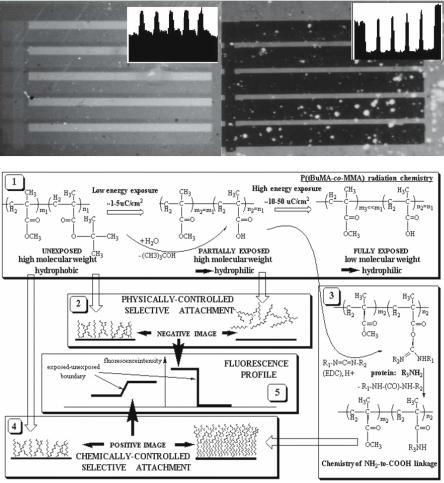
Covalent binding generally produces a higher concentration of protein than does adsorption. For instance, a study [29] compared the e ect of physical adsorption of a protein on Poly(tert–butyl–methacrylate), a highly hydrophobic surface, to the covalent binding of the same protein to a carboxylicfunctionalized surface (derived from the former via e-beam photolysis). Cova-
36 Kristi L. Hanson et al.
lent attachment resulted in significantly higher surface protein concentrations than adsorption, despite the fact that carboxylic functional groups result in a hydrophilic surface which tends to repel protein (Fig. 2.7).
2.4.3 Oriented Attachment
A variety of oriented immobilization techniques have been attempted, and have recently been summarized [30]. These techniques can be adsorptive, covalent or a combination of both. Some of the more common methods include:
1. use of antibody binding proteins to bind the Fc portion of antibodies leaving the binding sites exposed to solution [31, 32];
2.terminal biotinylation of genetically engineered proteins with subsequent end-specific attachment to a streptavidin coated surface [33];
3.terminal His–tag addition and subsequent attachment to a nitrilotriacetic acid-coated surface [34];
4.use of carbohydrate binding molecules to bind the carbohydrate moieties of antibodies [35]; and
5.cystine thiol production on the C–terminal (non-antigen binding) end of cleaved Fab regions, with subsequent attachment using the cystine thiol ‘handle’ [32, 33].
A recent study [19] explored the e ect of some of the above methods of antibody attachment on analyte binding capacity, and found that orientation increases analyte binding capacity up to 10–fold. When Fab’ fragments were specifically oriented in a dense monolayer, 90% of the adsorbed molecules were active, while randomly attached Fab fragments were packed at much lower density, and showed a much lower specific activity. Thus for applications requiring high sensitivity and low detection limits, such techniques are likely to greatly improve performance.
While the above discussion has outlined that there are, in fact, a variety of methods which are useful for the immobilisation of proteins and detection of target analytes in array format, this methodological variation also has a potential downside. Heterogeneous information from di erent laboratories may ultimately result in non-standardized datasets, di cult to compare and interpret, thus hindering the overall goal of a more complete understanding of proteomes.
2.5 Carbohydrate Immobilization
Carboydrate-based microarrays, which have appeared only recently, have recently been reviewed [36]. Applications of these arrays have enormous potential in microarray technology due to the structural diversity, specificity, and di erential expression of carbohydrates [37]. Further, these molecules are
2 |
Biomolecules and Cells on Surfaces – Fundamental Concepts |
37 |
(a) |
(b) |
|
(c)
Fig. 2.7. Comparison of protein concentration on the surface, following adsorption on a hydrophobic and covalent attachment on a carboxylic-functionalized surface [29]. The image on the top left shows fluorescently-labelled (FITC) avidin adsorbed on the hydrophobic surface of Poly(tert–butyl–methacrylate), with dark regions showing lack of protein on the carboxylic-rich (hydrophilic) surface of Poly(methacrylic acid), obtained by deep–UV patterning. The image on the top right shows a similar patterned surface with protein covalently bound on the carboxylicrich surface (lightest areas) and still adsorbed at lower concentrations on the hydrophobic surface (darker bands). (Reprinted with permission from 02Taguchi99. Copyright 1999 American Chemical Society Publications)
38 Kristi L. Hanson et al.
associated with a number of cell characteristics, including adhesion, carcinogenesis and immunity [38]. The ability to rapidly determine the presence and type of carboydrate molecules in a sample would therefore greatly increase our understanding of their in vivo functions.
Carbohydrates, like proteins, are structurally heterogeneous and require preservation of molecular conformation, 3–D structure, and topological configuration on a chip in order for molecular recognition to occur. As such, the existing array surfaces commonly used for DNA are generally not amenable to carbohydrate immobilization.
From an immobilization perspective, perhaps the most common theme to emerge from recent studies is that larger carbohydrate molecules are easily retained on relatively hydrophobic (e.g., nitrocellulose or treated polystyrene) surfaces [39, 40], but smaller carbohydrates show much lower binding e ciencies [39]. To overcome this problem, synthetic glycoconjugates have been used, allowing linkage of the carbohydrate to a protein, lipid or polyacrylamide chain which can then be easily immobilized on a nitrocellulose surface.
In general, this relatively simple means of attaching carbohydrates is associated with retention of the immunological properties of a variety of carbohydrates with distinct structural configurations and diverse sugar chain contents [39]. The authors note, however, that individual preparations must still be tested on such a substrate, given the wide structural diversity of carbohydrate antigens.
2.6 Immobilization of Cells on Surfaces
Cell-based microarrays are being developed for a number of applications, such as medical screening (where the capability of cells to selectively respond to di erent agents can be assessed) and the study of fundamental cell behaviors (such as cell–cell communication and cell spreading). The starting point for these techniques is the ability to pattern arrays of single–cells that can be perturbed and monitored individually. As a consequence, the impact of cell confinement on microsized areas (i.e. areas that have dimensions comparable to that of a single cell – a technology generally referred as ‘cell patterning’ [41]) is of extreme importance in the context of microarray technology.
On a molecular level, the immobilization of cells is far more complex than the immobilization of single biomolecules, and may therefore require situationspecific studies to determine proper surfaces for a particular application. The di culty of cell immobilization arises in the first instance from the complexity of the cell membrane, containing many types of molecules (membrane proteins, glycoproteins; lipid bilayer supramolecular structures; small molecules, etc.). These biomolecules could attach to a given surface based on the concepts described in the previous sections. Though each such interaction could be analyzed independently, it is likely that these interactions are cooperative or at least not fully independent. Furthermore, the cell is also very flexible,
2 Biomolecules and Cells on Surfaces – Fundamental Concepts |
39 |
which makes the attachment of the respective molecular patches, independently and collectively, dynamic. Finally, and most importantly, the cell is a living entity that responds to the stimuli presented by the surface. One mechanism of response, and in fact the simplest from a panoply of responses, is to secrete chemical species that will extend the ‘controlled’ environment of the cell beyond its cell wall.
Simplistically speaking, cell attachment should follow the same rules that govern the non-covalent biomolecular immobilization. For instance, electrostatic interactions can be used for cell immobilization, if the surface of the cell is charged, as is the case for neuronal cells (negatively charged) or some bacteria (most negatively, but some positively charged). Because cells are normally surrounded by a sheath of proteins which presents the hydrophilic face towards the exterior, hydrophobicity-driven immobilization is generally less e ective. The most powerful means of cell immobilization is by biomolecular recognition. Cells present proteins on the exterior of their walls that can be unique to a particular cell type or species and that can be recognized by complementary biomolecules (receptors). Alternatively, cells may present proteins with specific functions (including surface attachment) that can be supplied to immobilize cells. While the former mechanism has the propensity to be cell-specific, the latter is more general.
At the first instance, surfaces covered with cell specific proteins would be the natural technological path for cell immobilization. However, in the previous section we saw that the general behavior of proteins on surfaces is di cult to predict. The problems related to protein adsorption in the context of cell immobilization have been concisely described by Mrksich [42]. Briefly, it is di cult to know the density of ligands that are e ectively available for binding to cellular receptors, due to the distribution in conformation and orientation of adsorbed protein. Many studies aimed at investigating the role of ligand density in cell adhesion and migration have improperly assumed a linear correlation between the density of adsorbed protein and the concentration of protein used to coat the substrates [43]. Also, as expected, the activity of protein-coated substrates can show a dramatic dependence on the choice of substrate. For instance, culturing of myoblasts on two di erent types of polystyrene resulted in completely di erent outcomes, namely proliferation or di erentiation, even though both were coated with comparable densities of fibronectin (a cell adhesion protein) [44].
Because of the diversity of the response of di erent cells to surfaces, it is generally necessary to systematically and specifically test cell adhesion and preservation of bioactivity on substrates intended for microarray devices. For instance, one set of experiments [45,46] examined the attachment of neuronal cells on photosensitive polymers. The photoresists, when exposed to UV light, generate carboxylic-rich surfaces (with concentration modulated by exposure energy) that can be further functionalized with neuropeptides. Thermal processing was also used to manipulate the surface properties, either via polymer crosslinking and decarboxylation, or via di usion of silicon-rich species. It was
40 Kristi L. Hanson et al.
found that two pairs of partially independent antagonistic surface characteristics, namely (i) amino-rich vs. carboxylic-rich surfaces and (ii) hydrophilic vs. hydrophobic surfaces, controlled the cell attachment, with the former promoting adhesion (Fig. 2.8). This complex relationship means that one cannot predict the attachment of cells based only on hydrophobicity or hydrophilicity of the surface. However, surfaces designed for biomolecular recognition mechanisms (e.g. neuropeptide–functionalized) were the most e ective for attachment of neuronal cells, and those designed with very high hydrophobicity were the most e ective for repelling neuronal cells. This discussion is illustrative of the specific issues raised by cell attachment (i.e. neuronal cells) but these conclusions cannot, however, be extrapolated to other types of cells due to the diverse nature of cellular membranes types and receptors.
In addition to determining whether attachment will occur, it is also necessary to examine the e ect that cell confinement will have on cell behavior. Several studies (e.g. [47]) have studied this relationship. Microcontact printing of SAMs has been used to fabricate substrates with micrometer–scale islands of bovine and human endothelial cell extracellular matrix separated by nonadhesive regions. The size and geometry of the islands were found to control cell shape, with immediate impact on the control of apoptosis as well as growth. Progressive restriction of cell extension by culturing cells on smaller and smaller micropatterned adhesive islands regulated a transition
Fig. 2.8. Mechanisms of immobilization of neuronal cells on photoresist surfaces. The vertical bar on the left of each diagram represents the relative repelling e ect of the respective surface. Neuropeptide-functionalized surfaces are found to be the most e ective for immobilization of neuronal cells, while highly hydrophobic and negatively charged surfaces are the most repelling (Reprinted with permission from [46]. Copyright 1999 Academic Press Inc Elsevier Science)
2 Biomolecules and Cells on Surfaces – Fundamental Concepts |
41 |
from growth to apoptosis on a single continuum of cell spreading. This work showed that the size and geometry of the microarray pattern can have profound e ects on cellular behavior, which in turn can influence the performance of a cell-based microarray.
In addition to surface chemistry, size and geometry, topography also influences cell physiology [48]. Substratum topography was found to influence a number of cell behaviors, such as spreading, secretion, attachment, shape, growth, polarity and di erentiated functions [49]. The ability to e ectively immobilize cells for the development of microarrays thus relies on the ability to accurately design and control the microarray surface properties at the micro– and nano– scale level.
Another fundamental problem is the intrinsic limitation of culturing cells in an environment that lacks cells’ natural three–dimensional organization. The question of whether a cell patterned on a flat surface will behave the same way as when in a three–dimensional matrix (e.g. a gel) is still a matter of investigation, but evidence points to important di erences in cell behavior when grown in 2D versus 3D cultures [50–52]. This could mean that unpredictable and di erent behaviors might be obtained when cells are patterned over a 2D environment, such as a microarray. Moreover, cell behavior in vivo is modulated by interaction with the surrounding cells and by the environment, a heterogeneous medium which comprises gradients of nutrients and secreted factors. As a result, use of isolated cell populations in vitro may trigger di erent behavior from the ‘natural’ state. These issues are currently under extensive research and should be taken in great consideration when designing a cell-based microarray and evaluating its performance.
2.7 Conclusions
The immobilization of biomolecules and cells for microarray technology has three ‘dimensions’: (i) a biomolecule or cell to be immobilized on a surface, of which we have limited information and which generally cannot, and/or should not, be altered; (ii) an immobilization surface which can be partially tuned, and of which we have quasi-complete information, and (iii) a liquid environment which is fully controllable and of which we have complete information. This chapter addressed the fundamental concepts dictating the likely response of biomolecules and cells immobilized on surfaces, and their resulting bioactivity. Our approach was to present the general interrelationships between the input and output technological parameters, and then to qualify these general rules on several specific situations. The only certainty that we hope we transmitted to the reader, in a field littered with more exceptions than rules, is that, although general rules are relevant in all situations, nothing can replace experience and innovation.
42 Kristi L. Hanson et al.
References
1.Norde, W. 2003. Colloids and interfaces in life sciences. Marcel Dekker, Monticello, NY
2.Haynes, C. A. and W. Norde. 1994. Globular proteins at solid/liquid interfaces. Colloids and surfaces B: Biointerfaces 2:517–566
3.Derjaguin, B. V. and D. L. Landau. 1941. A theory of the stability of strongly charged lyophobic sols and the coalescence of strongly charged particles in electrolytic solution. Acta Physicochimica USSR 14:633–662
4.Verwey, E. J. W. and J. Th. G. Overbeek. 1948. Theory of stability of lyophobic colloids. Elsevier, Amsterdam
5.Schena, M., D. Shalon, R. W. Davis, and P. O. Brown. 1995. Quantitative monitoring of gene expression patterns with a complementary DNA microarray. Science 270:467–470
6.Chan, V., D. J. Graves, P. Fortina, and S. E. McKenzie. 1997. Adsorption and surface di usion of DNA oligonucleotides at liquid/solid interfaces. Langmuir 13:320–329
7.Chan, V., S. E. McKenzie, S. Surrey, P. Fortina, and D. J. Graves. 1998. E ect of Hydrophobicity and Electrostatics on Adsorption and Surface Di usion of DNA Oligonucleotides at Liquid/Solid Interfaces. Journal of Colloid & Interface Science 203:197–207
8.Ausubel, F. M., R. Brent, R. E. Kingston, D. D. Moore, J. G. Seidman, J. A. Smith, and K. Struhl. 1997. Current protocols in molecular biology. John Wiley and Sons, New York
9.Brett, A. M. O. and A.-M. Chiorcea. 2003. Atomic Force Microscopy of DNA Immobilized onto a Highly Oriented Pyrolytic Graphite Electrode Surface. Langmuir 19:3830–3839
10.Zhao, Y.-D., D.-W. Pang, S. Hu, Z.-L. Wang, J.-K. Cheng, and H.-P. Dai. 1999. DNA–modified electrodes; part 4: optimization of covalent immobilization of DNA on self–assembled monolayers. Talanta 49:751–756
11.Watterson, J. H., P. A. E. Piunno, C. C. Wust, and U. J. Krull. 2000. E ects of Oligonucleotide Immobilization Density on Selectivity of Quantitative Transduction of Hybridization of Immobilized DNA. Langmuir 16:4984–4992
12.Franssen-van Hal, N. L. W., O. Vorst, E. Kramer, R. D. Hall, and J. Keijera. 2002. Factors in sequencing cDNA microarray hybridization on silylated glass slides. Analytical Biochemistry 308:5–17
13.Stillman, B. A. and J. L. Tonkinson. 2001. Expression microarray hybridization kinetics depend on length of the immobilized DNA but are independent of immobilization substrate. Analytical Biochemistry 295:149–157
14.Norde, W. 1986. Adsorption of Proteins from Solution at the Solid–Liquid Interface. Advances in Colloid and Interface Science 25:267–340
15.Brash, J. L. and T. A. Horbett. 1987. Protein at interfaces: physicochemical and biochemical studies. American Chemical Society, Washington, D.C.
16.Horbett, T. A. and J. L. Brash. 1995. Protein at interfaces II: fundamentals and applications. American Chemical Society, Washington, D.C.
17.Angenendt, P., J. Glokler, D. Murphy, H. Lehrach, and D. J. Cahill. 2002. Toward optimized antibody microarrays: a comparison of current microarray support materials. Analytical Biochemistry 309:253–260
18.Wilchek, M. and T. Miron. 2003. Oriented versus random protein immobilization. Journal of Biochemical and Biophysical Methods 55:67–70
2 Biomolecules and Cells on Surfaces – Fundamental Concepts |
43 |
19.Peluso, P., D. S. Wilson, D. Do, H. Tran, M. Venkatasubbaiah, D. Quincy, B. Heidecker, K. Poindexter, N. Tolani, M. Phelan, K. Witte, L. S. Jung, P. Wagner, and S. Nock. 2003. Optimizing antibody immobilization strategies for the construction of protein microarrays. Analytical Biochemistry 312:113–124
20.Asthagiri, D. and A. M. Lenho . 1997. Influence of Structural Details in Modeling Electrostatically Driven Protein Adsorption. Langmuir 13:6761–6768
21.Ladam, G., P. Schaaf, F. J. G. Cuisinier, G. Decher, and J.-C. Voegel. 2001.
Protein adsorption onto auto–assembled polyelectrolyte films. Langmuir 17:878– 882
22.Lestelius, M., B. Liedberg, and P. Tengvall. 1997. In Vitro Plasma Protein Adsorption on –Functionalized Alkanethiolate Self–Assembled Monolayers. Langmuir 13:5900–5908
23.Sundaram, S., F. Lim, S. L. Cooper, and R. W. Colman. 1996. Role of leucocytes in coagulation induced by artificial surfaces: investigation of expression of Mac– 1, granulocyte elastase release and leucocyte adhesion on modified polyurethanes. Biomaterials 17
24.Nicolau, D. V. Biomolecular adsorption database. www.bionanoeng.com/BAD/
25.Nicolau, D. V. Jr. and D. V. Nicolau. 2002. A database comprising biomolecular descriptors relevant to protein adsorption on microarray surfaces. SPIE Proceedings 3:109–116
26.Nicolau, D. V. Jr. and D. V. Nicolau. 2002. A model of protein adsorption to solid surfaces from solution. In Biomedical Nanotechnology Architectures and Applications, Darryl J. Bornhop; David A. Dunn; Raymond P. Mariella; Catherine J. Murphy; Dan V. Nicolau; Shuming Nie; Michelle Palmer; Ramesh Raghavachari; Eds, Proc. SPIE Vol. 4626, 1–8, 2002
27.Nicolau, D. V. Jr., Fulga, F. and Nicolau, D. V. 2003 Impact of Protein Adsorption on the Geometry of Microfluidics Devices. Biomedical Microdevices 5:3, 227–233
28.Wong, S. H. 1991. Chemistry of protein conjugation and cross–linking. CRC Press, Boca Raton, FL
29.Nicolau, D. V., T. Taguchi, H. Taniguchi, and S. Yoshikawa. 1999. Positive and negative tone protein patterning using conventional deep–UV/e-beam resists. Langmuir 15:3845–3851
30.Seong, S. and C. Choi. 2003. Current status of protein chip development in terms of fabrication and application. Proteomics 3:2176–2189
31.Vijayendran, R. A. and D. E. Leckband. 2001. A Quantitative Assessment of Heterogeneity for Surface–Immobilized Proteins. Analytical Chemistry 73:471– 480
32.Nakanishi, K., H. Muguruma, and I. Karube. 1996. A novel method of immobilizing antibodies on a quartz crystal microbalance using plasma–polymerized films for immunosensors. Analytical Chemistry 68:1695–1700
33.Rowe, C. A., S. B. Scruggs, M. J. Feldstein, J. P. Golden, and F. S. Ligler. 1999. An Array Immunosensor for Simultaneous Detection of Clinical Analytes. Analytical Chemistry 71:433–439
34.Lu, Y. J., F. Zhang, and S. F. Sui. 2002. Specific binding of integrin to RGD peptide immobilized on a nitrilotriacetic acid chip: a surface plasmon resonance study. Biochemistry (Moscow) 67:1122–1129
35.Ho man, W. L. and D. J. O’Shannessy. 1988. Site-specific immobilization of antibodies by their oligosaccharide moieties to new hydrazide derivatized solid supports. Journal of Immunological Methods 112:113–120
44 Kristi L. Hanson et al.
36.Wang, D. 2003. Carbohydrate microarrays. Proteomics 3:2167–2175
37.Wang, D. and E. A. Kabat . 1996. Carbohydrate Antigens (Polysaccharides), p. 247–276. In M. H. V. Van Regenmortal (ed.), Structure of Antigens, Volume Three. CRC Press, Boca Raton, New York, London, Tokyo
38.Hirabayashi, J., Y. Arata, and K. Kasai. 2001. Glycome project: Concept, strategy and preliminary application to Caenorhabditis elegans. Proteomics 1:295–303
39.Wang, D., S. Liu, B. J. Trummer, C. Deng, and A. Wang. 2002. Carbohydrate microarrays for the recognition of cross–reactive molecular markers of microbes and host cells. Nature biotechnology 20:275–281
40.Willats, W. G. T., S. E. Rasmussen, T. Kristensen, J. D. Mikkelsen, and J.
P.Knox. 2002. Sugar–coated microarrays: A novel slide surface for the high– throughput analysis of glycans. Proteomics 2:1666–1671
41.Folch, A. and M. Toner. 2000. Microengineering of cellular interactions. Annual review of biomedical engineering 2:227-256
42.Mrksich, M. 2000. A surface chemistry approach to studying cell adhesion. Chemical Society Reviews 29
43.Palecek, S. P., J. C. Loftus, M. H. Ginsberg, D. A. Lau enburger, and A. F. Horwitz. 1997. Integrin–ligand binding properties govern cell migration speed through cell–substratum adhesiveness. Nature 385:537–540
44.Garcia, A. J., M. D. Vega, and D. Boettiger. 1999. Modulation of cell proliferation and di erentiation through substrate–dependent changes in fibronectin. Molecular biology of the cell 10:785–798
45.Nicolau, D. V., T. Taguchi, H. Tanigawa, and S. Yoshikawa. 1996. Control of the neuronal cell attachment by functionality manipulation of diazo–naphtho– quinone/novolak photoresist surface. Biosensors and Bioelectronics 11:1237– 1252
46.Nicolau, D. V., T. Taguchi, H. Taniguchi, H. Tanigawa, and S. Yoshikawa. 1999.
Patterning neuronal and glia cells on light–assisted functionalized photoresists. Biosensors & Bioelectronics 14:317–325
47.Chen, C. S., M. Mrksich, S. Huang, G. M. Whitesides, and D. E. Ingber. 1998.
Micropatterned surfaces for control of cell shape, position, and function. Biotechnology Progress 14
48.Flemming, R. G., C. J. Murphy, G. A. Abrams, S. L. Goodman, and P. F. Nealey. 1999. E ects of synthetic micro– and nano–structured surfaces on cell behavior. Biomaterials 20:573–588
49.Singhvi, R., G. Stephanopoulos, and D. I. C. Wang. 1994. Review: E ects of substratum morphology on cell physiology. Biotechnology and Bioengineering 43:764–771
50.Abbott, A. 2003. Biology’s new dimension. Nature 424:870–872
51.Weaver, V. M., O. W. Petersen, F. Wang, C. A. Larabell, P. Briand, C. Damsky, and M. J. Bissell. 1997. Reversion of the malignant phenotype of human breast cells in three–dimensional culture and in vivo by integrin blocking antibodies. The Journal of Cell Biology 137:231–245
52.Wang, F., V. M. Weaver, O. W. Petersen, C. A. Larabell, S. Dedhar, P. Briand,
R.Lupu, and M. J. Bissell. 1998. Reciprocal interactions between 1–integrin and epidermal growth factor receptor in three–dimensional basement membrane breast cultures: A di erent perspective in epithelial biology. Proceedings of the National Academy of Science 95:14821–14826