

Biomechanics Principles and Applications - Schneck and Bronzino
.pdf272 |
Biomechanics: Principles and Applications |
in Fig. 18.3, rather than one-half octave consistently shown in normal cochlear measurements of mechanical and neural response.
The significant nonlinear effect is the saturation of the active process at high amplitudes. This can be computed by letting the gain α be a function of the amplitude.
In the normal, active cochlea, it was first observed by Khanna and colleagues [1989] and subsequently by Gummer and colleagues [1996], that the tectorial membrane (TM in Fig. 18.1) has a substantially higher amplitude than the basilar membrane. Presumably, the electromotile expansion of the outer hair cells encounters less resistance from the tectorial membrane than the basilar membrane. This shows the importance of getting the correct stiffness and geometry into a model.
18.6 Fluid Streaming?
Békésy [1960] and many others have observed significant fluid streaming in the actual cochlea and in experimental models. Particularly for the high frequencies, it is tempting to seek a component of steady streaming as the significant mechanical stimulation of the inner hair cells [Lighthill, 1992]. Passive models indicated that such streaming occurs only at high sound intensity. Among the many open questions is whether or not the enhancement of amplitude provided by the feed-forward of energy by the outer hair cells and the mechanical nonlinearity at low amplitudes of displacement provided by the ciliary gating can trigger significant streaming. It is clear from the anatomy Fig. 18.1 that the motion and corresponding pressure in the fluid of the inner sulcus is the primary source of excitation for the inner hair cells. The DC pressure associated with DC streaming would be an important effect.
18.7 Clinical Possibilities
A better understanding of the cochlear mechanisms would be of clinical value. Auditory pathology related to the inner ear is discussed by Pickles [1988] and Gulick and colleagues [1989]. The spontaneous and stimulated emissions from the cochlea raise the possibility of diagnosing local inner ear problems, which is being pursued at many centers around the world. The capability for more accurate, physically realistic modeling of the cochlea should assist in this process. A significant step is provided by Zweig and Shera [1995], who find that a random distribution of irregularities in the properties along the cochlea explains much of the emissions.
A patient with a completely nonfunctioning cochlea is referred to as having “nerve deafness”. In fact, there is evidence that in many cases the nerves may be intact, while the receptor cells and organ of Corti are defective. For such patients, a goal is to restore hearing with cochlear electrode implants to stimulate the nerve endings directly. Significant progress has been made. However, despite electrode stimulation of nerves at the correct place along the cochlea for a high frequency, the perception of high frequency has not been achieved. So, although substantial advance in cochlear physiology has been made in the recent past, several such waves of progress may be needed to adequately understand the functioning of the cochlea.
References
Allen JB and Neely ST. 1992. Micromechanical models of the cochlea. Physics Today, 45:40–47. Ashmore JF. 1987. A fast motile response in guinea-pig outer hair cells: the cellular basis of the cochlear
amplifier. J. Physiol. 388:323–347.
Assad JA and Corey DP. 1992. An active motor model for adaptation by vertebrate hair cells. J. Neurosci. 12(9):3291–3309.
Békésy G von. 1960. Experiments in Hearing. McGraw-Hill, New York.
Böhnke F, von Mikusch-Buchberg J, and Arnold W. 1996. 3-D Finite Elemente Modell des cochleären Verstärkers. Biomedizinische Technik. 42:311–312.
Brownell WE, Bader CR, Bertrand D, and de Ribaupierre Y. 1985. Evoked mechanical responses of isolated cochlear outer hair cells. Science 227:194–196.

Cochlear Mechanics |
273 |
Canlon B, Brundlin L, and Flock Å. 1988. Acoustic stimulation causes tonotopic alterations in the length of isolated outer hair cells from the guinea pig hearing organ. Proc. Natl. Acad. Sci. USA 85:7033–7035.
Cabezudo LM. 1978. The ultrastructure of the basilar membrane in the cat. Acta Otolaryngol. 86:160–175. Dallos P. 1992. The active cochlea. J. Neurosci. 12(12):4575–4585.
De Boer E. 1991. Auditory physics. Physical principles in hearing theory. III. Phys. Rep. 203(3):126–231. Freeman DM and Weiss TF. 1990. Hydrodynamic analysis of a two-dimensional model for microme-
chanical resonance of free-standing hair bundles. Hearing Res. 48:37–68.
Fuhrmann E, Schneider W, and Schultz M. 1987. Wave propagation in the cochlea (inner ear): effects of Reissner’s membrane and non-rectangular cross-section. Acta Mechanica 70:15–30.
Furness DN, Zetes DE, Hackney CM, and Steele CR. 1997. Kinematic analysis of shear displacement as a means for operating mechanotransduction channels in the contact region between adjacent stereocilia of mammalian cochlear hair cells. Proc. R. Soc. Lond. B 264:45–51.
Geisler CD. 1993. A realizable cochlear model using feedback from motile outer hair cells. Hearing Res., 68:253–262.
Geisler CD and Sang C. 1995. A cochlear model using feed-forword outer-hair-cell forces, Hearing Res. 85:132–146.
Gulick WL, Gescheider GA, and Fresina RD 1989. Hearing: Physiological Acoustics, Neural Coding, and Psychoacoustics. Oxford University Press, London.
Gummer AW, Johnston BM, and Armstrong NJ. 1981. Direct measurements of basilar membrane stiffness in the guinea pig. J. Acoust. Soc. Am. 70:1298–1309.
Gummer AW Hemmert W, and Zenner HP. 1996. Resonant tectorial membrane motion in the inner ear: its crucial role in frequency tuning. Proc. Natl. Acad. Sci. USA, 93:8727–8732.
Hackney CM, Furness DN, and Katori Y. 1996. Stereociliary ultrastructure in relation to mechanotransduction: tip links and the contact region. In Diversity in Auditory Mechanics. University of California, Berkeley, 173–180.
Hemmert W, Schauz C, Zenner HP, and Gummer AW. 1996. Force generation and mechanical impedance of outer hair cells. In Diversity in Auditory Mechanics. University of California, Berkeley, 189–196.
Holley MD. 1990. Cell biology of hair cells. Seminars in the Neurosciences 2:41–47. Hubbard AE. 1993. A traveling wave-amplifier model of the cochlea. Science 259:68–71. Hudspeth AJ. 1989. How the ears work. Nature 34:397–404.
Iwasa KH, and Chadwick RS. 1992. Elasticity and active force generation of cochlear outer hair cells.
J. Acoust. Soc. Am. 92:3169–3173.
Jen DH and Steele CR. 1987. Electrokinetic model of cochlear hair cell motility. J. Acoust. Soc. Am. 82:1667–1678.
Kalinec F, Holley MC, Iwasa KH, Lim D, Kachar B. 1992. A membrane-based force generation mechanism in auditory sensory cells. Proc. Natl. Acad. Sci. USA 89:8671–8675.
Keidel WD and Neff WD Eds. 1976. Handbook of Sensory Physiology, Vol. V: Auditory System. SpringerVerlag, Berlin.
Kemp DT 1978. Stimulated acoustic emissions from within the human auditory system. J. Acoust. Soc. Am. 64:1386–1391.
Khanna SM, Flock Å, and Ulfendahl M. 1989. Comparison of the tuning of outer hair cells and the basilar membrane in the isolated cochlea. Acta Otolaryngol. [Suppl.] Stockholm 467:141–156.
Kolston PJ and Ashmore JF 1996. Finite element micromechanical modeling of the cochlea in three dimensions. J. Acoust. Soc. Am. 99:455–467.
Lighthill J. 1991. Biomechanics of hearing sensitivity. J. Vibration Acoust. 113:1–13. Lighthill J. 1992. Acoustic streaming in the ear itself. J. Fluid Mech. 239:551–606.
Miller CE. 1985. Structural implications of basilar membrane compliance measurements. J. Acoust. Soc. Am. 77:1465–1474.
Nobili R, Mommano F, and Ashmore J. 1998. How well do we understand the cochlea? TINS 21(4): 159–166.

274 Biomechanics: Principles and Applications
Olson ES, and Mountain DC. 1994. Mapping the cochlear partition’s stiffness to its cellular architecture.
J. Acoust. Soc. Am. 95(1):395–400.
Olson ES. 1998. Observing middle and inner ear mechanics with novel intracochlear pressure sensors.
J. Acoust. Soc. Am. 103(6): 3445–3463.
Pickles JO. 1988. An Introduction to the Physiology of Hearing, 2nd ed. Academic Press, London.
Preyer S, Renz S, Hemmert W, Zenner H, and Gummer A. 1996. Receptor potential of outer hair cells isolated from base to apex of the adult guinea-pig cochlea: implications for cochlear tuning mechanisms. Auditory Neurosci. 2:145–157.
Probst R. 1990. Otoacoustic emissions: an overview. Adv. Oto-Rhino-Laryngol. 44:1–9.
Raftenberg MN. 1990. Flow of endolymph in the inner spiral sulcus and the subtectorial space. J. Acoust. Soc. Am. 87(6):2606–2620.
Ranke OF. 1950. Theory of operation of the cochlea: a contribution to the hydrodynamics of the cochlea.
J. Acoust. Soc. Am. 22:772–777.
Rhode WS. 1971. Observations of the vibration of the basilar membrane in squirrel monkeys using the Mössbauer technique. J. Acoust. Soc. Am. 49:1218–1231.
Ruggero MA. 1993. Distortion in those good vibrations. Curr. Biol. 3(11):755–758.
Russell IJ and Sellick PM. 1977. Tuning properties of cochlear hair cells. Nature, 267:858–860.
Siebert WM. 1974. Ranke revisited—a simple short-wave cochlear model. J. Acoust. Soc. Am. 56(2):594–600.
Steele CR. 1987. Cochlear Mechanics. In Handbook of Bioengineering, R. Skalak and S. Chien, Eds., pp. 30.11–30.22. McGraw-Hill, New York.
Steele CR. 1992. Electroelastic behavior of auditory receptor cells. Biomimetics 1(1):3–22.
Steele CR, Baker G, Tolomeo JA, and Zetes DE. 1993. Electro-mechanical models of the outer hair cell. In Biophysics of Hair Cell Sensory Systems, H. Duifhuis, J.W. Horst, P. van Dijk, and S.M. van Netten, Eds., World Scientific, Singapore.
Strelioff D and Flock Å. 1984. Stiffness of sensory-cell hair bundles in the isolated guinea pig cochlea.
Hearing Res. 15:19–28.
Taber LA and Steele CR. 1979. Comparison of ‘WKB’ and experimental results for three-dimensional cochlear models. J. Acous. Soc. Am. 65:1007–1018.
Tolomeo JA and Steele CR. 1995. Orthotropic piezoelectric propeties of the cochlear outer hair cell wall.
J. Acous. Soc. Am. 95 (5):3006–3011.
Tolomeo JA, Steele CR, and Holley MC. 1996. Mechanical properties of the lateral cortex of mammalian auditory outer hair cells. Biophys. J. 71:421–429.
Watts L. 1993. Cochlear Mechanics: Analysis and Analog VLSI, Ph.D. thesis, California Institute of Technology. West CD. 1985. The relationship of the spiral turns of the cochlea and the length of the basilar membrane to the range of audible frequencies in ground dwelling mammals. J. Acoust. Soc. Am. 77(3):
1091–1101.
Zhang L, Mountain DC and Hubbard AE. 1996. Shape changes from base to apex cannot predict characteristic frequency changes. Diversity in Auditory Mechanics. University of California, Berkeley, 611–618.
Zhou G, Bintz L, Anderson DZ, and Bright KE. 1994. A life-sized physical model of the human cochlea with optical holographic readout. J. Acoust. Soc. Am. 93(3):1516–1523.
Zweig G and Shera CA. 1995. The origin of periodicity in the spectrum of evoked otoacoustic emissions.
J. Acoust. Soc. Am. 98(4):2018–2047.
Zwislocki JJ, and Cefaratti LK. 1989. Tectorial membrane II: Stiffness measurements in vivo. Hearing Res., 42:211–227.

Cochlear Mechanics |
275 |
Further Information
The following are workshop proceedings that document many of the developments:
De Boer E, and Viergever MA, Eds. 1983. Mechanics of Hearing. Nijhoff, The Hague.
Allen JB, Hall JL, Hubbard A, Neely ST, and Tubis A, Eds. 1985. Peripheral Auditory Mechanisms. Springer, Berlin.
Wilson JP and Kemp DT, Eds. 1988. Cochlear Mechanisms: Structure, Function, and Models. Plenum Press, New York.
Dallos P, Geisler CD, Matthews JW, Ruggero MA, and Steele CR, Eds. 1990. The Mechanics and Biophysics of Hearing. Springer-Verlag, Berlin.
Duifhuis H, Horst JW, van Kijk P, and van Netten SM, Eds. 1993. Biophysics of Hair Cell Sensory Systems. World Scientific, Singapore.
Lewis ER, Long GR, Lyon RF, Narins PM, Steele CR, and Hecht-Poinar E, Eds. 1997. Diversity in Auditory Mechanics. World Scientific, Singapore.


19
Vestibular Mechanics
|
19.1 |
Structure and Function.................................................... |
277 |
|
19.2 |
Otolith Distributed Parameter Model ............................ |
278 |
|
19.3 |
Nondimensionalization of the Motion |
|
|
|
Equations .......................................................................... |
280 |
|
19.4 |
Otolith Transfer Function................................................ |
281 |
Wallace Grant |
19.5 |
Otolith Frequency Response............................................ |
284 |
Virginia Polytechnic Institute and |
19.6 |
Semicircular Canal Distributed Parameter Model......... |
285 |
State University |
19.7 |
Semicircular Canal Frequency Response ........................ |
287 |
The vestibular system is responsible for sensing motion and gravity and using this information for control of postural and body motion. This sense is also used to control eyes position during head movement, allowing for a clear visual image. Vestibular function is rather inconspicuous and for this reason is frequently not recognized for its vital roll in maintaining balance and equilibrium and in controlling eye movements. Vestibular function is truly a sixth sense, different from the five originally defined by Greek physicians.
The vestibular system is named for its position within the vestibule of the temporal bone of the skull. It is located in the inner ear along with the auditory sense. The vestibular system has both central and peripheral components. This chapter deals with the mechanical sensory function of the peripheral end organ and its ability to measure linear and angular inertial motion of the skull over the frequency ranges encountered in normal activities.
19.1 Structure and Function
The vestibular system in each ear consists of the utricle and saccule (collectively called the otolithic organs) which are the linear motion sensors, and the three semicircular canals (SCCs) which sense rotational motion. The SCCs are oriented in three nearly mutually perpendicular planes so that angular motion about any axis may be sensed. The otoliths and SCCs consist of membranous structures which are situated in hollowed out sections of the temporal bone. This hollowed out section of the temporal bone is called the bony labyrinth, and the membranous labyrinth lies within this bony structure. The membranous labyrinth is filled with a fluid called endolymph, which is high in potassium, and the volume between the membranous and bony labyrinths is filled with a fluid called perilymph, which is similar to blood plasma.
The otoliths sit within the utricle and saccule. Each of these organs is rigidly attached to the temporal bone of the skull with connective tissue. The three semicircular canals terminate on the utricle forming a complete circular fluid path, and the membranous canals are also rigidly attached to the bony skull. This rigid attachment is vital to the roll of measuring inertial motion of the skull.
0-8493-1492-5/03/$0.00+$.50 © 2003 by CRC Press LLC
278 |
Biomechanics: Principles and Applications |
Each SCC has a bulge called the ampulla near the one end, and inside the ampulla is the cupula, which is formed of saccharide gel. The capula forms a complete hermetic seal with the ampulla, and the cupula sits on top of the crista, which contains the sensory receptor cells called hair cells. These hair cells have small stereocilia (hairs) which extend into the cupula and sense its deformation. When the head is rotated the endolymph fluid, which fills the canal, tends to remain at rest due to its inertia, the relative flow of fluid in the canal deforms the cupula like a diaphragm, and the hair cells transduce the deformation into nerve signals.
The otolithic organs are flat layered structures covered above with endolymph. The top layer consists of calcium carbonate crystals with otoconia which are bound together by a saccharide gel. The middle layer consists of pure saccharide gel, and the bottom layer consists of receptor hair cells which have stereocilia that extend into the gel layer. When the head is accelerated, the dense otoconial crystals tend to remain at rest due to their inertia as the sensory layer tends to move away from the otoconial layer. This relative motion between the octoconial layer deforms the gel layer. The hair cell stereocilia sense this deformation, and the receptor cells transduce this deformation into nerve signals. When the head is tilted, weight acting on the otoconial layer also will deform the gel layer. The hair cell stereocilia also have directional sensitivity which allows them to determine the direction of the acceleration acting in the plane of the otolith and saccule. The planes of the two organs are arranged perpendicular to each other so that linear acceleration in any direction can be sensed. The vestibular nerve, which forms half of the VIII cranial nerve, innervates all the receptor cells of the vestibular apparatus.
19.2 Otolith Distributed Parameter Model
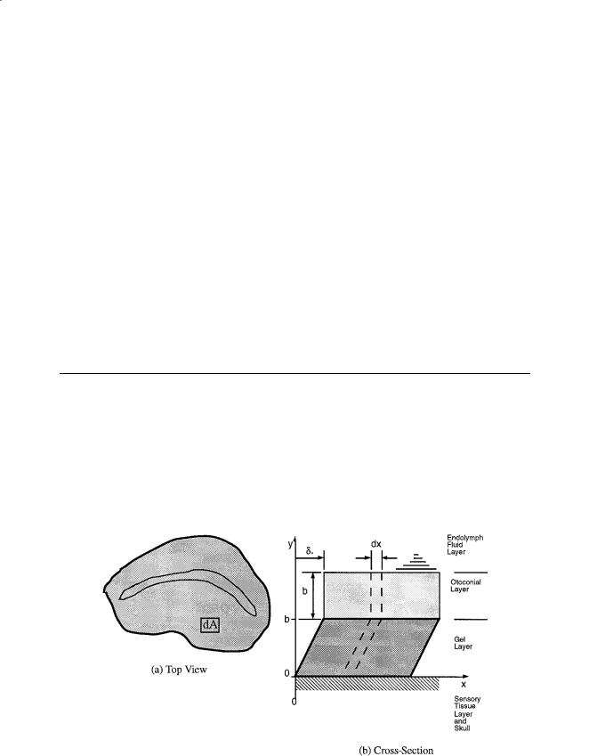
The otoliths are an overdamped second-order system whose structure is shown in Fig. 19.1. In this model the otoconial layer is assumed to be a rigid and nondeformable, the gel layer is a deformable layer of isotropic viscoelastic material, and the fluid endolymph is assumed to be a newtonian fluid. A small element of the layered structure with surface area dA is cut from the surface, and a vertical view of this surface element, of width dx, is shown in Fig. 19.2. To evaluate the forces that are present, free-body diagrams are constructed of each elemental layer of the small differential strip. See the nomenclature table for a description of all variables used in the following formulas, and for derivation details see Grant and colleagues [1984, 1991].
FIGURE 19.1 Schematic of the otolith organ: (a) Top view showing the peripheral region with differential area dA where the model is developed. (b) Cross-section showing the layered structure where dx is the width of the differential area dA shown in the top view at the left.
Vestibular Mechanics |
279 |
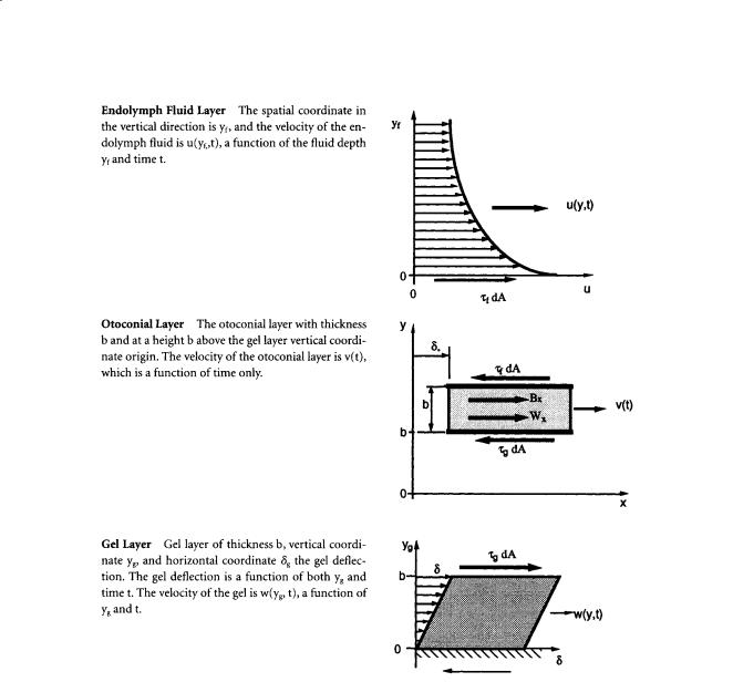
FIGURE 19.2 The free-body diagrams of each layer of the otolith with the forces that act on each layer. The interfaces are coupled by shear stresses of equal magnitude that act in opposite directions at each surface. The τg shear stress acts between the gel-otoconial layer, and the τf acts between the fluid-otoconial layer. The forces acting at these interfaces are the product of shear stress τ and area dA. The Bx and Wx forces are respectively the components of the buoyant and weight forces acting in the plane of the otoconial layer. See the nomenclature table for definitions of other variables.
In the equation of motion for the endolymph fluid, the force τf dA acts on the fluid, at the fluid–otoconial layer interface. This shear stress τf is responsible for driving the fluid flow. The linear Navier–Stokes equations for an incompressible fluid are used to describe this endolymph flow. Expressions for the pressure gradient, the flow velocity of the fluid measured with respect to an inertial reference frame, and the force due to gravity (body force) are substituted into the Navier–Stokes equation for flow in the x-direction yielding:
ρ |
|
∂u |
= µ |
|
∂2u |
(19.1) |
f |
|
f ∂y f |
||||
|
∂t |
|
||||
with boundary and initial conditions: u(0, t) = v(t); u(∞, t) = 0; u(yf , 0) = 0.

280 |
Biomechanics: Principles and Applications |
The gel layer is treated as a Kelvin–Voight viscoelastic material where the gel shear stress has both an elastic component and a viscous component acting in parallel. This viscoelastic material model is substituted into the momentum equation, and the resulting gel layer equation of motion is:
ρg |
∂w |
|
∫ |
t |
∂2w |
|
∂2w |
|
|
= G |
|
|
dt + µg |
|
(19.2) |
||
|
|
|
||||||
|
∂t |
|
|
2 |
|
2 |
|
|
|
|
0 |
|
∂yg |
|
∂yg |
|
with boundary and initial conditions: w(b,t) = v(t); w(0,t) = 0; w(yg,0) = 0; δg(yg,0) = 0. The elastic term in the equation is written in terms of the integral of velocity with respect to time, instead of displacement, so the equation is in terms of a single dependent variable, the velocity.
The otoconial layer equation was developed using Newton’s second law of motion, equating the forces that act on the otoconial layer—fluid shear, gel shear, buoyancy, and weight—to the product of mass and inertial acceleration. The resulting otoconial layer equation is:
ρob |
∂v |
+ (ρo |
− ρ f ) |
|
∂Vs |
|
|
|
|
− g x |
= µ f |
||||||
∂t |
∂t |
|||||||
|
|
|
|
|
|
|
|
|
∂u |
|
|
− G∫0 |
|
∂w |
|
|
|
|
∂w |
|
|
|
|
|||
|
|
|
|
|||||||||||||||
|
|
|
t |
|
|
|
|
|
|
|
|
|||||||
|
|
|
|
|
|
|
|
|
|
dt + µ g |
|
|
|
|
|
(19.3) |
||
∂y |
|
∂y |
|
∂y |
|
|||||||||||||
|
|
f |
|
y f =0 |
|
|
|
g |
|
|
y g=b |
|
|
g |
|
|
y g=b |
|
|
|
|
|
|
|
|
|
|
|
|
||||||||
with the initial condition v(0) = 0.
19.3 Nondimensionalization of the Motion Equations
The equations of motion are then nondimensionalized to reduce the number of physical and dimensional parameters and combine them into some useful nondimensional numbers. The following nondimensional variables, which are indicated by overbars, are introduced into the motion equations:
|
|
|
y f |
|
|
|
|
yg |
|
µ f |
|
|
|
u |
|
|
|
v |
|
|
|
w |
|
|
y f |
= |
yg |
= |
t = |
t u = |
v = |
w = |
(19.4) |
||||||||||||||||
b |
b |
ρob2 |
V |
V |
V |
|||||||||||||||||||
|
|
|
|
|
|
|
|
|
|
|
|
|
|
|
|
|||||||||
Several nondimensional parameters occur naturally as a part of the nondimensionalization process. These parameters are
R = |
ρf |
|
Gb2ρ |
µg |
|
|
|
|
ρ b2 |
|
||
|
= |
o |
M = |
|
g x |
= |
o |
g x |
(19.5) |
|||
ρo |
|
µ f |
||||||||||
|
|
µ2f |
|
|
|
Vµ f |
|
|
||||
where R is the density ratio, ε is a nondimensional elastic parameter, M is the viscosity ratio and represents a major portion of the system damping, and gx is the nondimensional gravity.
The governing equations of motion in nondimensional form are then as follows. For the endolymph fluid layer:
|
∂ |
|
|
= |
∂2 |
|
|
|
|
|
R |
u |
u |
|
(19.6) |
||||||
|
|
|
|
|
|
|
|
|||
|
∂t |
∂ |
|
f2 |
|
|
||||
|
y |
|
|
|||||||
with boundary conditions of u (0,t) = v(t) and u(∞,t) = 0 and initial conditions of u(yf ,0) = 0. For the otoconial layer:
Vestibular Mechanics
∂ |
|
|
∂ |
|
|
|
|
|
|
∂ |
|
|
|
|
t |
|
∂ |
|
|
|
|
|
|
∂ |
|
|
|
|
|
|||||||
|
|
|
|
|
|
|
|
|
|
|
|
|
||||||||||||||||||||||||
|
|
V |
|
|||||||||||||||||||||||||||||||||
v |
u |
|
w |
w |
||||||||||||||||||||||||||||||||
|
|
|
+ (1− R) |
|
s |
− g x = |
|
|
|
|
|
|
|
− ∫0 |
|
|
|
|
|
|
|
|
|
|
|
|
|
|
|
|
|
|
|
|||
∂t |
|
∂t |
∂ |
|
|
|
|
|
∂ |
|
|
|
|
|
dt − M |
∂ |
|
|
|
|
|
|
||||||||||||||
|
y |
|
|
y |
|
y |
|
|||||||||||||||||||||||||||||
|
|
|
|
|
|
|
|
|
|
|
|
|
|
f |
|
|
|
|
|
|
|
|
g |
|
|
|
|
|
|
|
|
g |
|
|
|
|
|
|
|
|
|
|
|
|
|
|
|
|
|
|
|
0 |
|
|
|
|
|
|
|
|
|
1 |
|
|
|
|
|
|
|
1 |
|||
|
|
|
|
|
|
|
|
|
|
|
|
|
|
|
|
|
|
|
|
|
|
|
|
|
|
|
||||||||||
with an initial condition of v(0) = 0. For the gel layer:
|
∂ |
|
|
|
∫ |
t |
∂2 |
|
|
|
|
∂2 |
|
|
|
||
|
w |
|
= |
w |
w |
|
|||||||||||
R |
|
|
|
|
|
|
|
|
|
dt + M |
|
|
|
|
|
||
|
|
|
|
|
|
|
|
|
|
|
|
|
|||||
|
∂t |
|
|
|
|
|
2 |
|
|
|
|
2 |
|
||||
|
|
|
|
|
|
||||||||||||
|
|
|
o |
∂yg |
|
|
∂yg |
|
|||||||||
281
(19.7)
(19.8)
with boundary conditions of w (1,t ) = v (t) and w (0,t ) = 0 and initial conditions of w (yg ,0) = 0 and
δg(yg ,0) = 0.
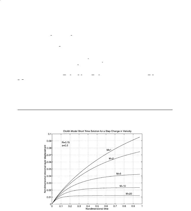
These equations can be solved numerically for the case of a step change in velocity of the skull. This solution is shown in Figure 19.3 for a step change in the velocity of the head and a step change in the acceleration of the head, and can be found in Grant and Cotton [1991].
19.4 Otolith Transfer Function
A transfer function of otoconial layer deflection related to skull acceleration can be obtained from the governing equations. For details of this derivation see Grant and colleagues [1994].
(a)
FIGURE 19.3 This figure shows the time response of the otolith model for various values of the nondimensional parameters. Parts (a) through (d) show the response to a step change in the velocity of the head. This response can be thought of as a transient response since it is an impulse in acceleration. The step change has a nondimensional skull velocity of magnitude one. Parts (e) and (f) show the response to a step change in the acceleration of the skull or simulates a constant acceleration stimulus. Again the magnitude of the nondimensional step change is one. (a) Short time response for various values of the damping parameter M. Note the effect on maximum displacement produced by this parameter.