

Biomechanics Principles and Applications - Schneck and Bronzino
.pdf252 |
Biomechanics: Principles and Applications |
to a variety of vasoactive stimuli [Ohhashi et al., 1978; Benoit, 1997], including signals which involve nitric oxide [Ohhashi and Takahashi, 1991; Bohlen and Lash, 1992; Yokoyama and Ohhashi, 1993]. None of these contractile features has been documented in the initial lymphatics.
The lymphatic endothelium has a number of similarities with the vascular endothelium. It forms a continuous lining. There are numerous caveolae, Weibel Palade bodies, but lymphatic endothelium has fewer interendothelial adhesion complexes and there is a discontinuous basement membrane. The residues of the basement membrane are attached to interstitial collagen via anchoring filaments [Leak and Burke, 1968] which provide relatively firm attachment of the endothelium to the interstitial structures.
Lymphatic Network Display
One of the interesting aspects regarding lymphatic transport in skeletal muscle is the fact that all lymphatics inside the muscle parenchyma are of the non-contractile initial type [Skalak et al., 1984]. Collecting lymphatics can only be observed outside the muscle fibers as a conduit to adjacent lymph nodes. The fact that all lymphatics inside the tissue parenchyma are of the initial type is not unique to skeletal muscle but has been demonstrated in other organs [Unthank and Bohlen, 1988; Yamanaka et al., 1995]. The initial lymphatics are positioned in the adventitia of the arcade arterioles surrounded by collagen fibers (Fig. 17.4). In this position they are in immediate proximity to the arteriolar smooth muscle and adjacent to myelinated nerve fibers and a set of mast cells that accompany the arterioles. The initial lymphatics are frequently sandwiched between arteriolar smooth muscles and their paired venules, and they in turn are embedded between the skeletal muscle fibers [Skalak et al., 1984]. The initial lymphatics are firmly attached to the adjacent basement membrane and collagen fibers via anchoring filaments [Leak and Burke, 1968]. The basement membrane of the lymphatic endothelium is discontinuous, especially at the interendothelial junctions, so that macromolecular material and even large colloidal particles enter the initial lymphatics [Casley-Smith, 1962; Bach and Lewis, 1973; Strand and Persson, 1979; Bollinger et al., 1981; Ikomi et al., 1996].
The lumen cross-section of the initial lymphatics is highly irregular in contrast to the overall circular cross-section of the collecting lymphatics (Fig. 17.4). The lumen cross-section of the initial lymphatics is partially or completely collapsed and may frequently span around the arcade arteriole. In fact, we have documented cases in which the arcade arteriole is completely surrounded by an initial lymphatic channel, highlighting the fact that the activity of the lymphatics is closely linked to that of the arterioles [Ikomi and Schmid-Schönbein, 1995]. Initial lymphatics in skeletal muscle have intra-luminal valves which consist of bileaflets and a funnel structure [Mazzoni et al., 1987]. The leaflets are flexible structures and are opened and closed by a viscous pressure drop along the valve funnel. In a closed position the leaflets are able to support considerable pressures [Eisenhoffer et al., 1995; Ikomi et al., 1997]. This arrangement serves to preserve normal valve function even in initial lymphatics with irregularly shaped lumen cross-sections.
The lymphatic endothelial cells are attenuated and have many of the morphological characteristics of vascular endothelium, including expression of P-selectin, von Willebrand factor [Di Nucci et al., 1996], and factor VIII [Schmid-Schönbein, 1990]. An important difference between vascular and lymphatic endothelium lies in the arrangement of the endothelial junctions. In the initial lymphatics, the endothelial cells lack tight junctions [Schneeberger and Lynch, 1984] and are frequently encountered in an overlapping but open position, so that proteins, colloid particles, and even chylomicron particles can readily pass through the junctions [Casley-Smith, 1962; Casley-Smith, 1964; Leak, 1970]. Examination of the junctions with scanning electron microscopy shows that there exists a periodic interdigitating arrangement of endothelial extensions. Individual extensions are attached via anchoring filaments to the underlying basement membrane and connective tissue, but the two extensions of adjacent endothelial cell resting on top of each other are not attached by interendothelial adhesion complexes. Mild mechanical stretching of the initial lymphatics shows that the endothelial extensions can be separated in part from each other, indicating that the membranes of two neighboring lymphatic endothelial cells are not attached to each other, but are firmly attached to the underlying basement membrane [Castenholz, 1984]. Lymphatic endothelium does not exhibit continuous junctional complexes, and instead has a “streak and dot”

Mechanics of Tissue and Lymphatic Transport |
253 |
FIGURE 17.4 Histological cross-sections of lymphatics (LYM) in rat skeletal muscle before (p. 253) and after (p. 254) contraction of the paired arcade arterioles (ART). The lymphatic channel is of the initial type with a single attenuated endothelial layer (curved arrows). Note, that in the dilated arteriole, the lymphatic is essentially compressed (p. 253) while the lymphatic is expanded after arteriolar contraction (p. 254) which is noticeable by the folded endothelial cells in the arteriolar lumen. In both cases the lumen cross-sectional shape of the initial lymphatic channels is highly irregular. All lymphatics within skeletal muscle (SKM) have these characteristic features. [Skalak et al., 1984.]
immunostaining pattern of VE-cadherin and associated intracellular proteins—desmoplakin and plakoglobulin [Schmelz et al., 1994]. But the staining pattern is not uniform for all lymphatics, and in larger lymphatics a more continuous pattern is present. This highly specialized arrangement has been referred to in the following as the lymphatic endothelial microvalves [Schmid-Schönbein, 1990].
Mechanics of Lymphatic Valves
In contrast to the central large valves in the heart, which are closed by inertial fluid forces, the lymphatic valves are small and the fluid Reynolds number is almost zero. Thus, since no inertial forces are available to open and close these valves, a different valve morphology has evolved. They form long funnel-shaped channels which are inserted into the lymph conduits and attached at their base and the funnel is prevented from inversion by attachment via a buttress to the lymphatic wall. The valve wall structure consists of a

254 |
Biomechanics: Principles and Applications |
FIGURE 17.4 (continued)
collagen layer sandwiched between two endothelial layers, and the entire structure is quite deformable under mild physiological fluid pressures. The funnel structure serves to create a viscous pressure gradient which is sufficient to generate, during forward fluid motion, a pressure drop to open the values and upon flow reversal to close the valves [Mazzoni et al., 1987].
Lymph Pump Mechanisms
One of the important questions which is fundamental in lymphology is: How do fluid particles in the interstitium find their way into the initial lymphatics? In light of the relative sparse display of the initial lymphatics, a directed convective transport is required—provided by either a hydrostatic or a colloid osmotic pressure drop [Zweifach and Silberberg, 1979]. The documentation of the exact mechanism has remained an elusive target. Several proposals have been advanced (which are discussed in detail in Schmid-Schönbein, 1990). Briefly, a number of authors have postulated that there exists a constant pressure drop from the interstitium into the initial lymph which may support a steady fluid motion into the lymphatics. But, repeated measurements with different techniques have uniformly failed to provide supporting evidence for a steady pressure drop to transport fluid into the initial lymphatics [Zweifach and Prather, 1975; Clough and Smaje, 1978]. Under steady-state conditions, a steady pressure drop does

Mechanics of Tissue and Lymphatic Transport |
255 |
not exist in the vicinity of the initial lymphatics in skeletal muscle within the resolution of the measurement (about 0.2 cm H2O) [Skalak et al., 1984]. An order of magnitude estimate of the pressure drop to be expected at the relatively slow flow rates of the lymphatics shows, however, that the pressure drop from the interstitium may be significantly lower [Schmid-Schönbein, 1990]. Furthermore, the assumption of a steady pressure drop is not in agreement with the substantial evidence that lymph flow rate is enhanced under unsteady conditions (see below). Some investigators have postulated an osmotic pressure in the lymphatics to aspirate fluid into the initial lymphatics [Casley-Smith, 1972] due to ultrafiltration across the lymphatic endothelium, a mechanism that has been referred to as “bootstrap effect” [Perl, 1975]. Critical tests of this hypothesis, such as the microinjection of hyperosmotic protein solutions, have not led to a uniformly accepted hypothesis for lymph formation involving an osmotic pressure. Others have suggested a retrograde aspiration mechanism, such that the recoil in the collecting lymphatics serves to lower the pressure in the initial lymphatics upstream of the collecting lymphatics [Reddy, 1986; Reddy and Patel, 1995], or an electric charge difference across lymphatic endothelium [O’Morchoe et al., 1984].
Tissue Mechanical Motion and Lymphatic Pumping
An intriguing feature of the lymphatic pressure is that lymphatic flow rates depend on tissue motion. In a resting tissue, the lymph flow rate is relatively small, but different forms of tissue motion serve to enhance the lymph flow. This was originally shown for pulsatile pressure in the rabbit ear. Perfusion of the ear with steady pressure (even at the same mean pressure) serves to stop most lymph transport, while pulsatile pressure promotes lymph transport [Parsons and McMaster, 1938]. In light of the paired arrangement of the arterioles and lymphatics, periodic expansion of the arterioles leads to compression of the adjacent lymphatics, and vice versa: a reduction of the arteriolar diameter during the pressure reduction phase leads to expansion of the adjacent lymphatics [Skalak et al., 1984] (Fig. 17.4). Vasomotion which is associated with a slower contraction of the arterioles, but with a larger amplitude than pulsatile pressure, serves to increase lymph formation [Intaglietta and Gross, 1982; Colantuoni et al., 1984]. In addition, muscle contractions, simple walking [Olszewski and Engeset, 1980], respiration, intestinal peristalsis, skin compression [Ohhashi et al., 1991], and other tissue motions are associated with an increase in the lymph flow rates. Periodic tissue motions are significantly more effective to enhance the lymph flow than, for example, elevation of the venous pressure [Ikomi et al., 1996], which is also associated with enhanced fluid filtration [Renkin et al., 1977].
A requirement for lymph fluid flow is the periodic expansion and compression of the initial lymphatics. Since initial lymphatics do not have their own smooth muscle, the expansion and compression of the initial lymphatics depends on the motion of tissue in which they are embedded. In skeletal muscle, the strategic location of the initial lymphatics in the adventitia of the arterioles provides the opportunity for the expansion and compression to be achieved via several mechanisms: (1) arteriolar pressure pulsations or vasomotion, (2) active or passive skeletal muscle contractions, or (3) external muscle compression. Direct measurements of the cross-sectional area of the initial lymphatics during arteriolar contractions or during skeletal muscle shortening support this hypothesis [Skalak et al., 1984; Mazzoni et al., 1990] (Fig. 17.5). The different lymph pump mechanisms are additive. Resting skeletal muscle has much lower lymph flow rates (provided largely by the arteriolar pressure pulsation and vasomotion) than skeletal muscle during exercise (produced by a combination of intramuscular pressure pulsations and skeletal muscle shortening).
Measurements of lymph flow rates in an afferent lymph vessel (diameter about 300 to 500 µm, proximal to the popliteal node) in the hind leg [Ikomi and Schmid-Schönbein, 1996] serve to demonstrate that lymph fluid formation can be influenced by passive or active motion of the surrounding tissue. The lymphatics in this tissue region drain muscle and skin of the hind leg, and the majority are of the initial type, whereas collecting lymphatics are detected outside the tissue parenchyma in the fascia proximal to the node. Without whole leg rotation, lymph flow remains at low non-zero values. If the pulse pressure is stopped, the lymph flow falls to values below the detection limit (less than about 10% of the values during pulse pressure). Introduction of whole leg passive rotation causes a strong, frequency-dependent
256 |
Biomechanics: Principles and Applications |
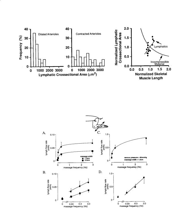
FIGURE 17.5 Histograms of initial lymphatic cross-sectional area in rat spinotrapezius m. before (left) and after (middle) contraction of the paired arteriole with norepinephrine; lymphatic cross-sectional area as a function of muscle length during active contraction or passive stretch (right). Cross-sectional area and muscle length are normalized with respect to the values in vivo in resting muscle. Note the expansion of the initial lymphatics with contraction of the arterioles or muscle stretch. [Skalak et al., 1984; Mazzoni et al., 1990.]
FIGURE 17.6 Lymph flow rates in a prenodal afferent lymphatic draining the hindleg as a function of the frequency of a periodic surface shear motion (massage) without (panels A, B) and with (panels C, D) elevation of the venous pressure by placement of a cuff. Zero frequency refers to a resting leg with a lymph flow rate which depends on pulse pressure. The amplitudes of the tangential skin shear motion were 1 cm and 0.5 cm (panels A, B) and 1 cm in the presence of the elevated venous pressure (panels C, D). Note that the ordinates in panels C and D are larger than those in panels A and B. [Ikomi and Schmid-Schönbein, 1996.]
lymph flow rate which increases linearly with the logarithm of frequency between 0.03 Hz and 1.0 Hz (Fig. 17.6). Elevation of the venous pressure, which serves to enhance fluid filtration from the vasculature and elevates the flow rates, does not significantly alter the dependency on periodic tissue motion [Ikomi et al., 1996].
Mechanics of Tissue and Lymphatic Transport |
257 |
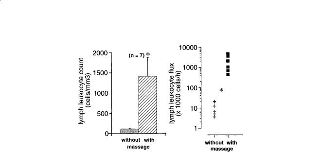
FIGURE 17.7 Lymph leukocyte count (left) and leukocyte flux (right) before and after application of periodic hind leg skin shear motion (massage) at a frequency of about 1 Hz and amplitude of 1 cm. The flux rates were computed from the product of lymph flow rates and the lymphocyte counts. *Statistically significant differences from case without massage. (Adapted from Ikomi et al. [1996].)
Similarly, application of passive tissue compression on the skin serves to elevate the lymph flow rate in a frequency-dependent manner. The lymph flow rates are determined to a significant degree by the local action of the lymph pump, since arrest of the heart beat and reduction of the central blood pressure to zero do not stop lymph flow, and instead reduces the lymph flow rates only about 50% during continued leg motion or application of periodic shear stress to the skin for several hours [Ikomi and SchmidSchönbein, 1996]. Periodic compression of the initial lymphatics also serves to enhance proteins and even lymphocyte counts in the lymphatics [Ikomi et al., 1996] (Fig. 17.7). Thus either arteriolar smooth muscle or parenchymal skeletal muscle activity serve to expand and compress the initial lymphatics in skeletal muscle. The mechanisms serve to adjust lymph flow rates according to organ activity, such that a resting skeletal muscle has low lymph flow rates. During normal daily activity or mild or strenuous exercise, the lymph flow rates as well as the protein and cell transport in the lymphatics increases [Olszewski et al., 1977].
In conclusion, the lymphatics are a unique transport system that is even present in primitive physiological systems. It likely carries out a multitude of functions, many of which have yet to be discovered. Details of its operation and its growth kinetics must await a more detailed bioengineering analysis, especially at the molecular level [Jeltsch et al., 1997].
Acknowledgments
We thank Karen Hutchinson for expert manuscript assistance. This work was supported by NASA grants 199-14-12-04 and 199-26-12-38 and NSF grant IBN-9512778.
Defining Terms
Capillary: The smallest blood vessel of the body which provides oxygen and other nutrients to nearby cells and tissues.
Colloid osmotic pressure: A negative pressure which depends on protein concentration (mainly of albumin and globulins) and prevents excess filtration across the capillary wall.
Edema: Excess fluid or swelling within a given tissue.
Interstitium: The space between cells of various tissues of the body. Normally fluid and proteins within this space are transported from the capillary to the initial lymphatic vessel.
Lymphatic system: The clear network of vessels which return excess fluid and proteins to the blood via the thoracic duct.

258 Biomechanics: Principles and Applications
References
Aratow M, Hargens AR, Meyer JU et al. 1991. Postural responses of head and foot cutaneous microvascular flow and their sensitivity to bed rest. Aviat. Space Environ. Med. 62:246.
Aukland K, Reed RK. 1993. Interstitial-lymphatic mechanisms in the control of extracellular fluid volume.
Physiol. Rev. 73:1.
Bach C, Lewis GP. 1973. Lymph flow and lymph protein concentration in the skin and muscle of the rabbit hind limb. J. Physiol. (Lond.) 235:477.
Benoit JN. 1997. Effects of alpha-adrenergic stimuli on mesenteric collecting lymphatics in the rat. Am. J. Physiol. 273:R331.
Bert JL, Pearce RH. 1984. The interstitium and microvascular exchange. In Handbook of Physiology: The Cardiovascular System: Microcirculation, Sec. 2, Vol. 4, Pt. 1, pp. 521–547, Bethesda, MD, American Physiological Society.
Bohlen HG, Lash JM. 1992. Intestinal lymphatic vessels release endothelial-dependent vasodilators. Am. J. Physiol. 262:H813.
Bollinger A, Jäger K, Sgier F et al. 1981. Fluorescence microlymphography. Circulation 64:1195. Casley-Smith JR. 1962. The identification of chylomicra and lipoproteins in tissue sections and their
passage into jejunal lacteals. J. Cell Biol. 15:259.
Casley-Smith JR. 1964. Endothelial permeability—the passage of particles into and out of diaphragmatic lymphatics. Quart. J. Exp. Physiol. 49:365.
Casley-Smith JR. 1972. The role of the endothelial intercellular junctions in the functioning of the initial lymphatics. Angiologica 9:106.
Casley-Smith JR. 1982. Mechanisms in the formation of lymph. Cardiovascular Physiology IV, International Review of Physiology, pp. 147–187, Baltimore, MD, University Park Press.
Castenholz A. 1984. Morphological characteristics of initial lymphatics in the tongue as shown by scanning electron microscopy. Scan. Electr. Microsc. 1984:1343.
Clough G, Smaje LH. 1978. Simultaneous measurement of pressure in the interstitium and the terminal lymphatics of the cat mesentery. J. Physiol. (Lond.) 283:457.
Colantuoni A, Bertuglia S, Intaglietta M. 1984. A quantitation of rhythmic diameter changes in arterial microcirculation. Am. J. Physiol. 246:H508.
Curry F-RE. 1984. Mechanics and thermodynamics of transcapillary exchange. In Handbook of Physiology: The Cardiovascular System: Microcirculation, Sec. 2, Vol. 4, Pt. 1, pp. 309–374, Bethesda, MD, American Physiological Society.
Di Nucci A, Marchetti C, Serafini S et al. 1996. P-selectin and von Willebrand factor in bovine mesenteric lymphatics: an immunofluorescent study. Lymphology 29:25.
Eisenhoffer J, Kagal A, Klein T et al. 1995. Importance of valves and lymphangion contractions in determining pressure gradients in isolated lymphatics exposed to elevations in outflow pressure.
Microvasc. Res. 49:97.
Hammel HT. 1994. How solutes alter water in aqueous solutions. J. Phys. Chem. 98:4196.
Hargens AR. 1986. Interstitial fluid pressure and lymph flow. Handbook of Bioengineering, 19:1–35, New York, McGraw-Hill.
Hargens AR, Akeson WH. 1986. Stress effects on tissue nutrition and viability. In Tissue Nutrition and Viability, pp. 1–24, New York, Springer-Verlag.
Hargens AR, Zweifach BW. 1977. Contractile stimuli in collecting lymph vessels. Am. J. Physiol. 233:H57. Hargens AR, Millard RW, Pettersson K et al. 1987. Gravitational haemodynamics and oedema prevention
in the giraffe. Nature 329:59.
Ikomi F, Schmid-Schönbein GW. 1995. Lymph transport in the skin. Clin. Dermatol. 13(5):419, Elsevier Science Inc.
Ikomi F, Schmid-Schönbein GW. 1996. Lymph pump mechanics in the rabbit hind leg. Am. J. Physiol. 271:H173.

Mechanics of Tissue and Lymphatic Transport |
259 |
Ikomi F, Hunt J, Hanna G et al. 1996. Interstitial fluid, protein, colloid and leukocyte uptake into interstitial lymphatics. J. Appl. Physiol. 81:2060.
Ikomi F, Zweifach BW, Schmid-Schönbein GW. 1997. Fluid pressures in the rabbit popliteal afferent lymphatics during passive tissue motion. Lymphology 30:13.
Intaglietta M, Gross JF. 1982. Vasomotion, tissue fluid flow and the formation of lymph. Int. J. Microcirc. Clin. Exp. 1:55.
Jain RK. 1987. Transport of molecules in the tumor interstitium: a review. Cancer Res. 47:3039.
Jeltsch M, Kaipainen A, Joukov V et al. 1997. Hyperplasia of lymphatic vessels in VEGF-C transgenic mice. Science 276:1423.
Lai-Fook SJ. 1986. Mechanics of lung fluid balance. Crit. Rev. Biomed. Eng. 13:171.
Leak LV. 1970. Electron microscopic observations on lymphatic capillaries and the structural components of the connective tissue-lymph interface. Microvasc. Res. 2:361.
Leak LV, Burke JF. 1968. Ultrastructural studies on the lymphatic anchoring filaments. J. Cell Biol. 36:129. Levick JR. 1984. Handbook of Physiology: The Cardiovascular System: Microcirculation, Sec. 2, Vol. 4, Pt. 1,
pp. 917–947, Bethesda, MD, American Physiological Society.
Mazzoni MC, Skalak TC, Schmid-Schönbein GW. 1987. The structure of lymphatic valves in the spinotrapezius muscle of the rat. Blood Vessels 24:304.
Mazzoni MC, Skalak TC, Schmid-Schönbein GW. 1990. The effect of skeletal muscle fiber deformation on lymphatic volume. Am. J. Physiol. 259:H1860.
Mizuno R, Dornyei G, Koller A et al. 1997. Myogenic responses of isolated lymphatics: Modulation by endothelium. Microcirculation 4:413.
Murthy G, Watenpaugh DE, Ballard RE et al. 1994. Supine exercise during lower body negative pressure effectively simulates upright exercise in normal gravity. J. Appl. Physiol. 76:2742.
O’Morchoe CCC, Jones WRI, Jarosz HM et al. 1984. Temperature dependence of protein transport across lymphatic endothelium in vitro. J. Cell Biol. 98:629.
Ohhashi T, Takahashi N. 1991. Acetylcholine-induced release of endothelium-derived relaxing factor from lymphatic endothelial cells. Am. J. Physiol. 260:H1172.
Ohhashi T, Kawai Y, Azuma T. 1978. The response of lymphatic smooth muscles to vasoactive substances.
Plügers Arch. 375:183.
Ohhashi T, Kobayashi S, Tsukahara S et al. 1982. Innervation of bovine mesenteric lymphatics: from the histochemical point of view. Microvasc. Res. 24:377.
Ohhashi T, Yokoyama S, Ikomi F. 1991. Effects of vibratory stimulation and mechanical massage on microand lymph-circulation in the acupuncture points between the paw pads of anesthetized dogs. In Recent Advances in Cardiovascular Diseases, pp. 125–133, Osaka, National Cardiovascular Center.
Olszewski WL., Engeset A. 1980. Intrinsic contractility of prenodal lymph vessels and lymph flow in human leg. Am. J. Physiol. 239:H775.
Olszewski WL, Engeset A, Jaeger PM et al. 1977. Flow and composition of leg lymph in normal men during venous stasis, muscular activity and local hyperthermia. Acta. Physiol. Scand. 99:149.
Parazynski SE, Hargens AR, Tucker B et al. 1991. Transcapillary fluid shifts in tissues of the head and neck during and after simulated microgravity. J. Appl. Physiol. 71:2469.
Parsons RJ, McMaster PD. 1938. The effect of the pulse upon the formation and flow of lymph. J. Exp. Med. 68:353.
Perl W. 1975. Convection and permeation of albumin between plasma and interstitium. Microvasc. Res. 10:83 Reddy NP. 1986. Lymph circulation: physiology, pharmacology, and biomechanics. Crit. Rev. Biomed.
Sci. 14:45.
Reddy NP, Patel K. 1995. A mathematical model of flow through the terminal lymphatics. Med. Eng. Phy. 17:134.
Reed RK, Johansen S, Noddeland H. 1985. Turnover rate of interstitial albumin in rat skin and skeletal muscle. Effects of limb movements and motor activity. Acta Physiol. Scand. 125:711.

260 Biomechanics: Principles and Applications
Renkin EM, Joyner WL, Sloop CH et al. 1977. Influence of venous pressure on plasma-lymph transport in the dog’s paw: convective and dissipative mechanisms Microvasc. Res. 14:191.
Schmelz M, Moll R, Kuhn C et al. 1994. Complex adherentes, a new group of desmoplakin-containing junctions in endothelial cells: II. Different types of lymphatic vessels. Differentiation 57:97.
Schmid-Schönbein GW. 1990. Microlymphatics and lymph flow. Physiol. Rev. 70:987. Schmid-Schönbein GW. Zweifach BW. 1994. Fluid pump mechanisms in initial lymphatics. News Physiol.
Sci. 9:67.
Schneeberger EE, Lynch RD. 1984. Tight junctions: their structure, composition and function. Circ. Res. 5:723.
Skalak TC, Schmid-Schönbein GW, Zweifach BW. 1984. New morphological evidence for a mechanism of lymph formation in skeletal muscle. Microvasc. Res. 28:95.
Skalak TC, Schmid-Schönbein GW, Zweifach BW. 1986. Lymph transport in skeletal muscle. In Tissue Nutrition and Viability, pp. 243–262, New York, Springer-Verlag.
Staub NC. 1988. New concepts about the pathophysiology of pulmonary edema. J. Thorac. Imaging 3:8. Staub NC, Hogg JC, Hargens AR. 1987. Interstitial-Lymphatic Liquid and Solute Movement, pp. 1–290,
Basel, Karger.
Strand S-E, Persson BRR. 1979. Quantitative lymphoscintigraphy I: Basic concepts for optimal uptake of radiocolloids in the parasternal lymph nodes of rabbits. J. Nucl. Med. 20:1038.
Taylor AE, Granger DN. 1984. Exchange of macromolecules across the microcirculation. Handbook of Physiology: The Cardiovascular System: Microcirculation, Sec. 2, Vol. 4, Pt. 1, pp. 467–520, Bethesda, MD, American Physiological Society.
Unthank JL, Bohlen HG. 1988. Lymphatic pathways and role of valves in lymph propulsion from small intestine. Am. J. Physiol. 254:G389.
Wiig H. 1990. Evaluation of methodologies for measurement of interstitial fluid pressure (Pi): physiological implications of recent Pi data. Crit. Rev. Biomed. Eng. 18:27.
Yamanaka Y, Araki K, Ogata T. 1995. Three-dimensional organization of lymphatics in the dog small intestine: a scanning electron microscopic study on corrosion casts. Arch. Hist. Cyt. 58:465.
Yokoyama S, Ohhashi T. 1993. Effects of acetylcholine on spontaneous contractions in isolated bovine mesenteric lymphatics. Am. J. Physiol. 264:H1460.
Zweifach BW, Silberberg A. 1979. The interstitial-lymphatic flow system. In International Review of Physiology—Cardiovascular Physiology III, pp. 215–260, Baltimore, MD, University Park Press.
Zweifach BW, Silverberg A. 1985. The interstitial-lymphatic flow system. In Experimental Biology of the Lymphatic Circulation, pp. 45–79, Amsterdam, Elsevier.
Zweifach BW, Lipowsky HH. 1984. Pressure-flow relations in blood and lymph microcirculation. In
Handbook of Physiology: The Cardiovascular System: Microcirculation, Sec. 2, Vol. 4, Pt. 1, pp. 251–307, Bethesda, MD, American Physiological Society.
Zweifach BW, Prather JW. 1975. Micromanipulation of pressure in terminal lymphatics of the mesentary.
J. Appl. Physiol. 228:1326.
Further Information
Drinker, C.K. and J.M. Yoffey. 1941. Lymphatics, Lymph and Lymphoid Tissue: Their Physiological and Clinical Significance. Harvard University Press, Cambridge, MA. This is a classic treatment of the lymphatic circulation by two pioneers in the field of lymphatic physiology.
Yoffey, J.M. and F.C. Courtice. 1970. Lymphatics, Lymph and the Lymphomyeloid Complex. Academic Press, New York. This is a classic book in the field of lymphatic physiology. The book contains a comprehensive review of pertinent literature and experimental physiology on the lymphatic system.

18
Cochlear Mechanics
|
18.1 |
Introduction ..................................................................... |
|
261 |
Charles R. Steele |
18.2 |
Anatomy............................................................................ |
|
262 |
Stanford University |
|
Components • Material Properties |
||
18.3 |
Passive Models |
|
263 |
|
Gary J. Baker |
|
|||
|
Resonators • Traveling Waves |
• |
One-Dimensional Model • |
|
Stanford University |
|
Two-Dimensional Model • Three-Dimensional Model |
||
Jason A. Tolomeo |
18.4 |
The Active Process............................................................ |
|
268 |
|
Outer Hair Cell Electromotility |
• |
Hair Cell Gating Channels |
|
Stanford University |
|
|||
18.5 |
Active Models |
|
271 |
|
Deborah E. Zetes-Tolomeo |
|
|||
18.6 |
Fluid Streaming?............................................................... |
|
272 |
|
Stanford University |
18.7 |
Clinical Possibilities.......................................................... |
|
272 |
18.1 Introduction
The inner ear is a transducer of mechanical force to appropriate neural excitation. The key element is the receptor cell, or hair cell, which has cilia on the apical surface and afferent (and sometimes efferent) neural synapses on the lateral walls and base. Generally for hair cells, mechanical displacement of the cilia in the forward direction toward the tallest cilia causes the generation of electrical impulses in the nerves, while backward displacement causes inhibition of spontaneous neural activity. Displacement in the lateral direction has no effect. For moderate frequencies of sinusoidal ciliary displacement (20 to 200 Hz), the neural impulses are in synchrony with the mechanical displacement, one impulse for each cycle of excitation. Such impulses are transmitted to the higher centers of the brain and can be perceived as sound. For lower frequencies, however, neural impulses in synchrony with the excitation are apparently confused with the spontaneous, random firing of the nerves. Consequently, there are three mechanical devices in the inner ear of vertebrates that provide perception in the different frequency ranges. At zero frequency, i.e., linear acceleration, the otolith membrane provides a constant force acting on the cilia of hair cells. For low frequencies associated with rotation of the head, the semicircular canals provide the proper force on cilia. For frequencies in the hearing range, the cochlea provides the correct forcing of hair cell cilia. In nonmammalian vertebrates, the equivalent of the cochlea is a bent tube, and the upper frequency of hearing is around 7 kHz. For mammals, the upper frequency is considerably higher, 20 kHz for man but extending to almost 200 kHz for toothed whales and some bats. Other creatures, such as certain insects, can perceive high frequencies, but do not have a cochlea nor the frequency discrimination of vertebrates.
Auditory research is a broad field [Keidel and Neff, 1976]. The present article provides a brief guide of a restricted view, focusing on the transfer of the input sound pressure into correct stimulation of hair cell cilia in the cochlea. In a general sense, the mechanical functions of the semicircular canals and the otoliths are clear, as are the functions of the outer ear and middle ear; however, the cochlea continues to elude a reasonably complete explanation. Substantial progress in cochlear research has been made in the past decade, triggered by several key discoveries, and there is a high level of excitement among workers
0-8493-1492-5/03/$0.00+$.50 © 2003 by CRC Press LLC