

Biomechanics Principles and Applications - Schneck and Bronzino
.pdf82 |
|
|
Biomechanics: Principles and Applications |
||
|
TABLE 4.1 Examples of Proposed Mechanisms and Studies of Synovial Joint Lubrication |
||||
|
|
|
|
|
|
|
Mechanism |
Author |
Date |
||
|
|
|
|
|
|
1. |
Hydrodynamic |
MacConnail |
1932 |
|
|
2. |
Boundary |
Jones |
1934 |
|
|
3. |
Hydrodynamic |
Jones |
1936 |
|
|
4. |
Boundary |
Charnley |
1959 |
|
|
5. |
Weeping |
McCutchen |
1959 |
|
|
6. |
Floating |
Barnett and Cobbold |
1962 |
|
|
7. |
Elastohydrodynamic (EHL) |
Tanner |
1966 |
|
|
|
|
|
Dowson |
1967 |
|
8. |
Thixotropic/elastic fluid |
Dintenfass |
1963 |
|
|
9. |
Osmotic (boundary) |
McCutchen |
1966 |
|
|
10. |
Squeeze-film |
Fein |
1966 |
|
|
|
|
|
Higginson et al. |
1974 |
|
11. |
Synovial gel |
Maroudas |
1967 |
|
|
12. |
Thin-film |
Faber et al. |
1967 |
|
|
13. |
Combinations of hydrostatic, |
Linn |
1968 |
|
|
|
|
boundary, and EHL |
|
|
|
14. |
Boosted |
Walker et al. |
1968 |
|
|
15. |
Lipid |
Little et al. |
1969 |
|
|
16. |
Weeping + boundary |
McCutchen and Wilkins |
1969 |
|
|
|
|
|
McCutchen |
1969 |
|
17. |
Boundary |
Caygill and West |
1969 |
|
|
18. |
Fat (or mucin) |
Freeman et al. |
1970 |
|
|
19. |
Electrostatic |
Roberts |
1971 |
|
|
20. |
Boundary + fluid squeeze-film |
Radin and Paul |
1972 |
|
|
21. |
Mixed |
Unsworth et al. |
1974 |
|
|
22. |
Imbibe/exudate composite model |
Ling |
1974 |
|
|
23. |
Complex biomechanical model |
Mow et al. |
1974 |
|
|
|
|
|
Mansour and Mow |
1977 |
|
24. |
Two porous layer model |
Dinnar |
1974 |
|
|
25. |
Boundary |
Reimann et al. |
1975 |
|
|
26. |
Squeeze-film + fluid film + boundary |
Unsworth, Dowson et al. |
1975 |
|
|
27. |
Compliant bearing model |
Rybicki |
1977 |
|
|
28. |
Lubricating glycoproteins |
Swann et al. |
1977 |
|
|
29. |
Structuring of boundary water |
Sokoloff et al. |
1979 |
|
|
30. |
Surface flow |
Kenyon |
1980 |
|
|
31. |
Lubricin |
Swann et al. |
1985 |
|
|
32. |
Micro-EHL |
Dowson and Jin |
1986 |
|
|
33. |
Lubricating factor |
Jay |
1992 |
|
|
34. |
Lipidic component |
LaBerge et al. |
1993 |
|
|
35. |
Constitutive modeling of cartilage |
Lai et al. |
1993 |
|
|
36. |
Asperity model |
Yao et al. |
1993 |
|
|
37. |
Bingham fluid |
Tandon et al. |
1994 |
|
|
38. |
Filtration/gel/squeeze film |
Hlavacek et al. |
1995 |
|
|
39. |
Surface-active phospholipid |
Schwarz and Hills |
1998 |
|
|
40. |
Interstitial fluid pressurization |
Ateshian et al. |
1998 |
|
|
|
|
|
|
|
|
A special note should be made concerning the doctoral thesis by Lawrence Malcom in 1976 [36]. This is an excellent study of cartilage friction and deformation, in which a device resembling a rotary plate rheometer was used to investigate the effects of static and dynamic loading on the frictional behavior of bovine cartilage. The contact geometry consisted of a circular cylindrical annulus in contact with a concave hemispherical section. It was found that dynamically loaded specimens in bovine synovial fluid yielded the more efficient lubrication based on friction measurements. The Malcom study is thorough and excellent in its attention to detail (e.g., specimen preparation) in examining the influence of type of
Joint Lubrication |
83 |
loading and time effects on cartilage friction. It does not, however, consider cartilage wear and damage except in a very preliminary way. And it does not consider the influence of fluid biochemistry on cartilage friction, wear, and damage. In short, the Malcom work represents a superb piece of systematic research along the lines of mechanical, dynamic, rheological, and viscoelastic behavior—one important dimension of synovial joint lubrication.
4.6 In Vitro Cartilage Wear Studies
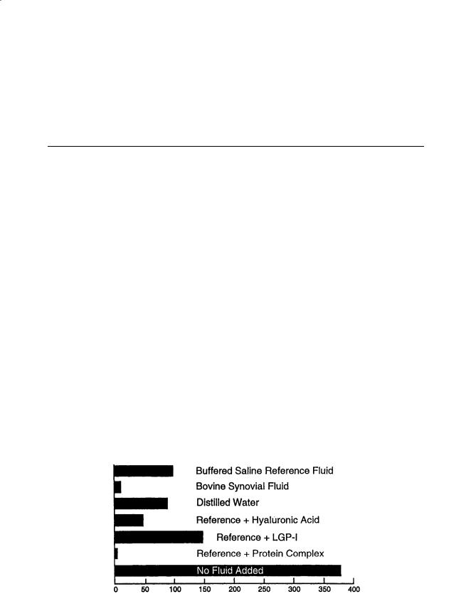
Over the past 15 years, studies aimed at exploring possible connections between tribology and mechanisms of synovial joint lubrication and degeneration (e.g., osteoarthritis) have been conducted by the author and his graduate and undergraduate students in the Department of Mechanical Engineering at Virginia Polytechnic Institute and State University. The basic approach used involved in vitro tribological experiments using bovine articular cartilage, with an emphasis on the effects of fluid composition and biochemistry on cartilage wear and damage. This research is an outgrowth of earlier work carried out during a sabbatical study in the Laboratory for the Study of Skeletal Disorders, The Children’s Hospital Medical Center, Harvard Medical School in Boston. In that study, bovine cartilage test specimens were loaded against a polished steel plate and subjected to reciprocating sliding for several hours in the presence of a fluid (e.g., bovine synovial fluid or a buffered saline reference fluid containing biochemical constituents kindly provided by Dr. David Swann). Cartilage wear was determined by sampling the test fluid and determining the concentration of 4-hydroxyproline—a constituent of collagen. The results of that earlier study have been reported and summarized elsewhere [37–40]. Figure 4.4 shows the average hydroxyproline contents of wear debris obtained from these in vitro experiments. These numbers are related to the cartilage wear which occurred. However, since the total quantities of collected fluids varied somewhat, the values shown in the bar graph should not be taken as exact or precise measures of fluid effects on cartilage wear.
The main conclusions of that study were as follows:
1.Normal bovine synovial fluid is very effective in reducing cartilage wear under these in vitro conditions as compared to the buffered saline reference fluid.
2.There is no significant difference in wear between the saline reference and distilled water.
3.The addition of hyaluronic acid to the reference fluid significantly reduces wear, but its effect depends on the source.
4.Under these tests conditions, Swann’s LGP-I (lubricating glycoprotein-I), known to be extremely effective in reducing friction in cartilage-on-glass tests, does not reduce cartilage wear.
FIGURE 4.4 Relative cartilage wear based on hydroxyproline content of debris (in vitro tests with cartilage on stainless steel).
84 |
Biomechanics: Principles and Applications |
FIGURE 4.5 Friction and wear are different phenomena.
5.However, a protein complex isolated by Swann is extremely effective in reducing wear—producing results similar to those obtained with synovial fluid. The detailed structure of this constituent is complex and has not yet been fully determined.
6.Last, the lack of an added fluid in these experiments leads to extremely high wear and damage of the articular cartilage.
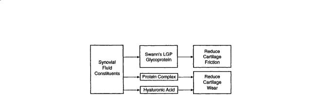
In discussing the possible significance of these findings from a tribological point of view, it may be helpful first of all to emphasize once again that friction and wear are different phenomena. Furthermore, as suggested by Fig. 4.5, certain constituents of synovial fluid (e.g., Swann’s lubricating glycoprotein) may act to reduce friction in synovial joints while other constituents (e.g., Swann’s protein complex or hyaluronic acid) may act to reduce cartilage wear. Therefore, it is necessary to distinguish between biochemical anti-friction and anti-wear compounds present in synovial fluid.
In more recent years, this study has been greatly enhanced by the participation of interested faculty and students from the Virginia-Maryland College of Veterinary Medicine and Department of Biochemistry and Animal Science at Virginia Tech. One major hypothesis tested is a continuation of previous work showing that the detailed biochemistry of the fluid-cartilage system has a pronounced and possibly controlling influence on cartilage wear. A consequence of the above hypothesis is that a lack or deficiency of certain biochemical constituents in the synovial joint may be one factor contributing to the initiation and progression of cartilage damage, wear, and possibly osteoarthritis. A related but somewhat different hypothesis concerns synovial fluid constituents which may act to increase the wear and further damage of articular cartilage under tribological contact.
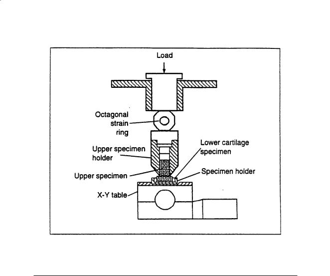
To carry out continued research on biotribology, a new device for studies of cartilage deformation, wear, damage, and friction under conditions of tribological contact was designed by Burkhardt [33] and later modified, constructed, and instrumented. A simplified sketch is shown in Fig. 4.6. The key features of this test device are shown in Table 4.2. The apparatus is designed to accommodate cartilage-on- cartilage specimens. Motion of the lower specimen is controlled by a computer-driven X–Y table, allowing simple oscillating motion or complex motion patterns. An octagonal strain ring with two full semiconductor bridges is used to measure the normal load as well as the tangential load (friction). An LVDT, not shown in the figure, is used to measure cartilage deformation and linear wear during a test. However, hydroxyproline analysis of the wear debris and washings is used for the actual determination of total cartilage wear on a mass basis.
In one study by Schroeder [41], two types of experiments were carried out, i.e., cartilage-on-stainless steel and cartilage-on-cartilage, at applied loads up to 70 N—yielding an average pressure of 2.2 MPa in the contact area. Reciprocating motion (40 cps) was used. The fluids tested included: (1) a buffered saline solution, (2) saline plus hyaluronic acid, and (3) bovine synovial fluid. In cartilage-on-stainless steel tests, scanning electron microscopy, and histological staining showed distinct effects of the lubricants on surface and subsurface damage. Tests with the buffered saline fluid resulted in the most damage, with large wear tracks visible on the surface of the cartilage plug, as well as subsurface voids and cracks. When hyaluronic acid, a constituent of the natural synovial joint lubricant, was added to the saline reference fluid, less severe damage was observed. Little or no cartilage damage was evident in tests in which the natural synovial joint fluid was used as the lubricant.
Joint Lubrication |
85 |
FIGURE 4.6 Device for in vitro cartilage-on-cartilage wear studies.
TABLE 4.2 Key Features of Test Device Designed for Cartilage Wear Studies [33]
Contact system |
Cartilage-on-cartilage |
Contact geometry |
Flat-on-flat, convex-on-flat, irregular-on-irregular |
Cartilage type |
Articular, any source (e.g., bovine) |
Specimen size |
Upper specimen, 4 to 6 mm diameter, lower specimen, ca. 15 to 25 mm diameter |
Applied load |
50–660 N |
Average pressure |
0.44–4.4 MPa |
Type of motion |
Linear, oscillating; circular, constant velocity; more complex patterns |
Sliding velocity |
0 to 20 mm/s |
Fluid temperature |
Ambient (20°C) or controlled humidity |
Environment |
Ambient or controlled humidity |
Measurements |
Normal load, cartilage deformation, friction; cartilage wear and damage, biochemical analysis of |
|
cartilage specimens, synovial fluid, and wear debris; sub-surface changes |
|
|
These results were confirmed in a later study by Owellen [42] in which hydroxyproline analysis was used to determine cartilage wear. It was found that increasing the applied load from 20 to 65 N increased cartilage wear by eightfold for the saline solution and approximately threefold for synovial fluid. Furthermore, the coefficient of friction increased from an initial low value of 0.01 to 0.02 to a much higher value, e.g., 0.20 to 0.30 and higher, during a normal test which lasted 3 hours; the greatest change occurred during the first 20 minutes. Another interesting result was that a thin film of transferred or altered material was observed on the stainless steel disks—being most pronounced with the buffered saline lubricant and not observed with synovial fluid. Examination of the film with Fourier transfer infrared microspectrometry shows distinctive bio-organic spectra which differ from that of the original bovine cartilage. We believe this to be an important finding since it suggests a possible biotribochemical effect [43].

86 |
Biomechanics: Principles and Applications |
FIGURE 4.7 Cartilage damage produced by sliding contact.
In another phase of this research, the emphasis is on the cartilage-on-cartilage system and the influence of potentially beneficial as well as harmful constituents of synovial fluid on wear and damage. In cartilage- on-cartilage tests, the most severe wear and damage occurred during tests with buffered saline as the lubricant. The damage was less severe than in the stainless steel tests, but some visible wear tracks were detectable with scanning electron microscopy. Histological sectioning and staining of both the upper and lower cartilage samples show evidence of elongated lacunae and coalesced voids that could lead to wear by delamination. An example is shown in Fig. 4.7 (original magnification of 500× on 35-mm slide). The proteoglycan content of the subsurface cartilage under the region of contact was also reduced. When synovial fluid was used as the lubricant, no visible wear or damage was detected [44]. These results demonstrate that even in in vitro tests with bovine articular cartilage, the nature of the fluid environment can have a dramatic affect on the severity of wear and subsurface damage.
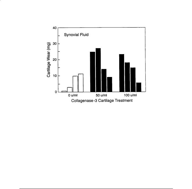
In a more recent study carried out by Berrien in the biotribology program at Virginia Tech, a different approach was taken to examine the role of joint lubrication in joint disease, particularly osteoarthritis. A degradative biological enzyme, collagenase-3, suspected of playing a role in a cartilage degeneration was used to create a physiologically adverse biochemical fluid environment. Tribological tests were performed with the same device and procedures described previously. The stainless steel disk was replaced with a 1-in. diameter plug of bovine cartilage to create a cartilage sliding on cartilage configuration more closely related to the in vivo condition. Normal load was increased to 78.6 N and synovial fluid and buffered saline were used as lubricants. Prior to testing, cartilage plugs were exposed to a fluid medium containing three concentrations of collagenase-3 for 24 h. The major discovery of this work was that exposure to the collagenase-3 enzyme had a substantial adverse effect on cartilage wear in vitro, increasing average wear values by three and one-half times those of the unexposed cases. Figure 4.8 shows an example of the effect of enzyme treatment when bovine synovial fluid was used as the lubricant. Scanning electron microscopy showed disruption of the superficial layer and collagen matrix with exposure to collagenase-3, where unexposed cartilage showed none. Histological sections showed a substantial loss of the superficial layer of cartilage and a distinct and abnormal loss of proteoglycans in the middle layer of collagenase-treated cartilage. Unexposed cartilage showed only minor disruption of the superficial layer [45].
This study indicates that some of the biochemical constituents that gain access to the joint space, during normal and pathological functions, can have a significant adverse effect on the wear and damage
Joint Lubrication |
87 |
FIGURE 4.8 Effect of collagenase-3 on cartilage wear.
of the articular cartilage. Future studies will include determination of additional constituents that have harmful effects on cartilage wear and damage. This research, using bovine articular cartilage in in vitro sliding contact tests, raises a number of interesting questions:
1.Has Nature designed a special biochemical compound that has as its function the protection of articular cartilage?
2.What is the mechanism (or mechanisms) by which biochemical constituents of synovial fluid can act to reduce wear of articular cartilage?
3.Could a lack of this biochemical constituent lead to increased cartilage wear and damage?
4.Does articular cartilage from osteoarthritic patients have reduced wear resistance?
5.Do any of the findings on the importance of synovial fluid biochemistry on cartilage wear in our in vitro studies apply to living or in vitro systems as well?
6.How does collagenase-3 treatment of cartilage lead to increased wear and does this finding have any significance in the in vivo situation? This question is addressed in the next section.
4.7Biotribology and Arthritis: Are There Connections?
Arthritis is an umbrella term for more than 100 rheumatic diseases affecting joints and connective tissue. The two most common forms are osteoarthritis (OA) and rheumatoid arthritis (RA). Osteoarthritis— also referred to as osteoarthrosis or degenerative joint disease—is the most common form of arthritis. It is sometimes simplistically described as the “wear and tear” form of arthritis. The causes and progression of degenerative joint disease are still not understood. Rheumatoid arthritis is a chronic and often progressive disease of the synovial membrane leading to release of enzymes which attack, erode, and destroy articular cartilage. It is an inflammatory response involving the immune system and is more prevalent in females. Rheumatoid arthritis is extremely complex. Its causes are still unknown.
Sokoloff defines degenerative joint disease as “an extremely common, noninflammatory, progressive disorder of movable joints, particularly weight-bearing joints, characterized pathologically by deterioration of articular cartilage and by formation of new bone in the sub-chondral areas and at the margins of the joint” [46]. As mentioned, osteoarthritis or osteoarthrosis is sometimes referred to as the “wear and tear” form of arthritis, but wear itself is rarely a simple process even in well-defined systems.
It has been noted by the author that tribological terms occasionally appear in hypotheses which describe the etiology of osteoarthritis (e.g., “reduced wear resistance of cartilage” or “poor lubricity of

88 |
Biomechanics: Principles and Applications |
synovial fluid”). It has also been noted that there is a general absence of hypotheses connecting normal synovial joint lubrication (or lack thereof) and synovial joint degeneration. Perhaps it is natural (and unhelpful) for a tribologist to imagine such a connection and that, for example, cartilage wear under certain circumstances might be due to or influenced by a lack of proper “boundary lubrication” by the synovial fluid. In this regard, it may be of interest to quote Swanson [12] who said in 1979 that “there exists at present no experimental evidence which certainly shows that a failure of lubrication is or is not a causative factor in the first stages of cartilage degeneration.” A statement made by Professor Glimcher [52] may also be appropriate here. Glimcher fully recognized the fundamental difference between friction and wear as well as the difference between joint lubrication (one area of study) and joint degeneration (another area of study). Glimcher said that wearing or abrading cartilage with a steel file is not osteoarthritis, and neither is digesting cartilage in a test tube with an enzyme. But both forms of cartilage deterioration can occur in a living joint and in a way which is still not understood. It is interesting that essentially none of the many synovial joint lubrication theories consider enzymatic degradation of cartilage as a factor whereas practically all the models of the etiology of degenerative joint disease include this as an important factor.
It was stated earlier that there are at least two main areas to consider, i.e., (1) mechanisms of synovial joint lubrication and (2) the etiology of synovial joint degeneration (e.g., as in osteoarthrosis). Both areas are extremely complex. And the key questions as to what actually happens in each have yet to be answered (and perhaps asked). It may therefore be presumptuous of the present author to suggest possible connections between two areas which in themselves are still not fully understood.
Tribological processes in a movable joint involve not only the contacting surfaces (articular cartilage), but the surrounding medium (synovial fluid) as well. Each of these depends on the synthesis and transport of necessary biochemical constituents to the contact region or interface. As a result of relative motion (sliding, rubbing, rolling, and impact) between the joint elements, friction and/or wear can occur.
It has already been shown and discussed—at least in in vitro tests with articular cartilage—that compounds which reduce friction do not necessarily reduce wear; the latter was suggested as being more important [10]. It may be helpful first of all to emphasize once again that friction and wear are different phenomena. Furthermore, certain constituents of synovial fluid (e.g., Swann’s lubricating glycoprotein) may act to reduce friction in synovial joints while other constituents (e.g., Swann’s protein complex or hyaluronic acid) may act to reduce cartilage wear.
A significant increase in joint friction could lead to a slight increase in local temperatures or possibly to reduce mobility. But the effects of cartilage wear would be expected to be more serious. When cartilage wear occurs, a very special material is lost and the body is not capable of regenerating cartilage of the same quality nor at the desired rate. Thus, there are at least two major tribological dimensions involved—one concerning the nature of the synovial fluid and the other having to do with the properties of articular cartilage itself. Changes in either the synovial fluid or cartilage could conceivably lead to increased wear or damage (or friction) as shown in Fig. 4.9.
A simplified model or illustration of possible connections between osteoarthritis and tribology is offered in Fig. 4.10 taken from Furey [53]. Its purpose is to stimulate discussion. There are other pathways to the disease, pathways which may include genetic factors.
FIGURE 4.9 Two tribological aspects of synovial joint lubrication.

Joint Lubrication |
89 |
FIGURE 4.10 Osteoarthritis–tribology connections?
In some cases, the body makes an unsuccessful attempt at repair, and bone growth may occur at the periphery of contact. As suggested by Fig. 4.10, this process and the generation of wear particles could lead to joint inflammation and the release of enzymes which further soften and degrade the articular cartilage. This softer, degraded cartilage does not possess the wear resistance of the original. It has been shown previously that treatment of cartilage with collagenase-3 increases wear significantly, thus supporting the idea of enzyme release as a factor in osteoarthritis. Thus, there exists a feedback process in which the occurrence of cartilage wear can lead to even more damage. Degradative enzymes can also be released by trauma, shock, or injury to the joint. Ultimately, as the cartilage is progressively thinned and bony growth occurs, a condition of osteoarthritis or degenerative joint disease may exist. There are other pathways to the disease, pathways which may include genetic factors. It is not argued that arthritis is a tribological problem. However, the inclusion of tribological processes in one set of pathways to osteoarthrosis would not seem strange or unusual.
A specific example of a different tribological dimension to the problem of synovial joint lubrication (i.e., third-body abrasion), was shown by the work of Hayes et al. [54]. In an excellent study of the effect of crystals on the wear of articular cartilage, they carried out in vitro tests using cylindrical cartilage subchondral bone plugs obtained from equine fetlock joints in sliding contact against a stainless steel plate. They examined the effects of three types of crystals (orthorhombic calcium pyrophosphate tetrahydrate, monoclinic calcium pyrophosphate dehydrate, and calcium hydroxyapatite) on wear using a Ringer’s solution as the carrier fluid. Concentration of cartilage wear debris in the fluid was determined by analyzing for inorganic sulphate derived from the proteoglycans present. Several interesting findings were made, one of them being that the presence of the crystals roughly doubled cartilage wear. This is an important contribution which should be read by anyone seriously contemplating research on the tribology of articular cartilage. The careful attention to detail and potential problems, as well as the precise description of the biochemical procedures and diverse experimental techniques used, set a high standard.
4.8 Recapitulation and Final Comments
It is obvious from the unusually large number of theories of synovial joint lubrication proposed, that very little is known about the subject. Synovial joints are undoubtedly the most sophisticated and complex tribological systems that exist or will ever exist. It will require a great deal more research—possibly very different approaches—before we even begin to understand the processes involved.

90 |
Biomechanics: Principles and Applications |
Some general comments and specific suggestions are offered—not for the purpose of criticizing any particular study but hopefully to provide ideas which may be helpful in further research as well as in the re-interpretation of some past research.
Terms and Definitions
First of all, as mentioned earlier in this chapter, part of the problem has to do with the use and misuse of various terms in tribology—the study of friction, wear, and lubrication. A glance at any number of the published papers on synovial joint lubrication will reveal such terms and phrases as “lubricating ability,” “lubricity,” “lubricating properties,” “lubricating component,” and many others, all undefined. We also see terms like “boundary lubricant,” “lubricating glycoprotein,” or “lubricin.” There is nothing inherently wrong with this but one should remember that lubrication is a process of reducing friction and/or wear between rubbing surfaces. Saying that a fluid is a “good” lubricant does not distinguish between friction and wear. And assuming that friction and wear are correlated and go together is the first pitfall in any tribological study. It cannot be overemphasized that friction and wear are different, though sometimes related, phenomena. Low friction does not mean low wear. The terms and phrases used are therefore extremely important. For example, in a brief and early review article by Wright and Dowson [55], it was stated that “Digestion of hyaluronate does not alter the boundary lubrication,” referring to the work of Radin, Swann, and Weisser [56]. In another article, McCutchen re-states this conclusion in another way, saying “… the lubricating ability did not reside in the hyaluronic acid” and later asks the question “Why do the glycoprotein molecules (of Swann) lubricate?” [57]. These statements are based on effects of various constituents of friction, not wear. The work of the present author showed that in tests with bovine articular cartilage, Swann’s lubricating glycoprotein (LGP-I) which was effective in reducing friction did not reduce cartilage wear. However, hyaluronic acid—shown earlier not to be responsible for friction reduction—did reduce cartilage wear. Thus, it is important to make the distinction between friction reduction and wear reduction. It is suggested that operational definitions be used in place of vague “lubricating ability,” etc. terms in future papers on the subject.
Experimental Contact Systems
Second, some comments are made on the experimental approaches that have been reported in the literature on synovial joint lubrication mechanisms. Sliding contact combinations in in vitro studies have consisted of (1) cartilage-on-cartilage, (2) cartilage-on-some other surface (e.g., stainless steel, glass), and (3) solids other than cartilage sliding against each other in X-on-X or X-on-Y combinations.
The cartilage-on-cartilage combination is of course the most realistic and yet most complex contact system. But variations in shape or macroscopic geometry, microtopography, and the nature of contact present problems in carrying out well-controlled experiments. There is also the added problem of acquiring suitable specimens which are large enough and reasonably uniform.
The next combination—cartilage-on-another material—allows for better control of contact, with the more elastic, deformable cartilage loaded against a well-defined hard surface (e.g., a polished, flat solid made of glass or stainless steel). This contact configuration can provide useful tribological information on effects of changes in biochemical environment (e.g., fluids), on friction, wear, and sub-surface damage. It also could parallel the situation in a partial joint replacement in which healthy cartilage is in contact with a metal alloy.
The third combination, which appears in some of the literature on synovial joint lubrication, does not involve any articular cartilage at all. For example, Jay made friction measurements using a latexcovered stainless steel stud in oscillating contact against polished glass [31]. Williams et al., in a study of a lipid component of synovial fluid, used reciprocating contact of borosilicate glass-on-glass [58]. And in a recent paper on the action of a surface-active phospholipid as the “lubricating component of lubricin,” Schwarz and Hills carried out friction measurements using two optically flat quartz plates in sliding contact [59]. In another study, a standard four-ball machine using alloy steel balls was used to examine

Joint Lubrication |
91 |
the “lubricating ability” of synovial fluid constituents. Such tests, in the absence of cartilage, are easiest to control and carry out. However, they are not relevant to the study of synovial joint lubrication. With a glass sphere sliding against a glass flat, almost anything will reduce friction—including a wide variety of chemicals, biochemicals, semi-solids, and fluids. This has little if anything to do with the lubrication of synovial joints.
Fluids and Materials Used as Lubricants
in in Vitro Biotribology Studies
Fluids used as lubricants in synovial joint lubrication studies have consisted of (1) “normal” synovial fluid (e.g., bovine), (2) buffered saline solution containing synovial fluid constituents (e.g., hyaluronic acid), and (3) various aqueous solutions of surface active compounds neither derived from nor present in synovial fluid. In addition, a few studies used synovial fluids from patients suffering from either osteoarthritis or rheumatoid arthritis.
The general comment made here is that the use of synovial fluids—whether derived from human or animal sources and whether “healthy” or “abnormal”—is important in in vitro studies of synovial joint lubrication. The documented behavior of synovial fluid in producing low friction and wear with articular cartilage sets a reference standard and demonstrates that useful information can indeed come from in vitro tests.
Studies that are based on adding synovial fluid constituents to a reference fluid (e.g., a buffered saline solution) can also be useful in attempting to identify which biochemical compound or compounds are responsible for reductions in frictions or wear. But if significant interactions between compounds exist, then such an approach may require an extensive program of tests. It should also be mentioned that in the view of the present author, the use of a pure undissolved constituent of synovial fluid, either derived or synthetic, in a sliding contact test is not only irrelevant but may be misleading. An example would be the use of a pure lipid (e.g., phospholipid) at the interface rather than in the concentration and solution form in which this compound would normally exist in synovial fluid. This is basic in any study of lubrication and particularly in the case of boundary lubrication where major effects on wear or friction can be brought on by minor, seemingly trivial, changes in chemistry.
The Preoccupation with Rheology and Friction
The synovial joint as a system—the articular cartilage and underlying bone structure as well as the synovial fluid as important elements—is extremely complex and far from being understood. It is noted that there is a proliferation of mathematical modeling papers stressing rheology and the mechanics of deformation, flow, and fluid pressures developed in the cartilage model. One recent example is the paper “The Role of Interstitial Fluid Pressurization and Surface Properties on the Boundary Friction of Articular Cartilage” by Ateshian et al. [21]. This study, a genuine contribution, grew out of the early work by Mow and connects also with the “weeping lubrication” model of McCutchen. Both McCutchen and Mow have made significant contributions to our understanding of synovial joint lubrication, although each approach is predominantly rheological and friction oriented with little regard for biochemistry and wear. This is not to say that rheology is unimportant. It could well be that, as suggested by Ateshian, the mechanism of interstitial fluid pressurization that leads to low friction in cartilage could also lead to low wear rates [60].
The Probable Existence of Various Lubrication Regimes
In an article by Wright and Dowson, it is suggested that a variety of types of lubrication operate in human synovial joints at different parts of a walking cycle: “At heel-strike a squeeze-film situation may develop, leading to elastohydrodynamic lubrication and possibly both squeeze-film and boundary lubrication, while hydrodynamic lubrication may operate during the free-swing phase of walking” [55].