

Biomechanics Principles and Applications - Schneck and Bronzino
.pdf262 |
Biomechanics: Principles and Applications |
in the area. It is evident that the normal function of the cochlea requires a full integration of mechanical, electrical, and chemical effects on the milli-, micro-, and nanometer scales. Recent texts, which include details of the anatomy, are by Pickles [1988] and Gulick and coworkers [1989]. A summary of analysis and data related to the macromechanical aspect up to 1982 is given by Steele [1987], and more recent surveys specifically on the cochlea are by de Boer [1991], Dallos [1992], Hudspeth [1989], Ruggero [1993], and Nobili and colleagues [1998].
18.2 Anatomy
The cochlea is a coiled tube in the shape of a snail shell (cochlea = schnecke = snail), with length about 35 mm and radius about 1 mm in humans. There is not a large size difference across species: the length is 60 mm in elephants and 7 mm in mice. There are two and one-half turns of the coil in humans and dolphins, and five turns in guinea pigs. Despite the correlation of coiling with hearing capability of land animals [West, 1985], no significant effect of the coiling on the mechanical response has yet been identified.
Components
The cochlea is filled with fluid and divided along its length by two partitions. The main partition is at the center of the cross-section and consists of three segments: (1) on one side—the bony shelf (or primary spiral osseous lamina), (2) in the middle, an elastic segment (basilar membrane) (shown in Fig. 18.1), and
(3) on the other side, a thick support (spiral ligament). The second partition is Reissner’s membrane, attached at one side above the edge of the bony shelf and attached at the other side to the wall of the cochlea. Scala media is the region between Reissner’s membrane and the basilar membrane, and is filled with endolymphatic fluid. This fluid has an ionic content similar to intracellular fluid, high in potassium and low in sodium, but with a resting positive electrical potential of around +80 mV. The electrical potential is supplied by the stria vascularis on the wall in scala media. The region above Reissner’s membrane is scala vestibuli, and the region below the main partition is scala tympani. Scala vestibuli and scala tympani are connected at the apical end of the cochlea by an opening in the bony shelf, the helicotrema, and are filled with perilymphatic fluid. This fluid is similar to extracellular fluid, low in potassium and high in sodium with zero electrical potential. Distributed along the scala media side of
FIGURE 18.1 Finite element calculation for the deformation of the cochlear partition due to pressure on the basilar membrane (BM). Outer hair cell (OHC) stereocilia are sheared by the motion of the pillars of Corti and reticular lamina relative to the tectorial membrane (TM). The basilar membrane is supported on the left by the bony shelf and on the right by the spiral ligament. The inner hair cells (IHC) are the primary receptors, each with about 20 afferent synapses. The inner sulcus (IC) is a fluid region in contact with the cilia of the inner hair cells.

Cochlear Mechanics |
263 |
the basilar membrane is the sensory epithelium, the organ of Corti. This contains one row of inner hair cells and three rows of outer hair cells. In humans, each row contains about 4000 cells. Each of the inner hair cells has about twenty afferent synapses; these are considered to be the primary receptors. In comparison, the outer hair cells are sparsely innervated but have both afferent (5%) and efferent (95%) synapses.
The basilar membrane is divided into two sections. Connected to the edge of the bony shelf, on the left in Fig. 18.1, is the arcuate zone, consisting of a single layer of transverse fibers. Connected to the edge of the spiral ligament, on the right in Fig. 18.1, is the pectinate zone, consisting of a double layer of transverse fibers in an amorphous ground substance. The arches of Corti form a truss over the arcuate zone, which consist of two rows of pillar cells. The foot of the inner pillar is attached at the point of connection of the bony shelf to the arcuate zone, while the foot of the outer pillar cell is attached at the common border of the arcuate zone and pectinate zone. The heads of the inner and outer pillars are connected and form the support point for the recticular lamina. The other edge of the recticular lamina is attached to the top of Henson cells, which have bases connected to the basilar membrane. The inner hair cells are attached on the bony shelf side of the inner pillars, while the three rows of outer hair cells are attached to the recticular lamina. The region bounded by the inner pillar cells, the recticular lamina, the Henson cells, and the basilar membrane forms another fluid region. This fluid is considered to be perilymph, since it appears that ions can flow freely through the arcuate zone of the basilar membrane. The cilia of the hair cells protrude into the endolymph. Thus the outer hair cells are immersed in perilymph at 0 mV, have an intracellular potential of –70 mV, and have cilia at the upper surface immersed in endolymph at a potential of +80 mV. In some regions of the ears of some vertebrates [Freeman and Weiss, 1990], the cilia are free standing. However, mammals always have a tectorial membrane, originating near the edge of the bony shelf and overlying the rows of hair cells parallel to the recticular lamina. The tallest rows of cilia of the outer hair cells are attached to the tectorial membrane. Under the tectorial membrane and inside the inner hair cells is a fluid space, the inner sulcus, filled with endolymph. The cilia of the inner hair cells are not attached to the overlying tectorial membrane, so the motion of the fluid in the inner sulcus must provide the mechanical input to these primary receptor cells. Since the inner sulcus is found only in mammals, the fluid motion in this region generated by acoustic input may be crucial to high frequency discrimination capability.
With a few exceptions of specialization, the dimensions of all the components in the cross section of the mammalian cochlea change smoothly and slowly along the length, in a manner consistent with high stiffness at the base, or input end, and low stiffness at the apical end. For example, in the cat the basilar membrane width increases from 0.1 to 0.4 mm while the thickness decreases from 13 to 5 µm. The density of transverse fibers decreases more than the thickness, from about 6000 fibers per µm at the base to 500 per µm at the apex [Cabezudo 1978].
Material Properties
Both perilymph and endolymph have the viscosity and density of water. The bone of the wall and the bony shelf appear to be similar to compact bone, with density approximately twice that of water. The remaining components of the cochlea are soft tissue with density near that of water. The stiffnesses of the components vary over a wide range, as indicated by the values of Young’s modulus listed in Table 18.1. These values are taken directly or estimated from many sources, including the stiffness measurements in the cochlea by Békésy [1960], Gummer and coworkers [1981], Strelioff and Flock [1984], Miller [1985], Zwislocki and Cefaratti [1989], and Olson and Mountain [1994].
18.3 Passive Models
The anatomy of the cochlea is complex. By modeling, one attempts to isolate and understand the essential features. Following is an indication of propositions and the controversy associated with a few such models.
264 |
|
Biomechanics: Principles and Applications |
|||
|
TABLE 18.1 Typical Values and Estimates for Young’s Modulus E |
||||
|
|
|
|
|
|
|
Compact bone |
|
20 |
GPa |
|
|
Keratin |
|
3 |
GPa |
|
|
Basilar membrane fibers |
|
1.9 |
GPa |
|
|
Microtubules |
|
1.2 |
GPa |
|
|
Collagen |
|
1 |
GPa |
|
|
Reissner’s membrane |
|
60 |
MPa |
|
|
Actin |
|
50 |
MPa |
|
|
Red blood cell, extended (assuming thickness = 10 nm) |
45 |
MPa |
||
|
Rubber, elastin |
|
4 |
MPa |
|
|
Basilar membrane ground substance |
|
200 |
kPa |
|
|
Tectorial membrane |
|
30 |
kPa |
|
|
Jell-O |
|
3 |
kPa |
|
|
Henson’s cells |
|
1 |
kPa |
|
|
|
|
|
|
|
Resonators
The ancient Greeks suggested that the ear consisted of a set of tuned resonant cavities. As each component in the cochlea was discovered subsequently, it was proposed to be the tuned resonator. The most well known resonance theory is Helmholtz’s. According to this theory, the transverse fibers of the basilar membrane are under tension and respond like the strings of a piano. The short strings at the base respond to high frequencies and the long strings toward the apex respond to low frequencies. The important feature of the Helmholz theory is the place principle, according to which the receptor cells at a certain place along the cochlea are stimulated by a certain frequency. Thus, the cochlea provides a real-time frequency separation (Fourier analysis) of any complex sound input. This aspect of the Helmholtz theory has since been validated, as each of the some 30,000 fibers exiting the cochlea in the auditory nerve is sharply tuned to a particular frequency. A basic difficulty with such a resonance theory is that sharp tuning requires small damping, which is associated with a long ringing after the excitation ceases. Yet, the cochlea is remarkable for combining sharp tuning with short time delay for the onset of reception and the same short time delay for the cessation of reception.
A particular problem with the Helmholtz theory arises from the equation for the resonant frequency for a string under tension:
f = |
1 |
|
T |
(18.1) |
2b |
|
ρh |
||
|
|
|
in which T is the tensile force per unit width, ρ is the density, b is the length, and h is the thickness of the string. In humans, the frequency range over which the cochlea operates is f = 200 to 20,000 Hz, a factor of 100, while the change in length b is only a factor of 5 and the thickness of the basilar membrane h varies the wrong way by a factor of 2 or so. Thus, to produce the necessary range of frequency, the tension T would have to vary by a factor of about 800. In fact, the spiral ligament, which would supply such tension, varies in area by a factor of only 10.
Traveling Waves
No theory anticipated the actual behavior found in the cochlea in 1928 by Békésy [1960]. He observed traveling waves moving along the cochlea from base toward apex which have a maximum amplitude at a certain place. The place depends on the frequency, as in the Helmholz theory, but the amplitude envelope is not very localized. In Békésy’s experimental models, and in subsequent mathematical and experimental models, the anatomy of the cochlea is greatly simplified. The coiling, Reissner’s membrane, and the organ of Corti are all ignored, so the cochlea is treated as a straight tube with a single partition.

Cochlear Mechanics |
265 |
(An exception is in Fuhrmann and colleagues [1986]). A gradient in the partition stiffness similar to that in the cochlea, gives beautiful traveling waves in both experimental and mathematical models.
One-Dimensional Model
A majority of work has been based on the assumption that the fluid motion is one dimensional. With this simplification the governing equations are similar to those for an electrical transmission line and for the long wavelength response of an elastic tube containing fluid. The equation for the pressure p in a tube with constant cross-sectional area A and with constant frequency of excitation is:
d2 p |
+ |
2ρω2 |
p = 0 |
(18.2) |
|
|
|||
d x2 AK |
|
|
||
in which x is the distance along the tube, ρ is the density of the fluid, ω is the frequency in radians per second, and K is the generalized partition stiffness, equal to the net pressure divided by the displaced area of the cross-section. The factor of 2 accounts for fluid on both sides of the elastic partition. Often K is represented in the form of a single degree-of-freedom oscillator:
K = k + iωd − mω2 |
(18.3) |
in which k is the static stiffness, d is the damping, and m is the mass density:
m = ρP |
h |
(18.4) |
|
b |
|||
|
|
in which ρP is the density of the plate, h is the thickness, and b is the width. Often the mass is increased substantially to provide better curve fits. A good approximation is to treat the pectinate zone of the basilar membrane as transverse beams with simply supported edges, for which
k = |
10Eh |
3c |
f |
(18.5) |
|
|
|||
b 5 |
|
|
||
|
|
|
|
in which E is the Young’s modulus, and cf is the volume fraction of fibers. Thus, for the moderate changes in the geometry along the cochlea as in the cat, h decreases by a factor of 2, cf decreases by a factor of 12, b increases by a factor of 5, and the stiffness k from Eq. (18.4) decreases by five orders of magnitude, which is ample for the required frequency range. Thus, it is the bending stiffness of the basilar membrane pectinate zone and not the tension which governs the frequency response of the cochlea. The solution of Eq. (18.2) can be obtained by numerical or asymptotic (called WKB or CLG) methods. The result is traveling waves for which the amplitude of the basilar membrane displacement builds to a maximum and then rapidly diminishes. The parameters of K are adjusted to obtain agreement with measurements of the dynamic response in the cochlea. Often all the material of the organ of Corti is assumed to be rigidly attached to the basilar membrane so that h is relatively large and the effect of mass m is large. Then the maximum response is near the in vacua resonance of the partition given by:
ω2 = |
b |
|
k |
(18.6) |
|
h ρ |
|||||
|
|
||||
The following are objections to the one-dimensional model [e.g., Siebert, 1974]: (1) The solutions of Eq. (18.2) show wavelengths of response in the region of maximum amplitude that are small in

266 |
Biomechanics: Principles and Applications |
comparison with the size of the cross section, violating the basic assumption of one-dimensional fluid flow. (2) In the drained cochlea, Békésy [1960] observed no resonance of the partition, so there is no significant partition mass. The significant mass is entirely from the fluid and therefore Eq. (18.6) is not correct. This is consistent with the observations of experimental models. (3) In model studies by Békésy [1960] and others, the localization of response is independent of the area A of the cross-section. Thus Eq. (18.2) cannot govern the most interesting part of the response, the region near the maximum amplitude for a given frequency. (4) Mechanical and neural measurements in the cochlea show dispersion which is incompatible with the one-dimensional model [Lighthill, 1991]. (5) The one-dimensional model fails badly in comparison with experimental measurements in models for which the parameters of geometry, stiffness, viscosity, and density are known.
Nevertheless, the simplicity of Eq. (18.2) and the analogy with the transmission line have made the one-dimensional model popular. We note that there is interest in utilizing the principles in an analog model built on a silicon chip, because of the high performance of the actual cochlea. Watts [1993] reports on the first model with an electrical analog of two-dimensional fluid in the scali. An interesting observation is that the transmission line hardware models are sensitive to failure of one component, while the two-dimensional model is not. In experimental models, Békésy found that a hole at one point in the membrane had little effect on the response at other points.
Two-Dimensional Model
The pioneering work with two-dimensional (2-D) fluid motion was begun in 1931 by Ranke, as reported in Ranke [1950] and discussed by Siebert [1974]. Analysis of 2-D and three-dimensional (3-D) fluid motion without the a priori assumption of long or short wavelengths and for physical values of all parameters is discussed by Steele [1987]. The first of two major benefits derived from the 2-D model is the allowance of short wavelength behavior, i.e, the variation in fluid displacement and pressure in the duct height direction. Localized fluid motion near the elastic partition generally occurs near the point of maximum amplitude and the exact value of A becomes immaterial. The second major benefit of a 2-D model is the admission of a stiffness-dominated elastic partition (i.e., massless) which better approximates the physiological properties of the basilar membrane. The two benefits together address all the objections the one-dimensional model discussed previously.
Two-dimensional models start with the Navier–Stokes and continuity equations governing the fluid motion, and an anisotropic plate equation governing the elastic partition motion. The displacement potential ϕ for the incompressible and inviscid fluid must satisfy Laplace’s equation:
ϕ, xx + ϕ,zz = 0 |
(18.7) |
where x is the distance along the partition and z the distance perpendicular to the partition, and the subscripts with commas denote partial derivatives. The averaged potential and the displacement of the partition are:
|
|
H |
|
ϕ = |
1 |
∫ϕdz w = ϕz (x, 0) |
(18.8) |
H |
|||
0 |
|
||
so Eq. (18.7) yields the “macro” continuity condition (for constant H):
ϕ, xx − w = 0 . |
(18.9) |
An approximate solution is
˜ |
[ |
] |
iωt |
(18.10) |
ϕ(x, z, t )= F (x)cosh n(x)(z − H ) e |
|
|||

Cochlear Mechanics |
267 |
where F is an unknown amplitude function, n is the local wave number, H is the height of the duct, t is time, and ω is the frequency. This is an exact solution for constant properties and is a good approximation when the properties vary slowly along the partition. The conditions at the plate fluid interface yield the dispersion relation:
n tanh(nH )= |
2ρω2 |
H |
(18.11) |
AK |
The averaged value Eq. (18.8) of the approximate potential Eq. (18.10) is:
ϕ˜ = F sinhnH
(18.12)
nH
so the continuity condition Eq. (18.9) yields the equation:
˜ |
+ n |
2 ˜ |
(18.13) |
ϕ, xx |
ϕ = 0 |
For small values of the wave number n, the system Eq. (18.11) and Eq. (18.13) reduce to the onedimensional problem Eq. (18.2).
For physiological values of the parameters, the wave number for a given frequency is small at the stapes and becomes large (i.e., short wave lengths) toward the end of the duct. With this formulation, the form of the wave is not assumed. It is clear that for large n the WKB solution will give excellent approximation to the solution of Eq. (18.13) in exponential form. So it is possible to integrate Eq. (18.13) numerically for small n for which the solution is not exponential and match the solution with the WKB approximation. This provides a uniformly valid solution for the entire region of interest without an a priori assumption of the wave form.
For a physically realistic model, the mass of the membrane can be neglected and K written as:
K = k(1+ iε) |
(18.14) |
in which k is the static stiffness. For many polymers, the material damping ε is nearly constant. If the damping comes from the viscous boundary layer of the fluid, then ε is approximated by
ε ≈ n |
µ |
(18.15) |
|
2ρω |
|||
|
|
in which µ is the viscosity. For water, ε is small with a value near 0.05 at the point of maximum amplitude. The actual duct is tapered, so H = H(x) and additional terms must be added to Eq. (18.13).
The best verification of the mathematical model and calculation procedure comes from comparison with measurements in experimental models for which the parameters are known. Zhou and coworkers [1994] provide the first life-sized experimental model, designed to be similar to the human cochlea, but with fluid viscosity 28 times that of water to facilitate optical imaging. Results are shown in Figs. 18.2 and 18.3. Eq. (18.13) gives rough agreement with the measurements.
Three-Dimensional Model
A further improvement in the agreement with experimental models can be obtained by adding the component of fluid motion in the direction across the membrane for a full 3-D model. The solution by direct numerical means is computationally intensive, and was first carried out by Raftenberg [1990],

268 |
Biomechanics: Principles and Applications |
FIGURE 18.2 Comparison of 3-D model calculations (solid curves) with experimental results of Zhou and coworkers [1994] (dashed curves) for the amplitude envelopes for different frequencies. This is a life-sized model, but with an isotropic BM and fluid viscosity 28 times that of water. The agreement is reasonable, except for the lower frequencies.
who reports a portion of his results for the fluid motion around the organ of Corti. Böhnke and colleagues [1996] use the finite element code ANSYS for the most accurate description to date of the structure of the organ of Corti. However, the fluid is not included and only a restricted segment of the cochlea considered. The fluid is also not included in the finite element calculations of Zhang and colleagues [1996]. A “large finite element method”, which combines asymptotic and numerical methods for shell analysis, can be used for an efficient computation of all the structural detail as shown in Fig. 18.1. Both the fluid and the details of the structure are considered with a simplified element description and simplified geometry by Kolston and Ashmore [1996], requiring some 105 degrees of freedom and hours of computing time (on a 66-MHz PC) for the linear solution for a single frequency. The asymptotic WKB solution, however, provides the basis for computing the 3-D fluid motion [Steele, 1987] and yields excellent agreement with older measurements of the basilar membrane motion in the real cochlea (with a computing time of 1 second per frequency). The 2-D analysis Eq. (18.13) can easily be extended to 3-D. The 3-D WKB calculations are compared with the measurements of the basilar membrane displacement in the experimental model of Zhou and coworkers [1994] in Figs. 18.2 and 18.3. As shown by Taber and Steele [1979], the 3-D fluid motion has a significant effect on the pressure distribution. This is confirmed by the measurements by Olson [1998] for the pressure at different depths in the cochlea, that show a substantial increase near the partition.
18.4 The Active Process
Before around 1980, it was thought that the processing may have two levels. First the basilar membrane and fluid provide the correct place for a given frequency (a purely mechanical “first filter”). Subsequently, the micromechanics and electrochemistry in the organ of Corti, with possible neural interactions, perform a further sharpening (a physiologically vulnerable “second filter”).
A hint that the two-filter concept had difficulties was in the measurements of Rhode [1971], who found significant nonlinear behavior of the basilar membrane in the region of the maximum amplitude at moderate amplitudes of tone intensity. Passive models cannot explain this, since the usual mechanical nonlinearities are significant only at very high intensities, i.e., at the threshold of pain. Russell and Sellick [1977] made the first in vivo mammalian intracellular hair cell recordings and found that the cells are as sharply tuned as the nerve fibers. Subsequently, improved measurement techniques in several laboratories revealed that the basilar membrane is actually as sharply tuned as the hair cells and the nerve fibers. Thus, the sharp tuning occurs at the basilar membrane. No passive cochlear model, even with
Cochlear Mechanics |
269 |
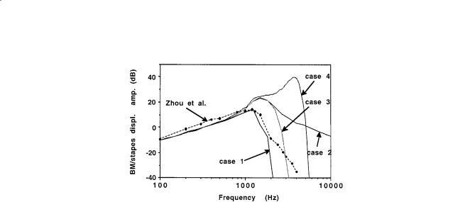
FIGURE 18.3 Comparison of 3-D model calculations with experimental results of Zhou and coworkers [1994] for amplitude at the place x = 19 mm as a function of frequency. The scales are logarithmic (20 dB is a factor of 10 in amplitude). Case 1 shows a direct comparison with the physical parameters of the experiment, with isotropic BM and viscosity 28 times that of water. Case 2 is computed for the viscosity reduced to that of water. Case 3 is computed for the BM made of transverse fibers. Case 4 shows the effect of active OHC feed-forward, with the pressure gain α = 0.21 and feed-forward distance ∆x = 25 microns. Thus, lower viscosity, BM orthotropy, and active feed-forward all contribute to higher amplitude and increased localization of the response.
physically unreasonable parameters, has yielded amplitude and phase response similar to such measurements. Measurements in a damaged or dead cochlea show a response similar to that of a passive model. Further evidence for an active process comes from Kemp [1978], who discovered that sound pulses into the ear caused echoes coming from the cochlea at delay times corresponding to the travel time to the place for the frequency and back. Spontaneous emission of sound energy from the cochlea has now been measured in the external ear canal in all vertebrates [Probst, 1990]. Some of the emissions can be related to the hearing disability of tinnitus (ringing in the ear). The conclusion drawn from these discoveries is that normal hearing involves an active process in which the energy of the input sound is greatly enhanced. A widely accepted concept is that spontaneous emission of sound energy occurs when the local amplifiers are not functioning properly and enter some sort of limit cycle (Zweig and Shera, 1995). However, there remains doubt about the nature of this process (Allen and Neely [1992], Hudspeth [1989], Nobili and coworkers [1998]).
Outer Hair Cell Electromotility
Since the outer hair cells have sparse afferent innervation, they have long been suspected of serving a basic motor function, perhaps beating and driving the subtectorial membrane fluid. Nevertheless, it was surprising when Brownell and colleagues [1985] found that the outer hair cells have electromotility: the cell expands and contracts in an oscillating electric field, either extraor intracellular. The electromotility exists at frequencies far higher than possible for normal contractile mechanisms [Ashmore, 1987]. The sensitivity is about 20 nm/mV (about 105 better than PZT-2, a widely used piezoelectric ceramic). It has not been determined if the electromotility can operate to the 200 kHz used by high frequency mammals. However, a calculation of the cell as a pressure vessel with a fixed charge in the wall [Jen and Steele, 1987] indicates that, despite the small diameter (10 µm), the viscosity of the intraand extracellular fluid is not a limitation to the frequency response. In a continuation of the work reported by Hemmert and coworkers [1996], the force generation is found to continue to 80 kHz in the constrained cell. In contradiction, however, the same laboratory (Preyer and coworkers [1996]) finds that the intracellular voltage change due to displacement of the cilia drops off at a low frequency.
The motility appears to be due to a passive piezoelectric behavior of the cell plasma membrane [Kalinec and colleagues, 1992]. Iwasa and Chadwick [1992] measured the deformation of a cell under pressure
270 |
Biomechanics: Principles and Applications |
loading and voltage clamping and computed the elastic properties of the wall, assuming isotropy. It appears that for agreement with both the pressure and axial stiffness measurements, the cell wall must be orthotropic, similar to a filament reinforced pressure vessel with close to the optimum filament angle of 38˚ (Tolomeo and Steele, 1995). Holley [1990] finds circumferential filaments of the cytoskeleton with an average nonzero angle of about 26°. Mechanical measurements of the cell wall by Tolomeo and coworkers [1996] directly confirm the orthotropic stiffness.
Hair Cell Gating Channels
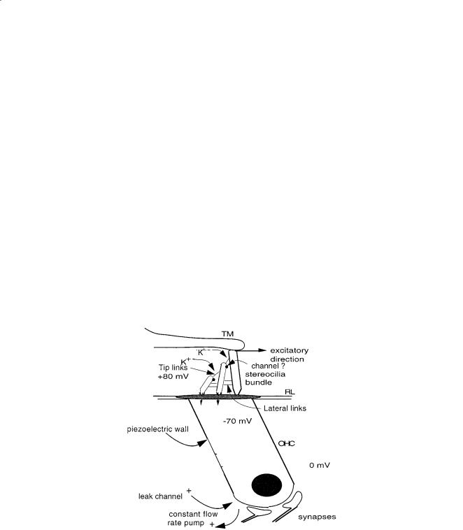
In 1984, Pickles and colleagues discovered tip links connecting the cilia of the hair cell, as shown in Fig. 18.4, that are necessary for the normal function of the cochlea. These links are about 6 nm in diameter and 200 nm long [Pickles, 1988]. Subsequent work by Hudspeth [1989] and Assad and Corey [1992] shows convincingly that there is a resting tension in the links. A displacement of the ciliary bundle in the excitatory direction causes an opening of ion channels in the cilia, which in turn decreases the intracellular potential. This depolarization causes neural excitation and, in the piezoelectric outer hair cells, a decrease of the cell length.
A purely mechanical analog model of the gating is in Steele [1992], in which the ion flow is replaced by viscous fluid flow and the intracellular pressure is analogous to the voltage. A constant flow-rate pump and leak channel at the base of the cell establish the steady-state condition of negative intracellular pressure, tension in the tip links, and a partially opened gate at the cilia through which there is an average magnitude of flow. The pressure drop of the flow through the gate has a nonlinear negative spring effect on the system. If the cilia are given a static displacement, the stiffness for small perturbation displacement is dependent on the amplitude of the initial displacement, as observed by Hudspeth [1989]. For oscillatory
FIGURE 18.4 Model of outer hair cell. The normal pumping of ions produces negative intracellular electrical potential. Displacement of the cilia in the excitatory direction causes an opening of the ion channels in the cilia. There is evidence that the channels are located at the point of closest proximity of the two cilia. The tip and lateral links are important to maintain the correct stiffness and position. The opening of the channels decreases the intracellular potential, causing a piezoelectric contraction of the cell and excitation of the neural synapses. This can be modeled by a constant flow rate pump, leak channel, and spring controlled gate. The mechanical effect of the flow on the gate is important. The inner hair cell also has cilia, no piezoelectric property, but about 20 afferent synapses.

Cochlear Mechanics |
271 |
forcing of the cilia, the fluid analog shows that a gain in power is possible, as in an electrical or fluidic amplifier, and that a modest change in the parameters can lead to instability.
Thus, it appears that amplification in the cochlea resides in the gating of the outer hair cell cilia, while the motility is due to passive piezoelectric properties of the cell wall. The flow through the gate has significant nonlinearity at small amplitudes of displacement of the cilia (10 nm). Sufficiently high amplitudes of displacement of the cilia will cause the tip links to buckle. We estimate that this will occur at around 70 dB sound pressure level, thereby turning off the active process for higher sound intensity.
There is evidence reported by Hackney and colleagues [1996] that the channels do not occur at either end of the tip links, but at the region of closest proximity of the cilia, as shown in Fig. 18.4. Mechanical models such as in Furness and colleagues [1997] indicate that the channels at such a location can also be opened by force on the cilia. More elaborate models show that the stiffness of the tip links remains important to the mechanism.
There may be a connection between the gating channels and the discovery by Canlon and coworkers [1988]. They found that acoustic stimulation of the wall of the isolated outer hair cell caused a tonic (DC) expansion of the cell over a narrow frequency band, which is related to the place for the cell along the cochlea. Khanna and coworkers [1989] observe a similar tonic displacement of the whole organ of Corti.
18.5 Active Models
De Boer [1991], Geisler [1993], and Hubbard [1993] discuss models in which the electromotility of the outer hair cells feeds energy into the basilar membrane. The partition stiffness K is expanded from Eq. (18.3) into a transfer function, containing a number of parameters and delay times. These are classed as phenomenological models, for which the physiological basis of the parameters is not of primary concern. The displacement gain may be defined as the ratio of ciliary shearing displacement to cell expansion. For these models, the gain used is larger by orders of magnitude than the maximum found in laboratory measurements of isolated hair cells.
Another approach [Steele and colleagues, 1993], which is physiologically based, appears promising. The outer hair cells are inclined in the propagation direction. Thus, the shearing of the cilia at the distance x causes a force from the hair cells acting on the basilar membrane at the distance x + ∆x. This “feed-forward” law can be expressed in terms of the pressure as:
[ |
] |
|
pohc (x + ∆x)= αp(x)= α 2 pf (x)+ pohc (x) |
(18.16) |
|
where p, the total pressure acting on the basilar membrane, consists of the effective pressure acting on
the basilar membrane from the hair cells pohc and the pressure from the fluid pf . The coefficient α is the force gain supplied by the outer hair cells. With this law, Eq. (18.11) is replaced by:
(1− ein∆x )n tanhnH = 2 ρω2 |
H |
(18.17) |
AK |
|
|
from which the local wave number n must be computed numerically. Only two new parameters are needed, the gain α and the spacing ∆x . With physiologically reasonable gain α = 0.18 and spacing ∆x = 20 µm, the result is an increase of the response of the basilar membrane for higher frequencies by a factor of 102 in a narrow sector, apical to the passive peak. The simple feed-forward addition in Eq. (18.17) enhances a narrow band of wave lengths without a closed control loop. At this time, it appears that much of the elaborate structure of the organ of Corti is for the purpose of such a “feed-forward”. This approach is also found to work well for the one-dimensional model by Geisler and Sang [1995].The results from the 3-D model for the effect of adding some feed-forward are shown in Fig. 18.3. One defect of the current feed-forward results is that the shift of the maximum response point is about one octave, as seen