

thin_section_microscopy
.pdfGuide
to
Thin Section Microscopy
Michael M. Raith, Peter Raase
& Jürgen Reinhardt
Raith, Raase, Reinhardt – January 2011
ISBN 978-3-00-033606-5 (PDF)
¤ 2011 by M.M. Raith (University of Bonn) , P. Raase (University of Kiel), J. Reinhardt (University of KwaZulu-Natal)
All rights reserved. No part of this e-book may be reproduced, in any form or by any means, without the prior written permission of the authors.
Mailing and E-Mail addresses:
Prof. Dr. Michael M. Raith, Roidestraße 19, 53359 Rheinbach. E-Mail: m.raith@uni-bonn.de Dr. Peter Raase, Steendiek 1, 24220 Schönhorst. E-Mail: pr@min.uni-kiel.de
Dr. Jürgen Reinhardt, School of Geological Sciences, University of KwaZulu-Natal, Private Bag X 54001, Durban 4000, SA. E-Mail: reinhardtj@ukzn.ac.za

Guide to Thin Section Microscopy |
Contents |
Contents
Preface…………………………………………………………………………………………………………………………………..…...I
Literature……………………………………………………………………………………………………………………..…………..II
Note on nomenclature and abbreviations…………………………………………………………………………….III
1.1 The petrographic microscope:
1.1Magnifying glass (loupe) and microscope
1.1.1Imaging by a converging lens (objective) .…....…………….................................. …….. 1-2
1.1.2Magnification with the loupe (ocular, eyepiece) ………........................................ …….. 3
1.1.3The compound microscope.…....…………….................................. ………………………… ….3-6
1.2Objectives and oculars
1.2.1Objective……………………………………….………….………………………………………………. 6-8
1.2.2Ocular……………………………………….………….…………………………………………...………. 8-9
1.2.3Tube, objective and ocular………………….…………………………………………...………. . 10
1.3Illumination
|
|
1.3.1 Aperture of illumination............................................................................................................... |
10-11 |
|
|
|
1.3.2 Light field…………...............................................................................................................................11 |
||
|
|
1.3.1 Glass diffusers and filters.......................................................................................................... |
11-13 |
|
|
1.4 |
Light paths in the microscope |
|
|
|
|
1.4.1 Köhler illumination………..…………................................................…………....................................14 |
||
|
|
1.4.2 Orthoscopic mode………..…………................................................………….......................................14 |
||
|
|
1.4.3 Conoscopic mode ………..…………................................................…………....................................15 |
||
|
1.5 |
Centring the microsope…………….…………………………………………...................................... |
15-18 |
|
|
1.6 |
Polarizer and analyzer……………………….………………………………………………………….. 18-20 |
||
|
1.7 |
Trouble-shooting |
|
|
2011 |
|
1.7.1 Optimising the image of the specimen……………………………................................ 20-21 |
||
|
1.7.2 Eliminating poor illumination………………………………………………................................ 21 |
|||
January |
|
|||
|
1.7.3 Sources of errors in the crossed-polarizers mode…………………................................ 22 |
|||
– |
|
|
|
|
Reinhardt |
2. Measuring angles, lengths and thicknesses: |
|
||
|
|
|||
Raase, |
2.1 |
Measurement of angles………….......................................................................................................... …23-24 |
||
2.2 |
Measurement of lengths……………………………………………………………………………….. 25-26 |
|||
Raith, |
||||
2.3 |
Determination of thin section thickness……………………………………………………… 27-29 |
|||
|
||||

Guide to Thin Section Microscopy |
Contents |
3. Morphological properties:
3.1 Grain shape and symmetry……………………………………………………………………………. 30-38
3.2 Cleavage and fracture.......................................................................................................................................... |
39 |
3.3Twinning……….................................................................................................................................................. 40-43
3.4Inclusions, intergrowths, alteration products ……………………………………………….. 44-48
4.Optical properties:
4.1Some basic principles
4.1.1Nature of light, refraction ………………………………………………………………….... 49-51
4.1.2 Isotropy and anisotropy.............................................................................................................. |
52-56 |
4.2 Optical characteristics used for mineral determination |
|
4.2.1 Colour and pleochroism.............................................................................................................. |
57-65 |
4.2.2 Light refraction (relief, chagrin, Becke line)............................................................. |
66-68 |
4.2.3 Double refraction (extinction behaviour, interference colour).................... |
69-79 |
4.2.4Extinction positions in birefringent crystal sections ………………………... 80-91
4.2.5Conoscopic methods (optical character)……………………......................................92-106
5. Routine mineral determination................................................................................................................................... |
107 |
Raith, Raase, Reinhardt – January 2011

Raith, Raase, Reinhardt – January 2011
Guide to Thin Section Microscopy |
Preface |
Preface
The microscopic investigation of materials such as minerals, rocks, ores, technical and other synthetic products in transmitted and reflected light remains one of the classic, and to this day indispensable, mineralogical methods of analysis. Polarized-light microscopy provides a nondestructive way to identify solid substances (whether crystalline or amorphous) with relatively high spatial resolution, while the phases can be studied within their textural framework. It allows an estimate of chemical compositions and provides clues to the history of formation of the material, using specific textural characteristics (structure, fabric, phase assemblage, phase relationships, reaction textures). Thus, in many respects polarized-light microscopy has distinct advantages over bulk-analytical methods that use sample powders for phase identification (XRD) or for the analysis of chemical composition (XRF, AAS). The limitations of polarized-light microscopy are obvious where the chemical composition of complex solid solutions has to be determined, or where the material is too fine-grained to allow the identification of single phases. Depending on the specific objectives and the nature of the material to be investigated, a modern study in material science will therefore aim to combine polarized-light microscopy with complementary non-destructive methods of high spatial resolution (EMPA, SEM-EDX, TEM).
There are numerous textbooks that provide detailed accounts of the determinative techniques of polarized-light microscopy as well as the crystal-optical background for examining transparent amorphous and crystalline phases (glasses, minerals, synthetic substances). Hence, there is no need for a comprehensive presentation of that material in the following introduction to mineral determination in thin section. For practical work at the polarized-light microscope it is sufficient to summarise the necessary facts such that even users without an in-depth knowledge of mineralogy can follow the instructions. Fundamentals of crystal optics and crystallography are included only where they are crucial for explaining the observed optical phenomena and the morphological properties of crystals.
The identification of minerals under the polarized-light microscope is based on optical and morphological properties. Books that contain extensive listings of such properties provide the data basis for the vast number of natural minerals and synthetic phases (see reference list).
This guide is based on a previously published text that has been widely used in the Germanspeaking world, but is now out of print: “Methoden der Dünnschliffmikroskopie” by G. Müller and M. Raith (Clausthaler Tektonische Hefte, vol.14). We adopted this text to a large degree, revising the figures using modern graphics software, and adding many more figures and photomicrographs to illustrate the various phenomena described in the text.
We hope that this guide will provide students with the necessary basics to master and successfully apply polarized-light microscopy.
Suggestions are always welcome!
January 2011 |
Michael M. Raith, Peter Raase & Jürgen Reinhardt |
I

Raith, Raase, Reinhardt – January 2011
Guide to Thin Section Microscopy |
Literature |
Literature
Optical crystallography and techniques in mineralogical and petrographic microscopy
Bloss, F.D. (1999): Optical Crystallography. Mineralogical Society of America, Washington, D.C. 239 p.
Dyar M.D., Gunter, M.E. & Tasa, D. (2008): Mineralogy and Optical Mineralogy. Mineralogical Society of America, Chantilly, Va. 708 p.
Ehlers, E.G. (1987): Optical Mineralogy, Vol. 1. Theory and Technique. Blackwell Scientific Publ., Palo Alto. 158 p.
Nesse, W.D. (2003): Introduction to Optical Mineralogy (3rd ed.). Oxford University Press, New York. 348 p.
Phillips, W.R. (1971): Mineral Optics – Principles and Techniques. Freeman and Company, San Francisco. 249 p.
Stoiber, R.E. & Morse, S.A. (1994): Crystal identification with the Polarizing Microscope. Chapman & Hall. 358 p.
Wahlstrom, E.E. (1979): Optical Crystallography (5th ed.). John Wiley & Sons, New York. 488 p.
Mineral determination
Deer, W.A., Howie, R.A. & Zussman, J. (1992): An Introduction to the Rock-Forming Minerals (2nd edition). Longman, London. 696 p. *
Ehlers, E.G. (1987): Optical Mineralogy, Vol. 2. Mineral Descriptions. Blackwell Scientific Publ., Palo Alto. 286 p. *
Heinrich, E.W. (1965): Microscopic Identification of Minerals. McGraw-Hill, New York. 414 p. *
Kerr, P.F. (1977): Optical Mineralogy (4th ed.). McGraw-Hill, New York. 492 p. *
MacKenzie, W.S. & Adams, A.E. (1994): A Colour Atlas of Rocks and Minerals in Thin Section. Manson Publ. 192 p.
MacKenzie, W.S. & Guilford, C. (1980): Atlas of Rock-Forming Minerals in Thin Section. Longman, London. 98 p.
Nesse, W.D. (2003): Introduction to Optical Mineralogy (3rd ed.). Oxford University Press, New York. 348 p. *
Perkins, D. & Henke, K.R. (2003): Minerals in Thin Section (2nd ed.). Prentice Hall, Upper Saddle River. 176 p. *
Phillips, W.R. & Griffen, D.T. (1981): Optical Mineralogy. The Nonopaque Minerals. W.H. Freeman, San Francisco. 677 p. *
Tröger, W.E., Bambauer, H.U., Taborszky, F. & Trochim, H.D. (1979): |
Optical |
Determination of Rock-Forming Minerals. Part 1: Determinative |
Tables. |
Schweizerbart, Stuttgart. 188 p. * |
|
* books with more or less extensive mineral data compilations |
II |
|

Guide to Thin Section Microscopy |
Terminology & abbreviations |
A note on terminology and some abbreviations used in this book
When using a polarized-light microscope, communicating directions in an unequivocal way is important. The cross-hairs in the ocular, the directions of light polarization and the microscope axis are the main reference directions. The four cardinal points (and intermediate directions derived from those) are commonly used to express and distinguish directions, with no geographical meaning, obviously. For a standard microscope set-up, "N-S" means parallel to the "vertical" cross-hair or parallel to a line running from brow to chin between the two eyes of the observer; "E-W" means parallel to the "horizontal" cross-hair or parallel to a line across the centre of both eyes. Diagonal directions are thus referred to as NW-SE and NESW.
The Greek letters Į, ȕ, Ȗ, İ, Ȧ are used by some authors as subscripts to refractive indices (n). In this text, we follow the terminology of Tröger et al. (1979) and others which is logical and intuitive in the sense that the refractive indices nx, ny and nz correlate with the axes X, Y and Z of the coordinate system in which the shape of the indicatrix of biaxial crystals is defined. Furthermore, Į, ȕ, Ȗ are crystallographic cell parameters, and hence any potential confusion with optical parameters should be avoided. In the same way as in nx, ny and nz, the subscripts o and e relate to refractive indices of O- and E-rays or O- and E-waves in uniaxial crystals.
In Optical Mineralogy the Greek letter delta is - unfortunately - used for two different, but related parameters, į for birefringence (e.g., nz–nx) and ǻ for retardation (ǻ = į*d; d = crystal plate thickness). We kept these abbreviations due to their common usage in Optical Mineralogy.
The Greek letter Ȝ is used for both wavelength and interference colour order (1Ȝ = first order red, 2Ȝ = second order red, etc.). We have tried to avoid reference to the latter, but the use of terms such as "Ȝ-plate" (meaning 1Ȝ or 551 nm retardation) and "Ȝ/4-plate" are somewhat entrenched.
Raith, Raase, Reinhardt – January 2011
III

Raith, Raase, Reinhardt – January 2011
Guide to Thin Section Microscopy |
Microscope |
1. The petrographic microscope
1.1 Magnifying glass (loupe) and microscope
In order to examine the microtextural and mineralogical features in the thin section of a rock with higher resolution than that of the naked eye, a microscope is used. A microscope has two systems of lenses. The first lens system (objective) produces a magnified image of the object. This real image is viewed by the second lens system (ocular or eyepiece) that also provides further magnification.
1.1.1 Imaging by a converging lens (objective)
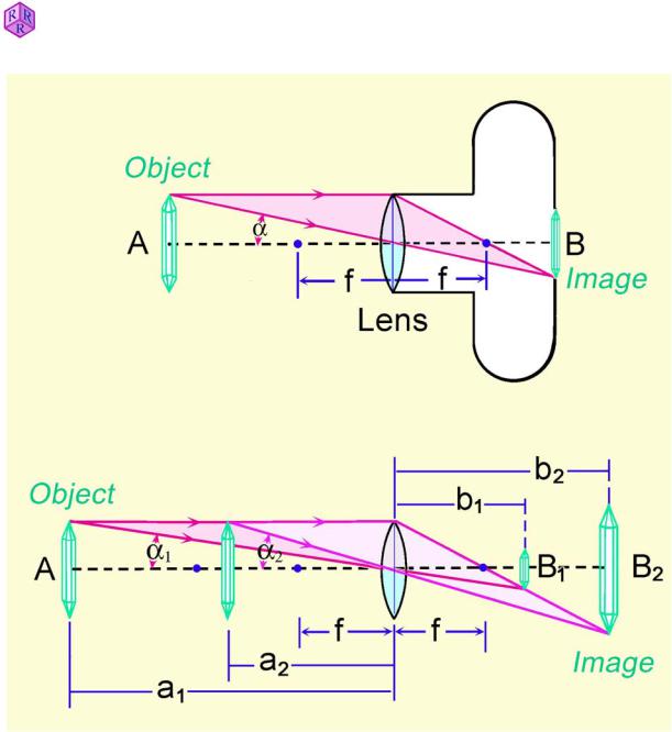
Optical images of an object are formed by converging lenses, i.e. spherical lenses with outward bulging surfaces (biconvex lenses). An inverted real image is formed if the object is placed beyond the focal length (f) of the lens. This image can be projected on a screen (which is the principle behind the camera and the human eye; Fig. 1.1-1, upper part).
The distances from the object to the lens (a) and from the lens to the image (b) are related to the focal lenghts (f) of the biconvex lens by the following equation (lens formula, Fig. 1.1-1, lower part):
1a 1b = 1f
The magnification of the lens is given by:
M |
B |
|
b |
|
b f |
|
f |
A |
|
a |
|
f |
|
a f |
|
|
|
|
|
The size of the real image (B) is larger than that of the object (A), if
b f |
! 1 i.e. 2f b resp. |
f |
! 1 i.e. a 2f. |
|
f |
a f |
|||
|
|
Example: If an object is placed at a distance of 33 mm in front of a biconvex lens with a focal length of 30 mm, the lens will produce a 10-times magnified image (M = 10:1) at a distance of 330 mm behind the lens.
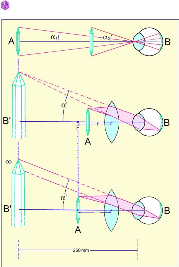
The human eye can modify the focal length by changing the curvature of its lens such that it can project sharp images on the retina of objects ranging in distance from about 250 mm to infinity.
As an object approaches the human eye from a greater distance it is seen at increasing visual angles (D) and, with the concomitant increasing magnification, the image on the retina becomes progressively larger (Fig. 1.1-2, upper part).
The shortest possible distance of focused vision varies individually. It has been set at 250 mm by the optical industry in order to standardize the calculation of magnification values.
1
Raith, Raase, Reinhardt – January 2011
Guide to Thin Section Microscopy |
Microscope |
|
|
|
|
Figure 1.1-1: Optical imaging of a crystal by a biconvex lens
Placed at the shortest distance of focused vision, the object is seen at the largest possible visual angle and thus with the highest possible magnification. Fine structures of an object, when observed at the shortest distance of vision, can only be resolved, however, if they appear at visual angles larger than 1’ (about 0.02°).
Structural details that remain below this limit of vision can still be resolved through further magnification with a magnifying glass (loupe) or microscope.
2

Raith, Raase, Reinhardt – January 2011
Guide to Thin Section Microscopy |
Microscope |
1.1.2 Magnification with the loupe (ocular, eyepiece)
The most simple device to increase the visual angle is a magnifying glass (loupe). It consists of a biconvex lens with a focal length that is smaller than the minimum distance of vision (<250 mm). If an object (A) is placed in the focal point F of the lens, the user sees an enlarged upright image (B) which appears to be located behind the loupe (i.e., on the object side) and appears to come from infinite distance (virtual image; Fig. 1.1-2, lower part).
The human eye now views the object at an increased visual angle D’. As a consequence, the image B projected on the retina is significantly enlarged.
The magnifying power of a loupe is defined as the ratio of the tangent values of the visual angles (or the sizes of the images) at which the object is seen with and without the lens at the minimum distance of vision of 250 mm (Fig. 1.1-2, upper und lower parts):
ML |
tanĮ' |
|
B'/250 |
|
A/f |
|
250 |
(loupe equation) |
tanĮ |
|
A/250 |
|
A/250 |
f |
|||
|
|
|
|
|||||
The magnification of a loupe ML is thus defined as the ratio of the minimum distance of vision (250 mm) and the focal length (f ) of the lens.
Example: A loupe with a focal length of 31.25 mm produces eightfold magnified images: 250/31.25 = 8 x.
If the object is not placed in the focal point of the loupe but within the focal length, (Fig. 1.1- 2, middle part), the magnification can vary up to a value of
250 ML f 1
ML values engraved on the casing of the loupe refer specifically to the magnification that applies if the object is placed in the focal point of the lens.
1.1.3 The compound microscope
In the most simple case, the magnification of an object in the microscope is achieved in two steps through a combination of two biconvex lenses (compound microscope; Fig. 1.1-3).
In a first imaging step (Fig. 1.1-3, upper part), a convex lens (objective) produces a real, enlarged and inverted image of the object at a magnification scale M.
In a second imaging step (Fig. 1.1-3, middle part), the real image is viewed by a magnifying glass (ocular, eyepiece), providing further magnification. To ensure that this virtual image can be viewed with a relaxed eye focused on infinity, the real image is placed in the focal plane of the ocular (Fig. 1.1-3, lower part).
The final image is created on the retina of the human eye.
3
Guide to Thin Section Microscopy |
Microscope |
Raith, Raase, Reinhardt – January 2011
Figure 1.1- 2: Magnification of a crystal by a biconvex lens (magnifying glass, loupe, ocular)
4