

Color.Atlas.of.Endodontics-ublog.tk
.pdfChapter One Diagnosis of Pulpal and Periradicular Pathosis |
5 |
FIGURE 1-8 An alternative method of thermal testing involves isolating individual teeth with a rubber dam and flooding the tooth with the appropriate hot or cold liquid. This method is especially useful when a patient complains of thermal sensitivity and traditional testing does not reproduce the patient's symptoms.
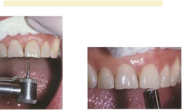
FIGURE 1-9 Electric pulp testing can be used to establish pulp vitality or confirm non responsiveness. In this case the failure of tooth #9 to respond confirms the results obtained with thermal testing.
stopping begins to soften, the clinician applies it to the lubricated tooth surface (Figure 1-7). A dry rubber prophylaxis cup can also be used to generate frictional heat. A more effective method of heat testing involves isolating individual teeth with a rubber dam and flooding the tooth with hot water (Figure 1-8). This method permits the application of a uniform temperature to each tooth and replicates the patient's normal activities. The technique is effective with full coverage restorations and can also be used with cold testing. Heat testing is the least valuable pulp test but is essential when the patient complains of sensitivity to heat.
Electric pulp testing stimulates the A-delta nerve fibers. The electric pulp test (EPT) indicates only whether the pulp is responsive or unresponsive. It does not provide information regarding the health of the pulp, nor can it differentiate degrees of pulp pathosis other than to indicate necrosis when no response occurs." It is often used to confirm the results of previous tests. The EPT requires an isolated dry field. Traditionally the electrode is coated with a conducting medium, usually toothpaste, and placed on the dry enamel labial or buccal surface of the tooth to be tested (Figure 1-9). Evidence indicates that the incisal edge is the optimal placement site for the electric pulp tester electrode to determine the lowest response threshold. 16 Contact with metallic restorations is to be avoided. The Analytical Technology (Analytic Endodontics, Sybron Dental Specialties, Orange, CA) pulp tester is recommended because it begins at zero current and increases the current gradually at a rate predetermined by the op-
erator.17 Patients are instructed to place a hand on the metal handle to begin the test and release the handle when they perceive a tingling sensation to stop the test. Having control of the test is reassuring to the patient. As with other tests, the clinician should test a normal tooth first to familiarize the patient with the procedure and sensation.
All pulp tests have a potential for false positive and false negative results. A false positive can occur when a tooth with a necrotic pulp nevertheless responds to testing. This can result from stimulation of adjacent teeth or the attachment apparatus, the response of vital tissue in a multirooted tooth with pulp necrosis in one or more canals, and patient interpretation. Furthermore, the clinician must keep in mind that the cell bodies of the neurons innervating the pulp lie in the Gasserian ganglion. Only the axons enter the pulp, so the nervous tissue can maintain vitality in a mass of necrotic pulp tissue. Neural elements have been shown to be more resistant to necrosis18 and C nerve fibers can function in a hypoxic environment. 19 Finally, pulp tests are not objective and require the patient to interpret the response, adding considerable subjectivity.
An example of a false negative in a pulp test is a tooth with a vital pulp that nevertheless does not respond to stimulation. False negatives can result from i nadequate contact with the stimulus, tooth calcification, immature apical development, traumatic in- j ury, and the subjective nature of the tests. They can also occur in elderly patients who have undergone regressive neural changes and in patients who have taken analgesics for pain. The neural elements develop after
6 |
Color Atlas o f Endodontics |
FIGURE 1-10 Direct dentinal stimulation is performed to eliminate the possibility of a false negative result with traditional testing. In this case no caries or restorations are present, leaving trauma as the only distinct etiology. Direct dentinal stimulation is employed when the clinician suspects that a tooth that does not respond is in fact vital.
FIGURE 1-11 Percussion can be performed with digital pressure, a mirror handle, or the Tooth Slooth. If the patient is symptomatic and complains of sensitivity to biting pressure, digital pressure may be all that is required to identify the offending tooth. In other cases, percussion with a mirror handle may be required to assess the periapical status.
eruption of the tooth ,20 and the aging of the dental pulp produces structural and neurochemical regressive changes that affect pulp innervation. 21 Traumatic in- j ury can damage the neural elements but leave the vascular supply to the tissue intact .22
DIRECT DENTINAL STIMULATION (TEST CAVITY). The test cavity is an invasive procedure that is often used to ensure that a negative response to previous pulp tests was accurate. Because this test is invasive and requires removal of tooth structure and/or restorative materials, it is used primarily to exclude false negative results. The test can be used in clinical cases in which a tooth does not respond to cold testing and EPT but lacks a distinct etiology for necrosis. In such cases direct dentinal stimulation can be used to reveal necrosis or establish vitality.
Direct dentinal stimulation involves removing enamel or restorative materials using a high-speed handpiece without local anesthesia (Figure 1-10). If the tooth is vital, the patient will experience a sharp, painful response when dentin is reached. Clinicians must caution patients that they will feel the sensations of vibration and pressure so that they can interpret the test correctly.
PERCUSSION. As pulp pathosis extends beyond the tooth into the supporting periodontal tissues and sur-
rounding bone, the patient's ability to localize the offending tooth increases. Proprioceptive fibers in the periodontal ligament are stimulated by force applied to the tooth and produce localized discomfort. Percussion is performed by applying force on the incisal or occlusal surface in an axial direction. This can be accomplished using digital pressure, tapping on the tooth with an instrument handle (Figure 1-11), or having the patient bite on a Tooth Slooth (Professional Results Inc., Laguna Niguel, CA) or cotton swab.
Although a positive response to percussion can indicate apical periodontitis secondary to pulp pathosis, other potential etiologies should also be considered. Tenderness to percussion can result from a variety of clinical problems such as a high restoration, traumatic injury, traumatic occlusion, a cracked tooth, a vertical root fracture, orthodontic treatment, a periodontal abscess, and maxillary sinusitis.
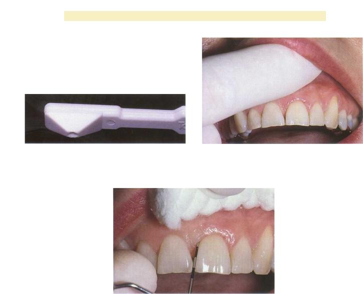
Clinicians can also use pressure to test for pulpal pathosis. Pressure can be applied by having the patient bite on a cotton swab or the Tooth Slooth (Figure 1-12), a device that permits the application of force to individual cusps and can be of value in the diagnosis of fractured or cracked teeth.
PALPATION. As periradicular inflammation extends through the cortical bone into the soft tissues, it can fre-
Chapter One Diagnosis o f Pulpal and Periradicular Pathosis |
7 |
FIGURE 1-12 The Tooth Slooth can be used to assess cracked teeth and incomplete cuspal fractures. The unique design allows the patient to exert pressure on individual cusps.
FIGURE 1-13 Palpation of the buccal and lingual soft tissues can detect areas of sensitivity and swelling, as well as determine the character of the swelling.
FIGURE 1-14 A limited periodontal assessment can be obtained by circumferential periodontal probing of the area. Often an isolated defect can be identified that is not otherwise apparent in the clinical and radiographic assessment.
quently be detected by digital palpation of the soft tissues over the apex of the root (Figure 1-13). When the mucoperiosteum is inflamed, the clinician will detect sensitivity in the involved area. As the inflammatory process progresses the operator may detect swelling of the soft tissues. The clinician should note the consistency of any swelling because not all swelling is the result of inflammatory disease. Palpation is not restricted to intraoral tissues. For example, palpation of extraoral structures can reveal lymphadenopathy.
MOBILITY. Tooth mobility can be assessed by moving the tooth in a facial or buccal-lingual direction. Mobility can be assessed by placing an index finger on the lingual surface and applying lateral force with an instrument handle from the buccal surface. The Miller Index of Tooth Mobility is commonly used to interpret the clini-
Class '1 is the first distinguishable sign of
greater-than-normal movement, Class 2 is movement of the crown as much as 1 mm in any direction, and Class 3 is movement of the crown more than 1 mm in any direction and/or vertical depression or rotation of the crown in its socket. Common causes of tooth mobility include periodontal disease, bruxism, clenching, traumatic occlusion, improper partial denture design, root fractures, and periradicular inflammation caused by pulp necrosis.
PERIODONTAL PROBING. Examination of the periodontal tissues is an essential component of the diagnostic process. Endodontic and periodontic lesions may mimic each other or occur concurrently. Because periodontal bone loss may not be detected radiographically and the gingival tissues may appear normal, probing is required ( Figure 1-14). Keeping a record of the probing depths aids in determining the patient's periodontal health and

8 |
Color Atlas o f Endodontics |
FIGURE 1-15 Transillumination is employed to evaluate teeth for fracture lines.
prognosis, and the pattern of probing also provides important information. To obtain adequate information when examining a specific tooth, the clinician should probe the entire circumference. Often a narrow probing defect can be detected with normal sulcular depths immediately adjacent to the defect. Common etiologies for isolated probing defects include periodontal disease, periapical pathosis forming a sinus-like trap through the periodontium, developmental defects such as a vertical groove defect, cracked teeth and vertical root fractures, and external root resorption.
TRANSILLUMINATION/DYE STAINING. The use of a fiberoptic light (Figure 1-15) is an excellent method o£ examining teeth for coronal cracks and vertical root fractures.24 The tooth or root should be examined in the presence of minimal background lighting. The fiberoptic light is then placed on the varied surfaces of the coronal tooth structure or on the root after flap reflection. Fracture lines can be visually detected when light fails to traverse the fracture line. The fractured segment near the light appears brighter than the segment away from the light.
Application of dyes to the tooth can also demonstrate fractures as the dye penetrates the fracture line. An ancillary technique is the application of dye to the internal surfaces of a cavity preparation or access opening; the clinician leaves the dye in place for a week before reexamining the tooth.
SELECTIVE ANESTHESIA/ANESTHETIC TEST. Because pain of pulpal origin is not referred beyond the midline, the administration of local anesthesia can help localize pain to a specific area in cases where patients exhibit referred pain that cannot be localized by the patient or by testing. Administration of a mandibular inferior alveolar nerve block will determine whether the pain is from the maxillary or mandibular teeth on the affected side. The pain will cease if it is from a mandibular tooth and persist if it is from a maxillary tooth. Although some clinicians feel that pain from an individual tooth can be isolated by administering local anesthetic with a periodontal ligament (PDL) injection, evidence suggests that this is inappropriate. PDL injections have been
shown to anesthetize teeth adjacent to the tooth being anesthetized.25
CARIES EXCAVATION. Caries excavation is a frequently used procedure to assess pulpal status. In patients exhibiting moderate to severe decay and normal responses to pulp testing, the clinician must remove the caries before deciding on a pulpal diagnosis. The initial response of the pulp to caries is chronic inflammation consisting of plasma cells and lymphocytes. This is a specific immune response to antigens leaching through the tubules. Excavation of caries and placement of a restoration remove the irritants and establish an environment for healing. As the dental pulp is exposed and bacteria invade, the existing chronic inflammatory response becomes acute as the host responds with polymorphonuclear leukocytes. This acute nonspecific inflammatory response results in the release of lysosomal enzymes and the destruction of host tissue as well as the invading bacteria. This is the crossover point from reversible to irreversible pulpitis. 26
Radiographic Examination
Radiographic examination of the hard tissues can often provide valuable information regarding caries and existing restorations, calcifications, internal and external resorptions, tooth and pulpal morphology, root fractures, the relationship of anatomic structures, and the architecture of the osseous tissues (Figure 1-16). In addition, radiographs can be used to trace sinus tracts ,27 demonstrate periodontal defects, and diagnose resorptive lesions (Figure 1-17). However, they do have many limitations and are of little value in assessing pulpal status. Vital and necrotic pulps cast the same image. Moreover, radiographs are only two-dimensional images of threedimensional structures.
Because radiography and some other imaging methods require ionizing radiation, during the clinical examination the clinician must prescribe the projection that will provide the most information at the lowest dose regarding the patient's problem. In most cases this is a periapical film or image, although bite-wing and extraoral films may be necessary.

Chapter One Diagnosis of Pulpal and Periradicular Pathosis |
9 |
FIGURE 1-16 Radiographic examination generally requires a periapical projection, although bite-wings and pantomographic projections are often useful. In this case the periradicular tissues appear normal; however, a comparison of the root canal space of #8 and #9 reveals that the space in tooth #9 is considerably larger. This is consistent with the clinical presentation, symptoms, and diagnostic testing results, which indicate necrosis.The radiographic appearance of the root canal system is caused by the lack of secondary dentin formation overtime.
FIGURE 1-17 Radiographs are useful in diagnosis. External resorptive defects such as the one depicted in the maxillary left central incisor are often irregular, with the root canal coursing through the lesion. Internal resorption such as that depicted in the maxillary left lateral incisor is often symmetric and exhibits destruction of the canal wall. In addition, internal resorptive lesions remain centered on angled radiographs.
Periapical radiographs and other images should be
exposed using a positioning device and a paralleling technique. This provides the most distortion-free image and accurate diagnostic information. Although great emphasis is often placed on the radiographic examination,
it is an imperfect diagnostic aid because of the varied techniques and methods for obtaining the film or image and the variable ability of practitioners to interpret the information correctly. 28-3 0 Subtle and moderate changes are often difficult to detect early in the pulpal and periradicular disease process. As the disease progresses, lesions become more distinct and easier to detect. Evidence suggests that a periapical lesion must erode the cortical plate to be visible on the film or image. 31 Making a second film using an angled projection can increase the diagnostic accuracy. 32
Periradicular lesions resulting from pulp necrosis have
a characteristic appearance. The radiolucency exhibits a "hanging drop" appearance, with the lesion beginning on the lateral osseous surfaces of the root and extending apically into the osseous tissues. The lamina dura is absent, and the lesion does not move when angled films are taken. In general, a radiolucent lesion associated with a tooth with a vital pulp is not of endodontic origin.
Condensing osteitis is a proliferative response of bone to periradicular inflammation. It is characterized by a diffuse appearance without distinct borders.
Radiographs and digital images appear to be equal in their diagnostic ability, although the astute clinician will use the radiographic examination to confirm the clinical examination. 28-30
DIAGNOSTIC CATEGORIES
The clinical diagnosis is based on the correlation of information. Because the information in the database is often incomplete or inconsistent, experience and the application of biologic principles allow for rational assessment.
Pulpal
NORMAL. The category of normal is used for teeth that are asymptomatic, respond normally to pulp testing, and are free of caries, deficient restorations, developmental defects, and cracks. Radiographically the periradicular tissues appear normal with an intact lamina dura.
The category of reversible pulpitis is used for teeth that respond normally to pulp testing.

1 0 |
Color Atlas o f Endodontics |
These teeth may be asymptomatic or have mild to moderate symptoms such as thermal sensitivity, sensitivity to sweets, pain to tactile stimulation, or pain when chewing. The pain generally subsides with removal of the irritant or stimulus, indicating A-delta nerve fiber activity. Common etiologies to consider are caries, deficient restorations, attrition, abrasion, erosion, cracks, or developmental defects that lead to exposed dentin. Dentinal hypersensitivity is a form of reversible pulpitis. Treatment may involve caries excavation, placing or replacing restorations, or sealing the dentin. If symptoms occur after a treatment procedure such as placement of a restoration or scaling and root planing, time may be required for symptoms to subside. The periradicular tissues appear normal.
IRREVERSIBLE PULPITIS. The etiologies for irreversible pulpitis are the same as those for reversible pulpitis, except that the symptoms are more severe and consistent with C nerve fiber activity. The tooth still responds to pulp testing. In general, the more intense the pain, the more likely that the pain is caused by irreversible pulpitis. Continuous or prolonged pain after a thermal stimulus is one of the first indications of irreversible pulpitis. Spontaneous pain is also associated with the condition. Pain that keeps the patient awake or awakens him or her is often indicative of irreversible pulpitis. A painful response to heat that is relieved by cold is a classic symptom. Root canal treatment, vital pulp therapy, or extraction is required. Generally the periradicular tissues appear normal, although in some cases the lamina dura appears widened or shows evidence of condensing osteitis.
NECROSIS. The positive response to cold and EPT occurs regardless of pulp status in normal, reversible, and irreversible pulpitis. Necrotic pulps do not respond. Teeth with necrotic pulps may or may not exhibit periradicular pathosis. Because teeth with necrotic pulps may exist within normal periradicular structures, the astute clinician performs pulp testing on all teeth before initiating restorative treatment. Pulp necrosis has two forms: dry and liquefactive. Dry necrosis is characterized by a root canal system devoid of tissue elements. This type of necrosis is most likely to produce periradicular pathosis. Liquefactive necrosis is characterized by pulp tissue with structure but lacking significant vascular elements. Liquefactive necrosis is more likely to produce symptoms and less likely to produce periradicular pathosis.
Periradicular
NORMAL. The category of normal is used to describe the periradicular status of teeth that are asymptomatic to percussion or palpation and exhibit normal-appearing osseous structures with an intact lamina dura.
ACUTE APICAL PERIODONTITIS. The category of acute apical periodontitis applies to teeth that exhibit normal periradicular structures but are painful to percussion because of the stimulation of proprioceptive fibers. The etiology can be pulp pathosis, but high restorations, traumatic occlusion, orthodontic treatment, cracked teeth and vertical root fractures, periodontal disease, and maxillary sinusitis may also produce this response. Treatment depends on the diagnostic findings. If pulp pathosis is the etiology, pulpectomy followed by root canal treatment or extraction is the most common treatment option.
CHRONIC APICAL PERIODONTITIS. Chronic apical periodontitis results from pulp necrosis and is characterized by the development of an asymptomatic periradicular lesion at the periapex and at the portal of exit in cases exhibiting lateral canals on the side of the root. Histologically this lesion is categorized as a granuloma or cyst. Root canal treatment or extraction are the treatment options.
CHRONIC PERIRADICULAR ABSCESS. Chronic periradicular abscess is similar to chronic apical periodontitis except that it is characterized by the presence of a draining sinus tract. The lesion is asymptomatic with an intermittent discharge of pus through the sinus tract. This lesion is also referred to as chronic suppurative apical periodontitis. Root canal treatment or extraction is required.
ACUTE PERIRADICULAR ABSCESS. Acute periradicular abscess is an inflammatory reaction resulting from pulp necrosis that is characterized by rapid onset, pain, and tenderness to percussion. Evidence of osseous destruction may or may not be present. A discharge of pus is evident, but swelling may or may not occur. The exudate can be confined to the alveolar bone, cause localized swelling of soft tissue, or extend into fascial spaces (cellulitis). The exacerbation of a previously asymptomatic chronic apical periodontitis has been termed a phoenix abscess.
The primary method of treating an acute periradicular abscess is to remove the irritants and provide drainage. This can be accomplished by initiating root canal treatment and debriding the radicular space or extracting the tooth. Antibiotics are not a substitute for definitive treatment procedures designed to remove the necrotic tissue and bacteria from the radicular space. Drainage can be accomplished through the tooth or through an incision of the involved soft tissues. This procedure relieves pressure, increases vascular flow, and evacuates the purulent exudate. In these cases, antibiotics serve a supportive role as adjuvants to treatment. Clinicians should prescribe antibiotics to medically compromised patients and patients with an increased temperature and systemic involvement.
CONDENSING OSTEITIS. Condensing osteitis is a proliferative inflammatory response to an irritant. The lesion

Chapter One Diagnosis o f Pulpal and Periradicular Patbosis |
11 |
is generally asymptomatic and is characterized radiographically by an increase in radiopacity.
SUMMARY
Clinicians must be knowledgeable and skilled in the process of diagnosis and treatment planning. They should be able to recognize that the patient has a problem, identify the etiology, establish a pulpal and periradicular diagnosis, and develop methods of treatment. Consultation with medical and dental specialists is often necessary during this process.
Pulpal and periradicular pathosis are inflammatory in nature. The accuracy of the clinical diagnosis is confirmed by resolution of the patient's signs and symptoms and healing of the involved tissues. Therefore periodic recall examination is an important part of the diagnostic process.
References
1.Bender IB: Pulpal pain diagnosis-a review, J Endodon 26:175, 2000.
2.Okeson JP, Falace DA: Nonodontogenic toothache, Dent Clin North Am 41:367, 1997.
3.Chang P: Evaluating imaging test performance: an introduction to Bayesian analysis for urologists, Monogr Urology 12:18, 1991.
4.Lipton JA, Ship JA, Larach-Robinson D: Estimated prevalence and distribution of reported orofacial pain in the United States, J Am Dent Assoc 124:115, 1993.
5.Falace DA, Reid K, Rayens MK: The influence of deep (odontogenic) pain intensity, quality, and duration on the incidence and characteristics of referred orofacial pain, J Orofac Pain 10:232, 1996.
6.Georgopoulou M, Kerani M: The reliability of electrical and thermal pulp tests. A clinical study, Stomatologia 46:317, 1989.
7.Peters DD, Baumgartner JC, Lorton L: Adult pulpal diagnosis. 1. Evaluation of the positive and negative responses to cold and electrical pulp tests, J Endodon 20:506, 1994.
8.Rickoff B et al: Effects of thermal vitality tests on human dental pulp, J Endodon 14:482, 1988.
9.Dummer PM, Tanner M, McCarthy JP: A laboratory study of four
electric pulp testers, Inter Endo.J 19:161, 1986.
10.Peters DD, Mader CL, Donnelly JC: Evaluation of the effects of carbon dioxide used as a pulpal test. 3. In vivo effect on human
enamel, J Endodon 12:13, 1986.
11.Ahlquist M et al: Dental pain evoked by hydrostatic pressures applied to exposed dentin in man: a test of the hydrodynamic theory of dentin sensitivity, J Endodon 20:130, 1994.
12.Narhi MV et al: The neurophysiological basis and the role of inflammatory reactions in dentine hypersensitivity, Arch Oral Biol 39(suppl):23S, 1994.
13.Hirvonen T, Narhi MV, Hakumaki MO: The excitability of dog pulp nerves in relation to the condition of dentin surface, J Endodon 10:294, 1984.
14.Rosenberg RJ: Using heat to assess pulp inflammation, J Am Dent Assoc 122(2):77, 1991.
15.Lado EA, Richmond AF, Marks RG: Reliability and validity of a digital pulp tester as a test standard for measuring sensory per-
ception, J Endodon 14:352, 1988.
16.Bender IB et al: The optimum placement-site of the electrode in electric pulp testing of the 12 anterior teeth, J Am Dent Assoc 118:305, 1989.
17.Kleier DJ, Sexton JR, Averbach RE: Electronic and clinical comparison of pulp testers, J Dent Res 61:1413, 1982.
18.Torneck CD: Changes in the fine structure of the human dental pulp subsequent to carious exposure, J Oral Pathol 6:82, 1977.
19.Narhi MV et al: Role of intradental A- and C-type nerve fibres in dental pain mechanisms, Proc Finn Dent Soc 88(suppl 1):507, 1992.
20.Johnsen DC, Karlsson UL: Development of neural elements in apical portions of cat primary and permanent incisor pulps, Anat Rec
189:29,1977.
21.Fried K: Aging of the dental pulp involves structural and neurochemical regressive changes in the innervation of the pulp, Proc
Finn Dent Soc 88:517, 1992.
22.Bhaskar SN, Rappaport HM: Dental vitality tests and pulp status, J Am Dent Assoc 86:409, 1973.
23.Miller SC: Textbook of periodontia, ed 3, Philadelphia, 1950,
Blackstone.
24.Schindler WG, Walker WA, III: Transillumination of the beveled root surface: an aid to periradicular surgery, J Endodon 20:408,
1994.
25.D'Souza JE, Walton RE, Peterson LC: Periodontal ligament injection: an evaluation of the extent of anesthesia and postinjection discomfort, J Am Dent Assoc 114:341, 1987.
26.Trowbridge HO: Pathogenesis of pulpitis resulting from dental caries, J Endodon 7:52, 1981.
27.Bonness BW, Taintor JF: The ectopic sinus tract: report of cases, J Endodon 6:614, 1980.
28.Goldman M, Pearson AH, Darzenta N: Reliability of radiographic interpretations, Oral Surg Oral Med Oral Patbol Oral Radiol Endod 38:287, 1974.
29.Gelfand M, Sunderman EJ, Goldman M: Reliability of radiographical interpretations, J Endodon 9:71, 1983.
30.Holtzmann DJ et al: Storage phosphor based computed radiography versus film based radiography in detection of pathologic periradicular bone loss in a cadaver model: an ROC study Oral Surg Oral Med Oral Patbol Oral Radiol Endod 86:90, 1998.
31.Bender IB: Factors influencing the radiographic appearance of bony lesions, J Endodon 23:5, 1997.
32.Brynolf 1: Roentgenologic periapical diagnosis. One, two or more roentgenograms? Swed Dent J 63:345, 1970.


14 |
Color Atlas o f Endodontics |
A great deal of frustration that many practitioners have with endodontic treatment stems from the difficulty of placing a 25-mm instrument in the mesiobuccal (MB) canal of a distally inclined maxillary second molar. Correct access design and straight-line access to facilitate instrument placement can greatly reduce frustration and dramatically decrease treatment time.
With the advent of hyperflexible NiTi instruments, clinicians might mistakenly conclude that minimizing instrument flexure is of lesser importance. In fact, straightline access and minimizing of instrument flexure is of increased importance in the use of NiTi instruments. Conventional stainless steel files can be precurved and "hooked" into canals. If a rotary NiTi file is curved or bent, it is ruined and must be discarded. In addition, straight-line access and reduced instrument flexure improve the clinician's ability to use the instruments as feeler gauges and improve control over the instruments' cutting action.
Specialists are often referred cases in which the general practitioner cannot find the canals. Most of the time the canals are in the chamber, but the access preparation precludes the practitioner from locating the canals. The problem is usually too small an access preparation with improper location and suboptimal shape. After the access has been reshaped, the canals are easily located. This is of particular importance with posterior teeth whose canals can be easily missed, leading to periapical pathosis or continued symptoms.
Unroo fing the Chamber
Unroofing the chamber and removing the coronal pulp facilitates the clinician's ability to visualize the chamber floor and aids in locating the canals. Complete removal of tissue and debris prevents discoloration and subsequent infection.
Unroofing the chamber and removing the coronal pulp (in vital cases) allow the clinician to see the pulpal floor. In cases of patent canals, most or all of the canal orifices may be easily located before the chamber is completely unroofed, but the clinician may nevertheless miss canals. In cases of calcification, performing these procedures increases the clinician's ability to visualize the pulpal floor and read the road map to the canal orifices detailed in the subtle color changes and patterns of calcification left by the receding pulp. This is extremely difficult or impossible to do through a "mouse hole" en-dodontic access.
Removal of the Coronal Pulp
Removal of the coronal pulp so that the canals may be located is necessary in cases with vital pulp. One advantage of removing the coronal pulp is that the radicular fragments may hemorrhage slightly, aiding in location of the canal orifices. This is especially useful in maxillary molar cases for locating the second mesiobuccal (MB2) canal.
Facilitation of Instrument Placement
Although contemporary endodontic techniques require fewer instruments, the overall thrust of endodontic cleaning and shaping continues to be the serial placement into the root canal system of variably sized, tapered, or shaped instruments. This serial placement of instruments is greatly facilitated by spending a few extra minutes on the access preparation. Access preparation becomes even more important with the use of rotary NiTi instruments. Placement of these instruments requires considerably more attention to gaining straight-line access.
With the use of traditional stainless steel hand files, the clinician has several advantages in instrument placement over rotary NiTi instruments. First, the stainless steel files may be pre-bent, allowing the clinician to hook the file into difficult-to-access canals. As stated before, a bent NITI rotary instrument is a discarded NiTi rotary instrument. Second, the stiffness of stainless steel provides the clinician with tactile feedback that can be used to drop the file through the orifice into the canal. The thin, flexible tips of the NiTi files impair the clinician's ability to feel obstacles and obstructions and locate the canal orifice. Further compounding this lack of tactile sensitivity, the NiTi files are used with a handpiece, which greatly decreases the tactile sensation of the sensitive and delicate pads of the fingertips.
Coronal and orifice access should act as a funnel to guide the instruments into the canal. Ideally, the line angles of the access preparation should smoothly guide the instrument into the correct canal. This funnel shape also facilitates the introduction of obturation instruments.
Minimizing of Instrument Flexure
With the greater emphasis on more conservative radicular shapes and the concomitant use of rotary NiTi files, the minimizing of instrument flexure has taken on a new importance. Two obvious reasons for reducing instrument flexure are to combat work hardening and decrease the stresses that the instruments undergo during preparation of the root canal system. This decreases fracture incidence and allows more of the energy applied to the instrument to be used for carving the preparation out of the radicular walls.
Locating Canals
With complete eradication of the radicular contents, obturation of the radicular space, and good coronal seal to prevent ingress of bacteria, endodontic treatment should approach 100% success. However, this does not occur in reality. The second most common error in access, one that is often not noticed until a recall film is taken or the patient complains of persistent symptoms, is missed canals. The greatest teacher of endodontic anatomy is the microscope. Clinicians have learned that all roots (not teeth) with the exception of #6 through #11 may have two or more canals .z The MB 2 canal of the maxil-

Chapter Two
lary first molar is commonly referred to as an "extra" canal, but this is not the case-the fifth and sixth canals are the "extras." Without obtaining adequate access in shape, size, and location, locating the exceedingly complex anatomy present in posterior teeth becomes an exercise in futility.
Many of these canals are hidden under dentin shelves, pulp stones, protrusions, and restorative materials. Successful treatment requires adequate access, knowledge of the radicular anatomy, determination, and the assumption of two canals per root until proven otherwise.
I NSTRUMENTS AND ARMAMENTARIUM
The endodontic tray setup should contain an assortment of round and fissure burs, tapered and round diamonds, and (for the adventurous) Mueller burs and ultrasonics. A sharp endodontic explorer is essential. Although they are often helpful in locating canals, hand files are generally not used during the access preparation.
Fissure Burs
in an uncrowned tooth exhibiting a patent canal, initial access is best accomplished by round or fissure carbide burs (Figure 2-1). Fissure burs such as the #558 produce less "chatter" when penetrating intact enamel or dentin
Endodontic Access |
1 5 |
compared with round carbide burs. In contrast, round carbide burs such as the #6 or #8 seem to be more controllable during the removal of carious dentin.
Round Diamond Burs
New round diamond burs in #4 and #6 sizes work predictably and quickly to cut through both porcelain- fused-to-metal (PFM) crowns and the new all-porcelain crowns (Figure 2-2). The clinician should use relatively new diamonds with abundant water and intermittent light pressure to avoid generating excessive heat. If dull diamonds are used, especially without water coolant, the clinician may be tempted to apply excessive pressure to accelerate the cutting process and thereby overheat the crown. This can result in craze lines and fractures, which may chip off during instrumentation (when they are easy to repair) or after treatment completion (when they are not). After removing the porcelain layer of the PFM, the clinician can then use a carbide fissure bur or specially designed metal cutting bur to perforate the metal substructure and underlying foundation.
Tapered Diamonds
Flame-shaped and round-ended tapered crown-prepa- ration style diamonds are excellent for endodontic access (Figure 2-3). They are unequaled for cutting with
FIGURE 2-1 From left to right, a #558 surgical length fissure bur followed by #1, #2, #4, #6, and #8 surgical length carbides.These are primarily used for cutting through natural tooth structure.
FIGURE 2-2 From left to right, round diamonds in sizes #4, #6, #8, and #10. Used with copious water and a very light touch, they can predictably and effortlessly cut through PFM and all-porcelain crowns without fracture.