

Color.Atlas.of.Endodontics-ublog.tk
.pdfChapter Ten Endodontic Surgery |
1 63 |
ANATOMIC CONSIDERATIONS. The anatomy and morphology of the tooth have significant implications for restorability, periodontal maintenance, and prognosis during treatment planning. The shape of the retained root may be difficult to restore and for the patient to maintain adequate oral hygiene.
MAXILLARY MOLARS. In maxillary molars the mesiobuccal root generally curves distally, is broad in the buccal lingual dimension with the furcation toward the lingual, and exhibits mesial and distal concavities. The root alignment is similar to that of the maxillary premolars, and the root exhibits a large surface area for support. In cross-section the root may have an hourglass appearance. This may complicate the restorative plan and make oral hygiene difficult. In addition, the presence of one or two small canals makes post placement difficult. The distobuccal root is relatively straight, but may exhibit an apical curvature. The root is round in crosssection. The palatal root is the most divergent, extending lingually in relationship to the crown. The root is wider in the mesiodistal dimension than it is buccolingually.
MANDIBULAR MOLARS. In mandibular molars the mesial root curves toward the distal, is wider in its buccolingual dimension than in its mesiodistal dimension, and exhibits the greatest surface area. The root has concavities on both the mesial and distal surfaces, giving the root an hourglass configuration in cross-section. The distal root is also wider in its buccolingual dimension than in its mesiodistal dimension. A concavity on the mesial surface gives the root a kidney bean configuration in cross-section.
Root Resection
When feasible, endodontic treatment should precede root removal. This permits adequate isolation for cleaning and shaping, assessment of the remaining tooth structure, and placement of a definitive restorative material in the coronal portion of the root to be resected. In addition, the tooth can be evaluated for occlusal interferences and a plan can be developed for provisional stabilization. To provide access and visibility and maintain adequate biologic width, flap reflection and osseous recontouring should be performed in conjunction with the root resection procedure.
Two primary methods are used for root removal (Figure 10-24): the horizontal cut and the vertical cut. A third, less popular, method involves presurgical crown contouring. The initial cut should be made in a manner that sacrifices tooth structure from the root to be removed while maintaining as much structure as possible on the segment to be retained. The retained segment can always be recontoured. After making the initial cut, the clinician should verify complete separation of the root before placing an elevator between the two segments and gently moving the two pieces apart. The root
FIGURE 10-24 A, A diagrammatic sketch of a mandibular first molar exhibiting periodontal bone loss involving the mesial root. B, A horizontal root resection that maintains the crown. C, A vertical resection removing the mesial root and associated portion of the crown.

164 |
Color Atlas o f Endodontics |
FIGURE 10-25 A, A clinical photograph of a mandibular first molar with a history of previous root canal treatment, retreatment, and restoration with an amalgam core and crown. Note the buccal area of inflammation. B, A preoperative radiograph displays radiolucent areas associated with the mesial and distal roots. C, Intentional replantation was selected as the treatment of choice because of the thickness of the buccal cortical bone. Root end resection was accomplished using a highspeed handpiece and water coolant. Root end fillings were placed in each root. D, A postoperative radiograph of the tooth. Note that the crown was dislodged during extraction. E, A 3-year recall radiograph. A deep mesial-buccal periodontal pocket was evident clinically. F, Examination on extraction revealed a vertical root fracture and external apical resorption. G and H, Scanning electron microscopic examination of the apical resorption.

Chapter Ten Endodontic Surgery |
1 65 |
should move independent of the crown. The area is irrigated and inspected after root removal to ensure the absence of overhangs or sharp edges and establish that the area is cleanable. Both the tooth and bone can be shaped to provide a cleanable area. Regardless of the technique employed, the clinician must exercise care to ensure that no overhanging tooth structure is left in the furcation and that 3 mm of circumferential tooth structure remains coronal to the osseous crest.
Success rates for root resection are 60% to 70%.60-62 Failures tend to occur for a variety of reasons after extended periods of time. Root resection is a predictable treatment option that may offer the opportunity to maintain a portion of a strategic tooth . 63
INTENTIONAL REPLANTATION
Intentional replantation is a procedure that offers the clinician the opportunity to maintain a tooth that cannot be managed nonsurgically. It can also be used in cases where root end surgery is not a viable option (Figure 10-25). Although intentional replantation is a treatment alternative, it should be used as a last resort because extraction of the tooth may lead to fracture, which renders replantation impossible. Success rates vary from 70% to 80%, with case selection being crucial . 64,65 Teeth with flared roots may fracture during extraction and must be evaluated closely when they are being considered for this type of procedure.
The success of intentional replantation primarily depends on the amount of time the tooth is out of the socket. Another factor is management of the root surface. After atraumatic extraction the periodontal tissue on the root or roots should be maintained in a moist environment. This can be accomplished by holding the tooth by the crown in a gauze soaked in physiologic saline or Hank's balanced salt solution. The root is kept moist with frequent saline irrigation.
If endodontic treatment is possible, the clinician should clean, shape, and fill the canal or canals and restore access before extraction and replantation. In cases where previous endodontic treatment has been undertaken or where the canals are calcified, the root end should be resected, a root end preparation performed, and a root end filling placed extraorally.
Immediately before replantation the socket should be irrigated with saline to remove the clot. The tooth is then replanted and stabilized if necessary. In posterior areas, splinting is often not necessary. If a tooth requires stabilization, an acid-etch physiologic splint or sutures can be used.
Success depends on precise and efficient performance of the procedure. Damage to the root surface during extraction and the length of time the tooth is out of the socket affect the prognosis. Maintaining the viability of the periodontal membrane reduces the incidence of replacement resorption and enhances healing.
References
1.Bergenholtz G et al: Retreatment of endodontic fillings, Scand J Dent Res 87:217, 1979.
2.Hartley F et al: The success rate of apicoectomy-a retrospective study of 1,016 cases, Br Dent J 129:407, 1970.
3.Milzman DP, Milzman JB: Patient assessment and preventive measures for medical emergencies in the dental office, Dent Clin North Am 43:383, 1999.
4.Morris WO: Informed consent and assault and battery. In Morris WO, editor: The dentist's legal advisor, St Louis, 1995, Mosby.
5.Denio D, Torabinejad M, Bakland LK: Anatomical relationship of the mandibular canal to its surrounding structures in mature mandibles, J Endodon 18:161, 1992.
6.Squier CA, Nanny D: Measurement of blood flow in the oral mucosa and skin of the Rhesus monkey using radiolabeled microspheres, Arch Oral Biol 30:313, 1985.
7.Canady JW, Johnson GK, Squier CA: Measurement of blood flow i n the skin and oral mucosa of the rhesus monkey (Maraca mulatta) using laser Doppler flowmetry, Comp Biochem & Physiol 106:61, 1993.
8.Johnson GK et al: Blood flow and epithelial thickness in different regions of feline oral mucosa and skin, J Oral Path 16:317, 1987.
9.Johnson WT et al: Measurement of blood flow to osseous tissue in dogs using the radiolabeled microsphere method, Comp Biochem
Physiol 106:649, 1993.
10.Cutright DE, Hunsuck EE: Microcirculation of the perioral regions in the Macaca rhesus part 1, Oral Surg 29:776, 1970.
11.Cutright DE, Hunsuck EE: Microcirculation of the perioral regions in the Macaca rhesus part 11, Oral Surg 29:926, 1970.
12.Kim S: Hemostasis in endodontic microsurgery, Dent Clin North Am 41:499,1997.
13.Buckley JA, Ciancio SG, McMullen JA: Efficacy of epinephrine concentration on local anesthesia during periodontal surgery, J
Periodontol 55:653, 1984.
14.Grung B: Healing of gingival mucoperiosteal flaps after marginal incision in apicoectomy procedures, Int J Oral Surg 2:20, 1973.
15.Harrison JW, Jurosky KA: Wound healing in the tissues of the periodontium following periradicular surgery. 1. The incisional wound, J Endodon 17:425, 1991.
16.Gutmann JL, Harrison JW: Posterior endodontic surgery: anatomical considerations and clinical techniques, Int Endod J 18:8, 1985.
17.Lang NP, Loe H: The relationship between the width of keratinized gingiva and gingival health, J Periodontol 43:623, 1972.
18.Kamper BJ, Kaminski EJ, Osetek EM: A comparative study of the wound healing of three types of flap design used in periradicular surgery, J Endodon 10:17, 1984.
19.Sanz M et al: Clinical enhancement of post-periodontal surgical therapy by a 0.12% chlorhexidine gluconate mouthrinse, J Peri-
odontoL 60:570, |
1989. |
20.Soulti A, Torbinejad M: Histologic study of healing periradicular lesions with and without curettage, J Endodon 20:188, 1994.
21.Lin LM, Gaengler P, Langeland K: Periradicular curettage, Int Endod J 29:220, 1996.
22.Rud J, Andreasen JO: Operative procedures in periapical surgery with contemporaneous root filling, Int J Oral Surg 1:297, 1972.
23.Rubenstein R: The anatomy of the surgical operating microscope and operating positions, Dent Clin North Am 41:391, 1997.
24.Carr GB: Ultrasonic root end preparations, Dent Clin North Am 41:541, 1997.
25.Gagliani M, Taschieri S, Molinari R: Ultrasonic root-end preparation: influence of cutting angle on the apical seal, J Endodon 24:626, 1998.
26.Hsu Y, Kim S: The resected root surface. The issue of canal isthmuses, Dent Clin North Am 41:529, 1997.
27.Gutmann JL et al: Ultrasonic root-end preparation. Part 1. SEM analysis, Int Endod J 27:318, 1994.

1 66 |
Color Atlas of Endodontics |
28.Gorman MC, Steiman HR, Gartner AH: Scanning electron microscopic evaluation of root-end preparations, J Endodon 21:113, 1995.
29.Engel TK, Steiman HR: Preliminary investigation of ultrasonic root end preparation, J Endodon 21:443, 1995.
30.Lin CP et al: The quality of ultrasonic root-end preparation: a quantitative study, J Endodon 24:666, 1998.
31.Mehlhaff DS, Marshall JG, Baumgartner JC: Comparison of ultrasonic and high-speed-bur root-end preparations using bilaterally matched teeth, J Endodon 23:448, 1997.
32.Saunders WP, Saunders EM, Gutrmann JL: Ultrasonic root-end preparation. Part 2. Microleakage of EBA root end fillings, Int Endod J 27:325, 1994.
33.von Arx T, Walker WA: Microsurgical instruments for root-end cavity preparation following apicoectomy: a literature review, Endod Dent Traumatol 16:47, 2000.
34.Layton CA et al: Evaluation of cracks associated with ultrasonic root-end preparation, J Endodon 22:157, 1996.
35.Abedi HR et al: Effects of ultrasonic root end cavity preparation on the root apex, Oral Surg Oral Med Oral Pathol Oral Radiol Endod 80:207, 1995.
36.Bader G, Lejeune S: Prospective study of two retrograde endodontic apical preparations with and without the use of CO2 laser, Endod Dent Traumatol 14:75, 1998.
37.von Arx T, Kurt B: Root-end cavity preparation after apicoectomy using a new type sonic and diamond-surfaced retrotip: a 1 year follow-up study, J Oral Maxillo fac Surg 57:656, 1999.
38.Withespoon DE, Gutmann JL: Haemostasis in periradicular surgery, Int Endod J 29:135, 1996.
39.Sauveur G et al: The control of haemorrhage at the operative site during periradicular surgery, Int Endod J 32:225, 1999.
40.Kim S, Rethnam S: Hemostasis in endodontic microsurgery, Dent Clin North Am 41:499, 1997.
41.Besner E: Systemic effects of racemic epinephrine when applied to the bone cavity during periapical surgery, Va Dent J 49:9, 1972.
42.Jeansonne BG, Boggs WS, Lemon RR: Ferric sulfate hemostasis: effect on osseous wound healing. II. With curettage and irrigation, J Endodon 19:174, 1993.
43.Ibarrola JL et al: Osseous reactions to three hemostatic agents, J Endodon 11:75, 1985.
44.Selden HS: Bone wax as an effective hemostat in periapical surgery, Oral Surg Oral Med Oral Pathol Oral Radiol Endod
29:262, 1970.
45.Craig K, Harrison JW: Wound healing following demineralization
of resected root ends in periradicular surgery, J Endodon 19:339, |
1993. |
46.Pantschev A, Carlsson AP, Andersson L: Retrograde root filling with EBA cement or amalgam. A comparative clinical study, Oral Surg Oral Med Oral Pathol Oral Radiol Endod 78:101, 1994.
47.Williams SS, Gutmann JL: Periradicular healing in response to Diaket root-end filling material with and without tricalcium phosphate, Int Endod J 29:84, 1996.
48.Dorn SO, Gartner AH: Retrograde filling materials a retrospective success-failure study of amalgam, EBA IRM, J Endodon 16:391, 1990.
49.Torabinejad M, Chivian N: Clinical applications of mineral trioxide aggregate, J Endodon 25:197, 1999.
50.Frank AL, Glick DH, Patterson SS: Long-term evaluation of surgically placed amalgam fillings, J Endodon 18:391, 1992.
51.Torabinejad M et al: Histologic assessment of mineral trioxide aggregate as a root-end filling in monkeys, J Endodon 23:225, 1997.
52.Saad AY, Clem WH: The use of radiographs in periapical surgery,
Oral Surg Oral Med Oral Pathol Oral Radiol Endod 69:361,
1990.
53.Selim HA, El Deeb ME, Messer HH: Blood loss during endodontic surgery, Endod Dent Traumatol 3:33, 1987.
54.Harrison JW Jurosky KA: Wound healing in the tissues of the periodontium following periradicular surgery. 2. The dissectional wound, J Endodon 17:544, 1991.
55.Pecora G et al: The guided tissue regeneration principle in endodontic surgery: one-year postoperative results of large periapical lesions, Int Endod J 28:41, 1995.
56.Bohning BP, Davenport WD, Jeansonne BG: The effect of guided tissue regeneration on the healing of osseous defects in rat calvaria,
J Endodon 25:81, 1999.
57.Seymour RA, Meechan JG, Blair GS: Postoperative pain after apicoectomy. A clinical investigation, Int Endod J 19:242, 1986.
58.Sumi Y et al: Ultrasonic root-end preparation: clinical and radiographic evaluation of results, J Oral Maxillo fac Surg 54:590,
1996.
59.Rubenstein RA, Kim S: Short-term observation of the results of endodontic surgery with the use of a surgical operation microscope and Super-EBA as a root end filling material, J Endodon 25:43,1999.
60.Langer B, Stein SD, Wagenberg B: An evaluation of root resections: a ten year study, J Periodontol 52:719, 1981.
61.Expenstein H: 3-year study of hemisectioned molars, J Clin Periodontol 10:1, 1983.
62.Buhler H: Evaluation of root resected teeth, results after 10 years,
J Periodontol 59:805, 1988.
63.Buhler H: Survival rates of hemisected teeth; an attempt to compare them with survival rates of alloplastic implants, J Perio Rest Dent 14:537, 1994.
64.Bender IB, Rossman LE: Intentional replantation of endodontically treated teeth, Oral Surg Oral Med Oral Pathol Oral Radiol Endod 76:623, 1993.
65.Ragboebar GM, Vissink A: Results of intentional replantation of molars, J Oral Maxillofac Surg 57:240, 1999.


168 |
Color Atlas o f Endodontics |
FIGURE 11-1 Detection of a horizontal root fracture may require increasing or decreasing the vertical angulation of the beam.
RADIOGRAPHIC EXAMINATION
Radiographs taken after traumatic injury may reveal tooth displacement, root fractures, osseous fractures, and foreign objects present in soft tissues. In cases of suspected root fractures, films exposed from several vertical angles may be required to reveal the fracture line9 (Figure 11-1). In lacerated areas a film of the soft tissues may reveal an embedded object. 10 Radiographs are also helpful in assessing the degree of apical root development in teeth exhibiting crown fractures. A pantomograph may be indicated in cases of suspected osseous fractures.
DOCUMENTATION
Documentation of findings from the history, clinical examination, and radiographic examination is important for an accurate diagnosis, justification of treatment, future diagnostic considerations, insurance issues, and defense against potential litigation.11
CROWN FRACTURES
Uncomplicated Fractures
Infraction is a term used to designate an incomplete fracture in enamel without loss of structure (see Figure 11-4, A). Enamel infractions result from direct impact, are best visualized with transillumination, and may be the only visible evidence that trauma has occurred.
Crown fractures result from a high-velocity impact. Fracture of the crown dissipates the force and minimizes injury to the supporting periodontal structures and bone. Coronal fractures can involve enamel or enamel and dentin. The primary symptom associated
with fractures involving enamel and dentin is thermal sensitivity. Although a fracture involving enamel often appears insignificant and can be managed by selective grinding or placement of an acid-etched composite restoration, the potential for pulpal damage exists. Pulp testing should be performed to assess vitality. The most severe pulp response is often seen in teeth exhibiting little or no physical damage. 12 In general, however, the pulpal prognosis for uncomplicated fractures of enamel
and dentin is favorable.13,14
Crown fractures involving both enamel and dentin expose dentinal tubules to saliva and bacteria (Figure 11-2). The treatment of choice involves placing a pro tective base and an acid-etch composite restoration. Recent advances in enamel-dentin bonding have enhanced the bonding of fractured segments.15 In cases with extensive damage, laminate veneers, porcelain-fused-to- metal crowns, or all-ceramic crowns may be required to restore form, function, and esthetics. With coronal fractures of enamel or enamel and dentin, recall evaluation is required to assess pulp vitality.
Complicated Crown Fractures
Fractures of enamel and dentin have the potential for pulp exposure. When a coronal fracture is complicated by pulp exposure in young patients, the extent of apical development must be determined. Teeth with incomplete root formation require apexogenesis for continued root development. Teeth that become nonvital before complete root formation require apexification.
Crown fractures that expose the dental pulp exhibit the best prognosis when treated immediately. The longer the pulp is exposed, the greater the bacterial contamination. This is especially important in teeth with incomplete apical development where maintaining vitality is
Chapter Eleven Traumatic Injuries to the Permanent Dentition |
169 |
A
B
C
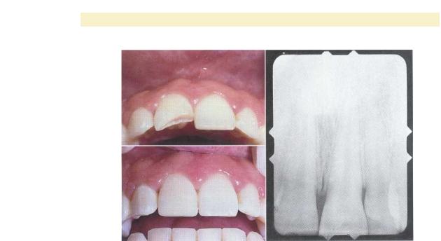
FIGURE 11-2 A, An uncomplicated crown fracture involving enamel and dentin. The patient was asymptomatic with the exception of sensitivity to cold. B, A periapical radiograph demonstrates the normal periradicular structures. C, Treatment consisted of placing a Class IV acid-etch composite restoration.
essential for continued root formation. If necrosis occurs, root development may cease.
APEXOGENESIS. Treatment procedures for teeth with exposed pulp include pulp capping, pulpotomy (Figure 11-3), and root canal treatment (Figure 11-4). Pulp capping is indicated for teeth with small exposures that can be treated soon after the injury. Capping can be accomplished with calcium hydroxide or a new material, mineral trioxide aggregate (MTA). Pulpotomy is indicated for more severe fractures or teeth in which treatment is delayed. Removal of several millimeters of pulp tissue provides space for placement of calcium hydroxide or MTA and is adequate to remove superficial inflamed tissue in cases where treatment is delayed. Removal of coronal pulp tissue is best accomplished with an abrasive diamond in a high-speed handpiece using a water coolant. 16 The amputation site should be clean of debris and tissue tags. Bleeding can be controlled with the application of a cotton pellet soaked with sterile saline. If calcium hydroxide is used, a glass ionomer or intermediate restorative material (IRM) base can be placed before restoration with composite resin. If MTA is used, the material should set for 1 week before the permanent restoration is placed. 17 An acid-etch composite restoration is placed to provide a coronal seal and restore esthetics and function.
With both pulp capping and pulpotomy, treatment consists of debridement of the wound and placement of calcium hydroxide or MTA. Although calcium hydroxide has been shown to be clinically effective over time, it pro-
duces a superficial layer of coagulation necrosis. The low-grade irritation of this layer induces the formation of a hard tissue barrier.18 MTA, however, has been shown to induce a hard tissue barrier without inflammation. 19
The time interval between exposure and treatment does not appear to be a significant factor affecting prognosis as long as the coronal inflamed tissue can be removed. Success rates for pulp capping and pulpotomy procedures after traumatic injury are high . 20 This is probably because of the acute nature of the injury and minimal bacterial contamination.
Recall evaluation should be performed after pulp capping and pulpotomy to assess the patient's response to treatment. Evaluation is often based on clinical and radiographic findings because pulp testing may not produce accurate information. In cases where root development is incomplete, verification of continued growth is evidence of successful treatment. Clinical studies indicate that successful pulpotomy procedures correlate with normal histopathologic findings in the pulp tissue . 20 Removal of the pulp after complete root development is contraindicated unless a post is required for coronal restoration. In cases where root development is complete and extensive coronal destruction has occurred, nonsurgical root canal treatment can be performed to facilitate placement of a post, core, and crown.
APEXIFICATION. In cases where necrosis occurs before complete root formation, apexification procedures are indicated to establish an apical barrier before obturation of the canal space with gutta-percha (Figure 11-5). In these

1 7 0

Chapter Eleven Traumatic Injuries to the Permanent Dentition |
171 |
FIGURE 11-4 A, A complicated root fracture with pulp exposure involving the maxillary right central incisor, tooth #8. Note the crown infraction associated with tooth #9. B, The radiographic examination reveals normal periradicular structures and complete apical development. C, Treatment consisted of placing an acid-etch composite as a provisional restoration and nonsurgical root canal treatment. Definitive treatment consisting of placement of a cast post and core was delayed until orthodontic treatment could be provided.
cases the clinician debrides the necrotic tissue remnants from the canal walls using traditional endodontic procedures. The canal is then packed with calcium hydroxide powder or a proprietary paste, which is left in place for 2 to 3 months. The location of the barrier depends on the level at which the calcium hydroxide contacts vital tissue, so it is important to place the material to the apex.
During the recall examination the calcium hydroxide is removed and the apical area is assessed radiographically and clinically for the development of a barrier. If no barrier is present, the procedure is repeated. The average time for barrier development is 6 to 12 months.21, 22 The success rates for apexification are high .21-23
CROWN ROOT FRACTURES
Crown root fractures involve enamel, dentin, and cementum and account for 5% of all injuries involving the
permanent dentition. 24 Because the fracture extends onto the root, esthetics and restorability are concerns in treatment planning (Figure 11-6). The fractures generally are oblique and extend into the gingival sulcus on either the buccal or lingual surface. Forces impacting on the tooth from a facial direction result in fractures that extend onto the lingual tooth surface. Forces from a lingual direction result in fractures that extend onto the facial surface of the tooth. Clinically the fractured segment is often held in place by the periodontal attachment apparatus. Symptoms are related to movement of the fractured segment. The fractures can be uncomplicated or complicated depending on whether the pulp is exposed. To assess restorability, the clinician must remove the fractured segment. Radiographic evidence regarding the extent of the fracture is often inconclusive.
Treatment options include crown lengthening and orthodontic extrusion to reestablish the biologic width in

1 72 |
Color Atlas o f Endodontics |
FIGURE 11-5 A, A clinical photograph of a patient with a history of a trauma (coronal fracture) to the maxillary left central incisor. The patient developed an apical abscess secondary to pulp necrosis while undergoing orthodontic treatment. B, A periapical radiograph demonstrating incomplete root development. Note the difference in canal size and root formation between #8 and #9. C, Although the radiolucent area appears to involve the maxillary left lateral incisor, this tooth was responsive to pulp testing. D, After initiating root canal treatment, the clinician packed the canal with dry calcium hydroxide powder. E, A calcific bridge could be detected with a file at the 3-month recall examination. F, Postoperative radiograph demonstrating apical bridge formation and osseous regeneration. G, A clinical photograph after treatment.